EFFECT OF ESTROGEN ON APOPTOTIC REGULATORY
MECHANISMS IN MESENCHYMAL STEM CELL
MAINTENANCE
A THESIS SUBMITTED TO
THE DEPARTMENT OF MOLECULAR BIOLOGY AND GENETICS AND THE INSTITUTE OF ENGINEERING AND SCIENCE OF
BILKENT UNIVERSITY
IN PARTIAL FULFILLMENT OF THE REQUIREMENTS FOR THE DEGREE OF MASTER OF SCIENCE
BY
ECE TERZİOĞLU SEPTEMBER 2006
I certify that I have read this thesis and that in my opinion it is fully adequate, in scope and in quality, as a thesis for the degree of Master of Science.
Assist. Prof. Dr. Can Akçalı
I certify that I have read this thesis and that in my opinion it is fully adequate, in scope and in quality, as a thesis for the degree of Master of Science.
Assist. Prof. Dr. Uygar Tazebay
I certify that I have read this thesis and that in my opinion it is fully adequate, in scope and in quality, as a thesis for the degree of Master of Science.
Prof. Dr. Meral Özgüç
Approved for the Institute of Engineering and Science
Director of Institute of Engineering and Science Prof. Mehmet Baray
ABSTRACT
EFFECT OF ESTROGEN ON APOPTOTIC REGULATORY MECHANISMS IN MESENCHYMAL STEM CELL MAINTENANCE
Ece Terzioğlu
M.S. in Molecular Biology and Genetics Supervisor: Assist. Prof. Dr. K. Can Akçalı
September, 91 Pages
Mesenchymal Stem Cells (MSCs) can both self-renew and differentiate into fat, bone, cartilage, and muscle. They have a high therapeutic value due to their differentiation potential and nonimmunogenic characteristics, however their rareness and duration of their culture are the main handicaps in their application in cell-based therapies. Therefore, our aim was to explore the possible mechanisms that are involved in MSC maintenance and proliferation by using rat MSCs as a model. We studied the effect of estrogen on MSCs due to its role in growth regulation, differentiation, and cellular proliferation. In MSCs isolated from both normal and ovariectomized animals, the number and the CFU activity were increased when cultured with estrogen. To reveal the mechanism of the action of estrogen on MSC maintenance, we investigated the apoptotic pathway since estrogen has been shown to have a detrimental effect on apoptosis in other systems. The number of apoptotic cells decreased when MSCs were cultured in the presence of estrogen. To elucidate the molecular mechanism of estrogen’s effect on MSC apoptosis, we examined the expression of the bcl-2 family of genes. The expression of anti- apoptotic Bcl-2 and Bcl-xL proteins increased in the presence of estrogen, whereas the expression of
pro-apoptotic Bak decreased. Our results clearly show that estrogen increases the number of the functional MSCs by differentially regulating the expression of the bcl-2 family of genes and inhibiting apoptosis. Therefore estrogen treatment of MSCs may offer a potential to increase the number of MSC for treatments.
ÖZET
ÖSTROJEN HORMONUNUN APOPTOZ YOLAKLARI ARACILIĞIYLA MEZENKİMAL KÖK HÜCRELERİNİN MUHAFAZA EDİLMELERİNDEKİ
ETKİSİ
Ece Terzioğlu
Moleküler Biyoloji ve Genetik Yüksek Lisans Tez Yöneticisi: Yard. Doç. Dr. K. Can Akçalı
Eylül 2006, 91 Sayfa
Mezenkimal Kök Hücreler (MKH), kendilerini yenileyebilmelerinin yanı sıra yağ, kemik, kıkırdak, ve kas hücrelerine farklılaşabilmektedirler. Farklı hücre tiplerine dönüşebilme kapasitelerinin yanı sıra alıcıda reddedilme sorunu olmaması, MKH’leri birçok hastalığın tedavisinde bir umut haline getirmiştir. Ancak sayıca az olmaları ve elde edilmelerindeki uzun süreç, MKH kullanılarak yapılacak olan hücre temelli tedavilere engel oluşturmaktadır. Bu sebeple amacımız MKH’lerin çoğalmaları ve muhafaza edilmelerindeki ana mekanizmaları, sıçan MKH’lerini model olarak kullanarak araştırmaktı. Östrojenin farklılaşmadaki, büyümedeki ve çoğalma kapasitesi üzerindeki etkisi göz önüne alınarak, MKH’ler üzerindeki rolü araştırıldı. Normal ve yumurtalıkları alınmış sıçanların MKH’lerinin sayılarının ve koloni oluşturma özelliklerinin bulundukları hücre kültürü ortamına östrojen eklenmesiyle beraber her 2 grupta da arttığı görüldü. Östrojenin MKH’lerin çoğalma katsayılarına etki eden mekanizmasını bulmak amacıyla, diğer sistemlerde östrojenin etkilediği bilinen apoptoz yolakları araştırmaya alındı. Apoptoza uğrayan MKH’lerin hem normal hem de yumurtalıkları alınmış gruplarda östrojen eklenmesiyle azaldığı gözlemlendi. Östrojenin MKH’ler üzerindeki etkisini sağlayan moleküler mekanizmayı açıklamak amacıyla östrojenin etkilediği bir molekül grubu olan bcl-2 gen ailesi üyelerinin ifadesi incelendi. Anti-apoptotik Bcl-2 ve Bcl-xL proteinlerinin
ifadeleri östrojenin varlığında artarken pro-apoptotik Bak proteininin ifadesi azaldı. Sonuçlarımıza göre östrojen, fonksiyonel MKH’lerin sayılarını bcl-2 gen ailesi üyelerinin ifadelerini düzenleyerek ve apoptozu engelleyerek artırmaktadır. Bu sebeple MKH’lerin östrojene maruz bırakılması, onların olası tedavilerde kullanılmaları açısından önemli bir potansiyel oluşturmaktadır.
TO MY FAMILY
FOR BEING MY SHOULDER TO CRY AND LAUGH
AND ALWAYS BEING THERE FOR ME,
AND TO DERYA KARA
ACKNOWLEDGEMENTS
I would like to express my deepest gratitude to my supervisor Assist. Prof. K. Can Akçalı. Without his endless support, encouragement and patience I would not be able to learn so much about life and my studies. His personal and academic advices besides his guidance and patience, was very valuable for me.
I would like to thank The Scientific and Technological Research Council of Turkey (TÜBİTAK) for their financial support throughout my studies.
I would also like to thank Fatma Ayaloğlu and Zeynep Tokcaer Keskin for their incredible help in my crisis moments and their support in the lab. I was very lucky to have such group mates and to be a part of Charlie’s Angels.
Thanks to Bala Gür Dedeoğlu, Hani AlOtaibi, and Iraz Toprak Aydın for being there with all their knowledge and patience whenever I had trouble with my studies and experiments.
I would also like to thank Ceren Sucularlı, and İrem Özel for their unconditional support and friendship. Life would not be so colourful without them.
In addition, I would like to thank MBG family members for their support, friendship, and help.
I would like to thank Derya Kara who always believed in me and supported me in every way to do my best.
Last but not least, I would like to thank my family for being there whenever I needed them and supporting me in every decision I gave. Without them and their unconditional love, nothing would be possible.
TABLE OF CONTENTS
SIGNATURE PAGE………...II ABSTRACT ...III ÖZET ...IV DEDICATION PAGE ...V ACKNOWLEDGEMENTS ...VITABLE OF CONTENTS... VII
LIST OF TABLES...X
LIST OF FIGURES ...XI
ABBREVIATIONS ... XIII
1. INTRODUCTION ...1
1.1. Stem Cells...3
1.1.1. Embryonic Stem Cells...4
1.1.2. Embryonic Germ Cells ...7
1.1.3. Adult Stem Cells ...9
1.1.3.1. Mesenchymal Stem Cells...11
1.2. Cell Death and Apoptosis...16
1.2.1. Apoptosis and its Evolutionary Conservation ...16
1.2.2. Regulation of Apoptosis ...18
1.2.2.1. Mitochondrial Apoptosis ...19
1.3. Estrogen...25
1.3.1. Estrogen, Apoptosis and Bcl-2 family of Proteins ...27
1.3.2. Estrogen Effects on Osteocytes and MSCs ...28
2.AIM OF STUDY ...29
3. MATERIALS AND METHODS ...30
3.1. Animals and Their Treatments ...30
3.1.1. The Maintenance of the Animals ...30
3.1.2. Ovariectomization of the Animals ...30
3.1.3. Isolation of the Bone Marrow from the Animals...31
3.2. Cell Culture...31
3.2.1. Cell Number Detection with Cell Count ...31
3.2.2. MSC Culture ...32
3.2.3. Estrogen Treatment ...32
3.3. Standard Solutions and Buffers ...32
3.4. MSC Count ...36
3.5. Colony Forming Assay...36
3.6. Determination of the Gene Expression ...36
3.6.1. Total RNA Isolation from the MSCs ...36
3.6.2. Concentration and Integrity of RNA...37
3.6.3. cDNA Synthesis and Polymerase Chain Reaction...37
3.6.3.1. cDNA Synthesis ...37
3.6.3.2. PCR...38
3.7. Protein Expression ...41
3.7.1. Total Protein Isolation from the MSCs...41
3.7.2. Protein Quantification...41
3.7.3. Western Blot ...42
3.7.3.1. SDS Polyacrylamide Gel Electrophoresis...42
3.7.3.2. Transfer of Proteins to the Membrane ...43
3.7.3.3. Immunological Detection of Immobilized Proteins ...43
3.7.3.4. Coomassie Blue Staining of the Gel and Membrane...44
3.7.4. Immunofluorescent Staining ...44
3.8. In Situ Cell Death Detection (TUNEL ASSAY) ...45
4. RESULTS...46
4.1. Effect of Estrogen on the Number of MSCs...46
4.2. Colony Forming Assay...49
4.3. Apoptotic Rate of MSCs ...53
4.4. Gene Expression ...56
4.4.1. Expression of the bcl-2 gene family in MSC Maintenance...56
4.4.1.1.mRNA Isolation and RT-PCR...56
4.4.1.2. Semi-quantitative PCR ...57
4.4.2. Expression of Bcl-2 family of proteins ...61
4.4.2.1. Immunofluorescent Staining (IF) ...61
4.4.2.2. Western Blot...68
5. DISCUSSION...73
6. FUTURE STUDIES ...79
LIST OF TABLES
Table 3.1 The sequences and the sizes of the primers used... 38
Table 3.2 The reaction ingredients used in PCRs ... 39
Table 3.3 The standards used for the derivation of the standard curve for protein concentration detection ... 42
Table 4.1 Cell counts on the 9th and 14th days of cell culture… ... 47
Table 4.2 The ratio of estrogen treated cells to the non-treated cells on the 9th day and 14th day cultures…....………..47
Table 4.3 Average number of colonies counted on the 9th and 14th day culture groups treated with and without estrogen………..50
LIST OF FIGURES
Figure 1.1 The differentiation of cells from the zygote...4
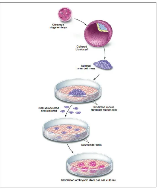
Figure 1.2 Tissue culture methods of ESCs...5
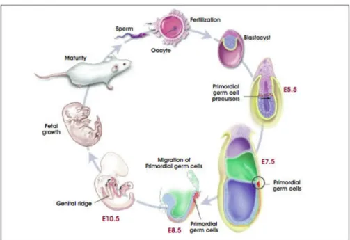
Figure 1.3 The role of primordial germ cells in life cycle ...8
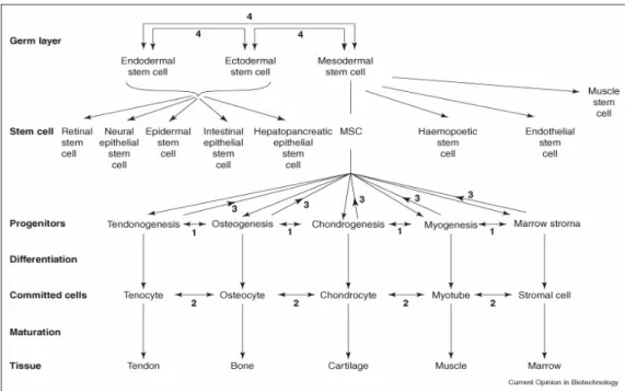
Figure 1.4 The differentiation of MSCs...12
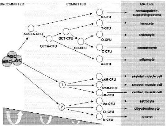
Figure 1.5 The differentiation and plasticity of MSC ...15
Figure 1.6 The conservation of the apoptotic mechanisms between species...18
Figure 1.7 Different mediations of the apoptotic pathways...19
Figure 1.8 Mitochondrial regulation of apoptosis ...21
Figure 1.9 Classification of the Bcl-2 family members...22
Figure 3.1 The isolation of the bone marrow of the rats...31
Figure 3.2 The cycle numbers and conditions of the PCRs...40
Figure 4.1 Cell number comparisons of the 9th day cell cultures...48
Figure 4.2 Cell number comparisons of the 14th day cell cultures...49
Figure 4.3 The number of colonies on the 9th day of the cell culture...50
Figure 4.4 The number of colonies on the 14th day of the cell culture...51
Figure 4.5 Normal and normal+estrogen treated MSC colonies on the 14th day of cell culture...52
Figure 4.6 Ovx and ovx+estrogen treated MSC colonies on the 14th day of cell culture ...52
Figure 4.7 A sample colony from the TUNEL assay ...53
Figure 4.8 The apoptotic cell ratios on the 9th day cell culture ...54
Figure 4.10 The isolation of mRNA and the optimization of PCR cycles ...57
Figure 4.11 The agarose gel picture of PCR products of the bcl-2 family of genes ..58
Figure 4.12 The expression graphs of bcl-2 family of genes on the 9th day cell culture groups...58-59 Figure 4.13 The expression graphs of bcl-2 family of genes on the 14th day cell culture groups...60-61 Figure 4.14 Expression of Bcl-2 on 9th day and 14th day culture groups ...63
Figure 4.15 Expression of Bcl-xL on 9th and 14th day culture groups ...64
Figure 4.16 Expression of Bak on 9th and 14th day culture groups ...65
Figure 4.17 Expression of Bax on 9th day and 14th day culture groups...66
Figure 4.18 CD90 expression in MSC colonies...67
Figure 4.19 Negative sample of MSC colonies...67
Figure 4.20 Bcl-2 expression on the 9th day of cell culture ...69
Figure 4.21 Bcl-xL expression on the 9th day of cell culture ...69
Figure 4.22 Bak expression on the 9th day of cell culture ...70
Figure 4.23 Bax expression on the 9th day of cell culture ...70
Figure 4.24 Bcl-2 expression on the 14th day of cell culture ...71
Figure 4.25 Bcl-xL expression on the 14th day of cell culture ...71
Figure 4.26 Bak expression on the 14th day of cell culture ...72
ABBREVIATIONS
AIF Apoptosis Inducing Factor AML Acute Myeloid Leukemia AP1 Activator Protein 1
APAF1 Pro-Apoptotic Protease-Activating Factor-1 ASC Adult Stem Cell
bFGF Basic Fibroblast Growth Factor
bp Base Pairs
BH Bcl-2 homology
BMP4 Bone Morphogenic Protein 4 BSA Bovine Serum Albumin
cDNA Complementary Deoxyribonucleic Acid ddH2O Double distilled water
DEPC Diethylpyrocarbonate dH2O Distilled Water
DIABLO Direct IAP binding protein with low pI DMEM Dulbecco’s Modified Eagle Medium DNA Deoxyribonucleic Acid
DNase Deoxyribonuclease
EB Embryoid Bodies
EGC Embryonic Germ Cells EGF Epidermal Growth Factor ER Estrogen Receptor
ERE Estrogen Response Elements ESC Embryonic Stem Cell GSC Germ-line Stem Cell HGF Hepatocyte Growth Factor HSC Hematopoietic Stem Cell IAP Inhibitor of Apoptosis
µg Microgram
µl Microliter
LIF Leukemia Inhibitory Factor
mM miliMolar
M Molar
MEF Mouse Embryonic Fibroblast MetOH Methyl Alcohol
MSC Mesenchymal Stem Cell NaCl Sodium Chloride
NGF Nerve Growth Factor OD Optical Density
OVX Ovariectomy
PBS Phosphate Buffered Saline PCR Polymerase Chain Reaction PFA Paraformaldehyde
PGC Primordial Germ Cell RNA Ribonucleic Acid
RT Room Temperature
RT-PCR Reverse-Transcriptase Polymerase Chain Reaction SDS Sodium Dodecyl Sulfate
Smac Second Mitochondria-Derived Activator of Caspases TGF-ß1 Transforming Growth Factor-Beta 1
1. INTRODUCTION
In the last century, the lifespan of humans has increased due to better life conditions. Advanced technology led to progressive research results for improved clinical applications and treatments. This longer life phenomenon has brought together the concept of a better life quality, since the body functions and health conditions decline with time. The recent research areas mainly focus on different intrinsic and extrinsic factors, which might help in the treatments of diseases and disorders which are results of a longer life time; such as cancer, osteoporosis, stroke and heart attack, in addition to neurodegenerative diseases like Parkinson’s disease.
In this respect, estrogen which is one of the most crucial hormones influencing the life quality of both men and women is a strong candidate in the maintenance of life standards. It is mainly responsible for female sexual features such as breast development and the menstrual cycle, but it is also involved in the regulation of several other mechanisms. Estrogen has an important effect on blood vessels, neurotransmitter systems and other aspects of the central nervous system (http://www.isoflavones.info/estrogen.php), sexual behaviour, bone structure, cognitive function (Simpson, 2003), and spermatogenesis (Simpson et al, 1999). When the estrogen levels decrease and menopause starts in women, osteoporosis and arteriosclerosis become the main problems due to estrogen’s role in bone mineralization and prevention of osteoporosis. After menopause, osteoporosis is more common in women than in men. Estrogen also decreases the possibility of becoming an Alzheimer’s patient (Yaffe et al, 1998). Alzheimer’s disease incidence is greater among women than among men mainly due to the decrease in the levels of estrogen after menopause (Simpson et al, 1999). Coronary heart disease increases double to threefold in women after menopause and it increases even more in case of the removal of ovaries prior to menopause (Skafar et al., 1997). These data drive attention to the importance of estrogen and due to this reason doctors tend to prescribe hormone based products as therapies for menopause
(http://www.isoflavones.info/estrogen.php). This indicates that estrogen has a physiologic significance throughout adult life and our understanding of its importance should not be limited only to the reproduction system.
Stem cells, which are an evolving research topic, are another future hope for therapeutic applications. They are important for living organisms since they carry a potential for differentiating into a wide variety of cells under certain circumstances. Stem cells give rise to multiple specialized cell types which make up the heart, lung, skin, and other tissues; besides replacing cells that are damaged (http://stemcells.nih.gov). Stem cells can also be used in regenerative medicine, gene therapy, tissue repair, and patient specific stem cell therapy (Ivanova et al, 2002). There are many disorders, injuries, and diseases which can be cured or at least has the potential to be cured by the use of stem cells; such as Alzheimer’s disease, spinal cord injuries, Parkinson’s disease, diabetes, Huntington’s disease, and different types of cancer (Fuchs et al, 2004). Each day new discoveries are being made on the subject.
Estrogen and stem cells, both being key factors in the preservation of life standards, are related with each other. Considering the protective effect of estrogen against severe heart, bone, and neuronal diseases combined with the refining and repairing characteristics of stem cells; they arefuture promise for many diseases. To understand the mechanism of their interaction is vital for obtaining the maximum efficiency from this collaboration. Apoptotic pathways, which are conserved throughout evolution, are strong candidates for this cooperation due to their estrogen responsive genes and their role in stem cell maintenance.
In this context, first stem cells will be explained with their features, types, and importance. Then the next part of the thesis will be based on cell death, apoptosis, and their mechanism of regulation. Finally estrogen will be explained together with its effects on stem cells, apoptotic molecules, and its role in regulating stem cell maintenance. Particularly MSCs which are vital in the formation of several tissues
1.1. Stem Cells
Stem cells are unspecialized cells which are capable of self-renewal and give rise to differentiated cells (Till and Mcculloch 1961). It is still not clear how they remain undifferentiated in their microenvironment. The internal signals which are regulated by genes, and the external signals which consist of secreted factors, and interactions among cells are the main factors stimulating the differentiation of stem cells. They are found in various sources such as the embryo, bone marrow, blood, cornea and retina of the eye, brain, skeletal muscle, dental pulp, liver, skin, lining of the gastrointestinal tract, and pancreas. Although self-renewal and differentiation to other cells are their common features; stem cells vary in their potential to differentiate, duration and pathways of self-renewal, places they are mostly found in, and division properties (Morrison et al, 1997).
Stem cells can be classified due to their source of origin and their potential to differentiate to different cell types. There are three main types of stem cells: ESCs, GSCs, and ASCs. When the oocyte is fertilized, it becomes a zygote which has the capacity to give rise to the embryo and extraembryonic membranes. The zygote is the only totipotent cell, which can differentiate to any type of cell and form the embryo. When the embryo becomes 4-5 days old, ESCs are derived from the inner cell mass of the embryo (Brook and Gardner, 1997). They are pluripotent and can give rise to cells from all three germ layers: endoderm, mesoderm, ectoderm (Thomson et al, 1998). GSCs are found in the gonadal ridge of the 5 -10 week fetus as primordial germ cells. They are pluripotent when cultured in vitro and form EGCs (Xi et al, 2005). ASCs on the other hand are found in specific tissues and organs. They are multipotent and give rise to more than a single cell type but are not capable of differentiating into tissues of all germ-layers (Figure 1.1) (Serakinci and Keith, 2006).
Figure 1.1 The differentiation of cells from the zygote (http://stemcells.nih.gov)
1.1.1. Embryonic Stem Cells
ESCs were first isolated from the inner cell mass of the mouse in 1981 by Kaufman and Martin, and from humans in 1998 by Thomson et al. ESCs are not embryos themselves but they can form cells from all three germ-layers and undergo an unlimited number of symmetrical divisions without differentiating in vitro (Burdon et al, 2002). In order to generate cultures of mouse and human ESCs, the inner cell mass is removed from the trophectoderm and is transferred to the top of MEFs on the culture medium. These MEFs which are called feeder layer are
and human ESCs can be grown both with and without a feeder layer (Xu et al, 2001; Reunbinoff et al, 2000). A single ESC can give rise to a colony of genetically identical cells, or clones under appropriate conditions (Sell, 2003) (Figure 1.2).
ESCs can stay undifferentiated for a long period of time in vitro, and there are several examples to prove the pluripotentiality of ESCs. When mouse ESCs derived from one blastocyst are injected to another blastocyst and is transferred to the uterus of a pseudopregnant mouse in vivo, chimeras form which are a mixture of tissues and organs derived both from the host and the donor blastocyst (Prelle et al, 1999). Also when ESCs are injected into adult immune-deficient mice, they develop into tumors called teratomas which contain cells from all three germ layers (Martin, 1981). In vitro when the culture conditions are changed, ESCs form embryoid bodies (EB) which have a similar structure to teratomas. They form large structures which contain partially differentiated cells from all three germ layers in a disorganized manner (Evans and Kaufman, 1981). Both mouse and human ESCs express oct-4 which is an important gene in the maintenance of pluripotency. Also with the induction of certain growth factors, ESCs differentiate into different types of cells. In order to induce ectodermally derived cells: retinoic acid, EGF, BMP4, and bFGF are added; to induce mesodermally derived cells: activin-A, and TGF-ß1 are added; and in order to induce differentiation into all three germ layers including the endoderm: HGF, and NGF are added to culture (Bishop et al, 2002). These indicate that ESCs are pluripotent and have the potential of contributing to the formation of cells from all germ layers both in vitro and in vivo.
ESCs also have high telomerase activity, which adds telomere repeats to the ends of chromosomes resulting in long telomeres (Armstrong et al, 2000; Xu et al, 2001). They have stable karyotypes, and X inactivation does not occur in the undifferentiated ESCs (Reunbinoff et al, 2000). Unlike differentiated cells, ESCs do not require any external stimulus in order to initiate DNA replication. ESCs lack the G1 checkpoint in the cell cycle and spend most of their time in the S phase of the cell cycle synthesizing DNA. When they start to differentiate, the G1 phase of the cell cycle becomes longer with the increase in Cyclin D expression and the rate of cell division slows (Burdon et al, 2002).
development. Understanding the genetic and molecular control mechanisms behind the stem cell regulation may provide an understanding on how genetic and growth abnormalities arise and suggest new strategies and methods for their therapy.
In addition to all the positive aspects of ESCs, there are several handicaps in their research and applications. Since there are many different cellular pathways regulating them, it is hard to figure out the interactions between them. ESCs can not give rise to a full embryo but the fact that they are isolated from embryos creates problems, therefore there are ethical problems arising. In addition, when injected to immunodeficient mice, they form tumors. This indicates that due to their high differentiation potential, they can form any type of cell so they have to be controlled very strictly and all the factors affecting the differentiation of ESCs should be figured out before using them in any clinical application. Also ESCs have a potential to cause immune rejection, which is another obstacle. Until the big gaps are filled in the area of stem cell regulation and differentiation besides the ethical issues being solved, ESCs can not be used in therapeutic applications.
1.1.2. Embryonic Germ Cells
EGCs were first identified in 1998 by John Gearheart and they were cultured from PGCs obtained from the gonadal ridge and mesentery of the 5th to 9th week of fetal tissue (Figure 1.3). They are pluripotent cells and express oct-4 besides other pluripotency markers similar to ESCs, and they have the capacity for long-term self-renewal. In addition they have a normal stable karyotype (Shamblott et al, 1998). The location and maintenance of germ-line stem cells are among the most studied areas and are clearly identified. Especially Drosophila is the best studied model in this area both in male and in female (Gilboa and Lehmann, 2004).
Figure 1.3 The role of primordial germ cells in life cycle (http://stemcells.nih.gov/info/scireport/appendixA.asp)
PGCs are diploid germ cell precursors which transiently exist in the embryo before they are committed as germ cells. In order to obtain EGCs, PGC cultures are grown in fetal bovine serum supplemented media. The PGCs are plated on a feeder layer consisting of STO fibroblasts which are non-dividing. They are cultured in a growth medium which includes the cytokine, LIF, bFGF, and forskolin. At the end of three weeks, the PGCs form dense, multilayered colonies of cells resembling EGCs. Although both are pluripotent, cultures derived from embryoid bodies generated from human EGCs have less capacity for proliferation compared to ESCs. EGCs will proliferate for 40 population doublings while human ESCs can proliferate for two years through 300 population doublings or even 450 population doublings. Another major difference between the pluripotency of ESC and EGC is that EGC do not form teratomas when injected to immunodeficient mouse (Pedersen, 1999).
Although both are pluripotent, EGCs do not have the same proliferation and differentiation capacity as ESCs do, which makes EGCs less appealing for
immunosuppressed mouse, and they do not participate in the formation of chimeras in viv0; which suggest that EGC’s differentiation potential is not as wide as ESC’s (Shamblott et al, 1998). In addition there is not much known about their telomerase activity either, which might be a reason why their doublings are shorter than ESCs (http://stemcells.nih.gov/).
1.1.3. Adult Stem Cells
In the pioneering experiments with ASCs in 1940-1950’s, the researchers were able to repopulate the blood cells of a mouse by the infusion of bone marrow cells from a different mouse. In 1960; two different kinds of stem cell population have been identified in the bone marrow; hematopoietic stem cells (HSCs) which form all types of blood cells and bone marrow stromal cells which generate bone, cartilage, fat, and fibrous connective tissue. In the 1990’s neural stem cells were discovered which are able to generate the brain's three major cell types: astrocytes, oligodendrocytes, and neurons.
ASCs are found in tissues and organs, and their primary role is to maintain homeostasis. They are important for the organ or tissue to fulfill their required task besides supplying them with the sufficient amount of stem cells for a life-time (Serakinci and Keith, 2006). ASCs are mostly found in the bone marrow, blood, cornea and retina of the eye, brain, skeletal muscle, dental pulp, liver, skin, the lining of the gastrointestinal tract, and pancreas. They usually divide to generate progenitor or precursor cells, which then give rise to specialized cells with specific shapes and functions (Weissman, 2000).
The number of ASCs is rare compared to other types of cells and they do not divide very often unless there is a stimulus such as tissue injuries or diseases. ASCs are scattered throughout the tissues of the mature organism and have very different roles depending on their local environment. Currently, cell surface markers and observations on stem cell differentiation patterns in test tubes and culture dishes are being used for the characterization of adult stem cells. It is also hard to expand ASCs
in culture in an undifferentiated state. Many factors including growth factors, extracellular matrix contacts, cell-cell interactions, and intrinsic cell kinetics (Sherley, 2002) effect their maintenance and differentiation.
Most of the ASCs are stored in specific cell compartments called niches, which are the microenvironments where ASCs are located with the same type of stem cells and differentiated cell types. These niches provide an extracellular matrix, adherens junctions, and integrins besides allowing physical interactions between cells. This enables stem cells to stay in an undifferentiated state and give rise to differentiated cell types when necessary. Niches are crucial by means of affecting the properties of stem cells but are not irreversible. They are also effective in the determination of symmetric division (Fuchs et al, 2004). In addition to differentiating into cells which they are programmed to, ASCs may also form the specialized cell types of other germlayers, which is known as plasticity (Horwitz, 2003). If the mechanisms beneath adult stem cell plasticity can be identified and controlled, existing stem cells from different sources might be used in the repair of diseased tissues and cells of other origins.
The most commonly studied ASCs are HSCs, MSCs, neural stem cells, epithelial stem cells, and skin stem cells. HSCs give rise to red blood cells, B lymphocytes, T lymphocytes, natural killer cells, neutrophils, basophils, eosinophils, monocytes, macrophages, and platelets. MSCs give rise to osteocytes, chondrocytes, adipocytes, and other kinds of connective tissue cells such as those in tendons. Neural stem cells in the brain give rise to three major cell types: nerve cells (neurons) and two categories of non-neuronal cells: astrocytes and oligodendrocytes. Epithelial stem cells in the lining of the digestive tract give rise to absorptive cells, goblet cells, paneth cells, and enteroendocrine cells. Skin stem cells are found in the basal layer of the epidermis and at the base of hair follicles. They give rise to keratinocytes, hair follicle, and epidermis (http://stemcells.nih.gov/).
compared to the available supply. ASCs offer the possibility of a renewable source for replacement of cells and tissues to treat several diseases. ASCs are potential future treatments for various diseases such as cardiac disorders, diabetes, Alzheimer disease and many others (http://stemcells.nih.gov/).
On the other hand there are still many unknowns and questions to be answered such as how they stay undifferentiated in a differentiated environment, the signals regulating their differentiation and proliferation, the stimuli leading them to sites of injury, and how they can be increased in number in order to reach an adequate number to heal injuries. In addition, due to the large set of unknowns in this area, it is hard to identify, isolate, and maintain ASCs (Badge, 2001). Also there are big gaps in the detection of the differentiation factors affecting their specialization, which prevent the in vitro tissue and organ synthesis. Mainly, the signals affecting ASC self-renewal and differentiation should be revealed in order to consider ASCs for possible treatments.
1.1.3.1 Mesenchymal Stem Cells
MSCs are adult stem cells mostly found in the stromal part of the bone marrow. They were first identified in 1966 by Friedenstein and Patrakova by the isolation of bone-forming progenitor cells from the rat bone marrow (Barry and Murphy, 2004). They can self-renew as all stem cells do, and have a multilineage differentiation potential. They can differentiate into osteoblasts, chondrocytes, and adipocytes, myocytes which form the bone, cartilage, tendon, muscle, and fat. They are thought to differentiate into certain types of endodermal and ectodermal tissues as well such as neurons and endothelial cells. In addition they are a part of the hematopoietic stem cell niche (Figure 1.4) (Baksh et al, 2004; Caplan and Bruder, 2001).
Figure 1.4 The differentiation of MSCs (Raghunath et al, 2005)
Although the main source of MSCs is the bone marrow, they can be isolated from other sources as well such as the trabecular bone (Noth et al, 2002), adipose tissue, synovium, skeletal muscle, lung, placenta, deciduous teeth (Tuan et al, 2003), human umbilical cord perivascular cells (Sarugaser et al, 2004), and periosteum (Minguell et al, 2001). Among the MSCs obtained from different sources, there is not much difference by means of yield, growth kinetics, cell senescence, multi-lineage differentiation capacity, and gene transduction capacity (De Ugarte et al, 2003). Although it is known that MSCs exist in many sources, their niche and the structure of their niche is not clear. HSC and MSC niche can be thought to be the same since both of them are found mostly in the bone marrow, but this is not the case because they are very different from each other and need to remain in different environments with different signals in order to stay undifferentiated. Also MSCs are found in many different sources, so the niches of HSCs and MSCs have to be different (Baksh et al, 2004).
MSCs are studied in various model organisms such as human, canine, rabbit, rat, and mouse and it is shown that MSCs isolated mostly from the bone marrow differentiate to different types of cells both in vitro and in vivo (Barry and Murphy, 2004). The MSC isolation from every animal is from different sites of the bone marrow using different methods. In humans it is mostly from the pelvis (Digirolamo et al, 1999), tibia and femur. In larger animals it is from the same site but in rodents it is usually harvested from the mid-diaphysis of the tibia and femur (Barry and Murphy, 2004).
MSCs are a very small fraction of the nucleated cells in the bone marrow; only %0.01- %0.1 of the total population (Pittenger et al, 1999). They can be isolated and expanded in tissue culture conditions with high efficiency and they can be induced to differentiate under specific conditions. They have fibroblastic morphology and they are adherent spindle-shaped cells. Under appropriate media conditions, MSCs adhere to the culture plate leading to the formation of colonies. Usually basal mediums with serum support are used to expend MSCs in tissue culture and growth factors are added in order for MSCs to differentiate (Barry and Murphy, 2004)
MSCs have a highly variable expansive potential in subculturing. Some MSC cultures can expand more than fifteen cell doublings, while others can replicate only about four cell doublings. There are several reasons for this difference such as the procedure to harvest the marrow, the low frequency of MSCs in marrow harvests, and the age or condition of the donor. Although MSCs have a high ex vivo expansion potential, they do not loose their normal karyotype and telomerase activity. However, in extensive subcultivation, signs of senescence and apoptosis are observed (Minguell et al, 2001).
There is no in vivo marker to distinguish MSCs from other cell types but in vitro STRO-1 (Minguell et al, 2001), SB-1, SH2, SH3, SH4 (Baksh et al, 2004) identify MSCs in humans and CD71, CD90, CD106, and CD117 identify them in rodents (Mangi et al, 2003). In general, it is hard to find specific markers for MSCs since they have common characteristics with other types of cells such as endothelia,
epithelial cells, and muscle cells. It is known though that they do not express CD45, CD34, and CD14 which are HSC markers (Baksh et al, 2004), and they mostly express a large spectrum of cell adhesion molecules which are important for cell binding and homing such as integrin markers alpha1, alpha5, and beta1 (Barry and Murphy, 2004).
Although MSCs might be thought as a homogeneous population which has a multilineage potential, they are a very heterogeneous population due to the complex regulatory mechanisms which are not fully revealed at the moment. Only a small proportion of MSCs are actively proliferating, and approximately 10% of them are at S + G2 + M stages. However the majority of them are at the Go/G1 phase of the cell cycle, which suggests that MSCs have a high potential to differentiate (Conget and Minguell, 1999). There are many molecules taking part in MSC regulation such as cytokines, growth factors, adhesion molecules, and extracellular matrix components (Kratchmarova et al, 2005). The master switches regulating the transitions from stem cells to differentiated cells, and the genes active in specific differentiation patterns are still not known. The reason for the difficulty to find these regulatory genes is their temporal activity, the reaction given to inductive molecules, and the varying differentiation pathways between organisms (Baksh et al, 2004; Caplan et al, 2001). MSC markers and colony forming assay are accepted currently to be sufficient to identify MSCs.
MSCs are already used in therapies for osteogenesis imperfecta, hematopoietic recovery, bone tissue regeneration (Baksh et al, 2004), cardiovascular repair, treatment of lung fibrosis, spinal cord injury, coronary artery disease (Minguell et al, 2001), local repair, and regeneration of bone cartilage and tendon. They can form intervertebral disc cartilage, bone, cardiomyocytes, articular cartilage at knee joints, besides neurons, skin epithelia, lung, liver, kidney, intestine, and spleen (Figure 1.5) (Baksh et al, 2004).
Figure 1.5 The differentiation and plasticity of MSC (Minguell et al, 2001)
Aside from their differentiation potential and immunesuppression, there are other aspects which make MSCs valuable for therapeutics. MSCs actively inhibit T-cell proliferation and due to this reason they are considered as nonimmunogenic or hypoimmunogenic which is important in host response to allogeneic MSC therapy. The only problem of this aspect is a potential disease transmission from donor to recipient. Another important feature of MSCs is long-term homing and engraftment of implanted cells to various tissues even after the development of immunocompetence. The implanted MSCs can migrate to the specific site of injury by the help of factors that are released from the wound. This has been observed in cases like bone fractures, myocardial infarction, meniscus, and ischemic cerebral injury. Also at specific sites it has been observed that the stem cells injected differentiate according to the local signals at the local site of injury (Miguell et al, 2001). MSCs have a high potential for regenerative medicine and tissue engineering besides being used as a gene delivery method, which makes their therapeutic value
very high. Researchers are looking for different ways to use specialized cells derived from MSCs in targeting specific cancerous cells and delivering treatments that will destroy them or make them benign.
Besides these advantages of MSC in therapeutic applications, there are some unknowns like long-term effect and safety which require more toxicology studies. In addition, the efficiency is not clear. There are some evidences on homing and differentiation, but they are insufficient. Also large-scale culture, storage and distribution are important for their applications of MSCs (Miguell et al, 2001).
1.2. Cell Death and Apoptosis
Cell death occurs through two main pathways: necrosis which is accidental cell death and apoptosis which is programmed cell death. These two pathways vary by means of their morphological and biochemical features besides the stimuli to induce them. Necrosis is non-programmed cell death which occurs abruptly when the cell is damaged. The cells swell, organelles dissolve, the plasma membrane ruptures, and the cytoplasmic content gets released which causes an inflammatory response (Desagher and Martinou, 2000). Apoptosis on the other hand, is a gradual and programmed cell death. The cells undergo morphological changes such as nuclear and cytoplasmic hypercondensation, cell shrinkage, plasma membrane blebbing, DNA fragmentation, and cleavage of chromosomes into nucleosomes. Apoptotic bodies form at the end of this process which contains components of dying cell and are removed by phagocytosis preventing the release of cytoplasmic and nuclear components to the extracellular space (Desagher and Martinou, 2000; Shi, 2001). Apoptosis is crucial for homeostasis and is involved in the regulation of several pathways.
genetic defaults or threaten the organism’s survival (Ashkenazi and Dixit, 1998). It can be triggered by external and internal death signals and it participates in diverse mechanisms such as embryonic development, tissue morphology determination, and creation of the neural and vascular network. The regulation of apoptosis is complicated and since it is conserved throughout evolution, is preserved very strictly. Any disruption in the apoptotic pathways cause many disorders such as autoimmune diseases, neurodegenerative diseases like Alzheimer’s disease, cancer, viral infections, osteoporosis, and AIDS (Green and Martin, 1995; Thompson, 1995).
The apoptotic regulation pathway was first discovered in the nematode Caenorhabditis Elegans (Ellis and Horvitz, 1986). The apoptotic interactions cause the death of approximately 131 cells out of 1090 somatic cells at different times and different locations during the development of C. Elegans (Steller, 1995). The main apoptotic genes in C. Elegans are ced4, ced3, and ced9. CED-4 binds to CED-3 promoting the activation of CED-3, while CED-9 binds to CED-4 inhibiting the activation of 3 (Hengartner and Horvitz, 1994). Under normal conditions CED-9 stays bound to CED-4 keeping CED-3 inactive, but apoptotic stimuli cause CED-CED-9 to dissociate by EGL-1 which allows the activation of CED-3 leading to apoptosis (Shi, 2001). A similar system also exists in other organisms. The mammalian homolog of CED-4 is Apaf-1, CED-3 is Caspase 3, Bid and Bcl-2, which are members of Bcl-2 family of proteins are similar to CED-9 and EGL-1 (Figure 1.6) (Zou et al, 1997; Shi, 2001).
Figure 1.6 The conservation of the apoptotic mechanisms between species (Shi, 2001)
1.2.2. Regulation of Apoptosis
Since apoptosis takes part in such a variety of physiological and non-physiological conditions, understanding the molecular mechanism of the regulation of apoptosis is important. Apoptosis can be induced by either intrinsic pathways which are activated as a result of intracellular stress like DNA damage, or extrinsic pathways which are triggered by extracellular factors acting on the death receptors such as Fas and TNFR-1 (Shi, 2001). In the intrinsic or mitochondrial pathway; cytochrome c is released, apoptosome which consists of cytochrome c is formed, and APAF1, pro-caspase-9, and caspase-9 are activated. Endoplasmic reticulum stress triggers the ER-specific pathway which results in the activation of caspase-12 in
activated by extracellular ligand binding to death receptors and results in either caspase activation or need further amplification through the mitochondrial pathway, according to the cell type. Fas and TNFR1 can be given as examples of the most commonly studied death receptors. (Figure 1.7)(Holcik and Sonenberg, 2005; Liston et al, 2003; Gebauer and Hentze, 2004; Ashkenazi and Dixit, 1998)
Figure 1.7 Different mediations of the apoptotic pathways (Holcik and Sonenberg, 2005)
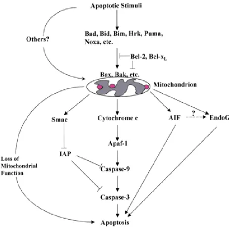
1.2.2.1. Mitochondrial Apoptosis
Mitochondrial apoptosis which is also the intrinsic pathway, is crucial for homeostasis and animal development. After receiving apoptotic stimuli, cytochrome c is released to the cytoplasm from the intermembrane space of mitochondria. The release of cytochrome c triggers the activation of a specific set of proteins called caspases. Caspases are a group of cysteine proteases which cleave selected proteins
at their aspartate residues. Fourteen caspases are identified in mammals and at least eight of them have critical roles in apoptotic regulation (Budihardjo et al, 1999). Caspases are categorized as the initiator caspases (caspase-2, caspase-8, caspase-9, caspase-10), and the effector caspases (caspase-3, caspase-6, caspase-7). The effector caspases are activated by initiator caspases while initiator caspases activate themselves. Initiator caspases are strictly regulated requiring an assembly of multicomponent complex since they activate a downstream of caspases. Caspase activity is inhibited by the conserved IAP family of proteins. Eight different mammalian IAPs which include XIAP, c-IAP1, c-IAP2, and ML-IAP/Livin have been identified (Shi, 2002). They target caspase-9, caspase-3 and caspase-7 but not other caspases (Holcik and Sonenberg, 2005).
After the release of cytochrome c from mitochondria, it binds to Apaf-1 and activates it by changing its confirmation (Jiang and Wang, 2000; Purring et al, 1999). This complex then binds to dATP or ATP, and forms a multimeric complex which is called apoptosome which activates procaspase-9 (Saleh et al, 1999) After being activated, caspase-9 forms a holoenzyme with the apoptosome, targeting primarily procaspase-3 which is one of the most important effector caspases (Jiang and Wang, 2000; Shi, 2001). Active caspase-3 cleaves a wide range of proteins including caspase-2, caspase-6, DFF45, DNA-dependent protein kinase, and nuclear lamin (Thornberry and Lazebnik, 1998). The degradation of DFF45 stops inhibiting DFF40 (CAD), which in return degrades chromatin into nucleosomal fragments, one of the main characteristics of apoptosis.
In cells which have not received any apoptotic stimuli, the enzymatic activity of mature caspases is inhibited by IAPs (Deveraux and Reed, 1999; Miller, 1999). However this inhibition must be supressed in cells which undergo apoptosis and this is regulated by smac also called DIABLO, which is a 25-kD mitochondrial protein (Verhangen et al, 2000). It is synthesized in the cytoplasm and is released together with cytochrome c to the cytosol upon apoptotic stimuli. While cytochrome c activates Apaf-1 and caspase-9, smac interacts with IAP enabling the activation of
Bcl-2 family members which effect cytochrome c release and apoptotic regulation (Adams and Cory, 1998; Adams and Cory, 2001). The importance of this pathway is clear by the observation that Apaf-1 is frequently inactivated in cancers such as malignant melanoma (Figure 1.8) (Soengas et al, 2001).
Figure 1.8 Mitochondrial regulation of apoptosis (Wang, 2001)
1.2.2.2. Bcl-2 Family of Proteins
Bcl-2 family of proteins is among the best characterized protein family which takes place in the regulation of apoptosis. It consists of both pro-apoptotic and anti-apoptotic members. They function by forming homo- and heterodimers. The rate of
the pro-apoptotic to anti-apoptotic molecules determines the susceptibility to death stimuli (Chao and Korsmeyer, 1998).
The prototype of this family, Bcl-2, was first identified in B-cell leukemia cell line as an oncogene which inhibited cell death rather than promoting proliferation (Cory and Adams 2002, Bakhshi et al, 1985; Cleary and Sklar, 1985). The anti-apoptotic activity of Bcl-2 was proven first by inducing apoptosis with IL-3 deprivation (Vaux et al, 1988) and then it was shown that Bcl-2 prevented apoptosis induced by several stimuli such as serum deprivation, chemotherapeutic agents, and heat shock (Tsujimoto, 1989). In higher eukaryotes at least 20 homologs of Bcl-2 have been identified, sharing at least one conserved BH domain (Vaux et al, 1992; Hengartner and Horvitz, 1994). These can be categorized into three groups: Bcl-2 like survival factors, Bax-like death factors, and BH-3 only death factors (Figure 1.9).
The pro-survival family consists of Bcl-2 and its closest homologs Bcl-xL,
and Bcl-w besides Mcl-1, A1/Bfl-1, and Diva. This is a new class of proto-oncogenes that block cell death without promoting cell proliferation and it also has a role in cell cycle (Conradt and Horvitz, 1998). Bcl-2 prevents cell death by inhibiting free radical production, suppressing caspase activation, regulating calcium sequestration, and by preventing the pro-apoptotic factors from inducing apoptosis (Merry and Korsmeyer, 1997). Bcl-2 deficient T cells show increased apoptosis following cell cycle progression. Bcl-2 is widely expressed during embryogenesis but it decreases and becomes much more restricted postnatally in the nervous system (Susin et al, 1996). Bcl-2 may be important in maintaining cell survival during interactions between epithelium and mesenchyme (Thornberry and Lazebnik, 1998; Tsujimoto, 1989). In addition, Bcl-2 and Bcl-xL have a reciprocal pattern of
expression in B cell compartments. Bcl-2 itself is required for the survival of kidney and melanocyte stem cells and mature lymphocytes. Bcl-2 and Bcl-xL prevent
apoptosis either by segmenting caspases or by preventing the release of mitochondrial apoptogenic factors such as cytochrome c and AIF into the cytoplasm. They contain BH domains BH1, BH2, BH3, BH4, and a hydrophobic C-terminal tail which functions as a membrane anchor (Janiak et al, 1994). BH1-3 form a hydrophobic groove, with which BH3-only death factors interact through their BH3 domain, while the BH4 domain stabilizes this pocket (Zha et al, 1996; Luo et al, 1998). The core three-dimensional structure is well conserved between Bcl-xL, Bcl-2,
and Bcl-w. They are targeted to the outer mitochondrial membrane, endoplasmic reticulum, and nuclear envelope. It is becoming clear that every nucleated cell needs the protection of at least one Bcl-2 homologue, and these homologs regulate tissue homeostasis. Bcl-2 is required for kidney survival, melanocyte stem cells, and mature lymphocytes; Bcl-xL for neuronal and erythroid cells; and Bcl-w for sperm
progenitors in adult mice (Cory and Adams, 2002).
The Bax-like pro-apoptotic death factors consist of Bax, Bak, and Mtd/Bok. These pro-apoptotic members lack the BH4 domain. The presence of either Bax or Bak, which are thought to function mainly at the mitochondria, seem to be crucial for apoptosis in many cell types since most death stimuli converge on their activation
(Gross et al, 1999; Vander Heiden and Thompson, 1999). They stimulate the release of caspases by heterodimerization and also by inducing the release of mitochondrial apoptogenic factors into the cytoplasm by acting on mitochondrial permeability transition pore, which leads to caspase activation (Tsujimoto, 1998). Bax heterodimerizes with Bcl-2, and homodimerizes with itself. When Bax is overexpressed in cells, apoptotic death in response to a death signal accelerates, but when Bcl-2 is overexpressed, it heterodimerizes with Bax and apoptosis is repressed (Kluck et al, 1997). Thus, the ratio of Bcl-2 to Bax is important in determining the tendency to go through apoptosis. In healthy cells Bax is a cytosolic monomer and is either loosely attached to the mitochondrial membrane or remains in the cytosol. Upon receiving death stimulus, cytosolic Bax goes through a conformational change and is oligomerized and translocated to the mitochondria where it becomes an integral membrane protein (Wolter et al, 1997). Also with the induction of the death stimuli the membrane associated Bax changes the exposition of its N-terminal BH3 domain (Zong et al, 2001) whereas in the absence of death signals the N-terminal domain does not allow the transmembrane anchor to function. This explains the reason for Bax failing to insert in the mitochondrial membrane in healthy cells. In addition, when Bax is activated, it binds to and antagonizes anti-apoptotic Bcl-2 proteins (Puthalakath et al, 1999). Bak on the other hand is an oligomeric integral mitochondrial membrane protein even in healthy cells, but changes conformation during apoptosis as well to form larger aggregates (Griffiths et al, 1999). The Bax-like proteins act in mitochondrial disruption.
The BH-3 only group consists of Bad, Bid, Bik/Biklk, Bim, Hrk, and Bnip3. They have only the BH3 domain which enables dimerization and is sufficient for their killing (Shi, 2001). They induce apoptosis mostly in response to developmental signals or intracellular damage (Huang and Strasser, 2000). They are upstream sensors and mediators of apoptotic cell death. BH3 domains of several BH3-only proteins either directly activate Bax/Bak proteins or bind to the hydrophobic pocket of anti-apoptotic Bcl-2 family members (Letai et al, 2002). The programmed death
eight or more mammalian BH3 proteins homologs allow a more-refined control over cell death.
There are a few models on how Bcl-2 family of proteins work. In one of the most accepted ones Bax and Bak are responsible for the release of mitochondrial proteins and are negatively regulated by Bcl-2 and Bcl-xL (Korsmeyer, 2000,
Antonsson et al, 2000). Bax forms a channel either by itself or with the help of other factors and in apoptotic cells this channel is responsible for the release of the proteins from the intermembrane space of mitochondria to the cytosol. However the binding of Bcl-2 to Bax closes this channel. Upon apoptotic stimuli, Bid or PUMA is activated and interacts with the Bcl-2 protein to relieve the inhibition of the Bax-dominated channel. This model is in consistency with many biological and biochemical observations. This model also differentiates the functions of the two apoptotic subfamilies, the BH3-only (Bid or PUMA) and the Bax-like pro-apoptotic members. Bcl-2 family of proteins control the permeability of mitochondria but it is not clear whether they are the primary components of the pores or they regulate the open-close status of the pores. How the three subfamilies of Bcl-2 proteins work in vivo is also unclear (Shi, Bcl-2001). In the overall picture though, the Bcl-2 family of proteins act as a critical life–death decision point within the common pathway of apoptosis.
1.3. Estrogen
Estrogen is a hormone which is produced by the ovaries and testes. It stimulates the development of secondary sexual characteristics, induces menstruation, and regulates growth, differentiation, cell proliferation, metabolic activities, reproduction, homeostasis, cardiovascular health, bone integrity, cognition, and behavior. It targets many organs like the brain, heart, bone, breast, uterus, and prostate. This wide range of organs, which estrogen is involved in the functioning of, makes estrogen a very important hormone for therapy and therefore it is crucial to understand the imbalances of estrogen, gene networks controlled by estrogen, and regulation of its targets (Deroo and Korach, 2006; Paech et al, 1997).
Estrogen regulation occurs through several different mechanisms but mostly it induces cellular changes through estrogen receptor (ER). ER exists in 2 forms: ERα and ERß which have different expression patterns according to the organism and organ they are found in. In this common regulation pathway estrogen diffuses to the cell, binds to an ER and results in the conformational change of the ER. Then this estrogen-ER complex binds to specific DNA sequences called estrogen response elements (ERE) directly or indirectly with AP1. The ligand which binds to ER makes a conformational change, which promotes the binding to ERE. When ERα and ERß form complexes with estrogen from an AP1 site, they signal in different ways depending on the ligand and response element, and they inhibit or activate transcription accordingly (Deroo and Korach, 2006; Smith and O’Maley, 2004). The ligand bound receptor also forms a complex with coactivator proteins which activates target gene expression through chromatin remodeling. This suggests that depending on the complex formed, ERs play different roles in gene regulation. (Weitzmann and Pacifici, 2006; Deroo and Korach, 2006; Paech et al, 1997).
Estrogen exerts its effects through its target genes. Estrogen affects hundreds of genes in different tissues and the expression profiles of these genes change with developmental stages and diseases. The coordination of these genes is mainly by estrogen but there are also other factors like type of ER, co-regulators, estrogen exposure time, and estrogen amount (Deroo and Korach, 2006; Tang et al, 2004). The regulatory groups which these genes affect can be classified as cytokines/immune response, signal transduction, cell motility/cytoskeleton regulation, growth factors/hormones, apoptosis/cell proliferation, housekeeping, nucleic acid processing, and other/unknown (Stossi et al, 2004). Due to these diverse mechanisms, estrogen responsive genes are very crucial for research and clinical purposes.
Estrogen regulation is affected by agonists and antagonists which exhibit tissue-specific estrogenic activity. The same molecule might act both as an antagonist and an agonist depending on the organism and tissue. For example
Raloxifene resembles tamoxifen and in addition it is used in the prevention of osteoporosis. These estrogen receptor modulators also affect the ER signaling (Deroo and Korach, 2006; Paech et al, 1997).
A very tight correlation has been established between estrogen and several diseases such as breast cancer, ovarian cancer, colorectal cancer, prostate cancer, endometrial cancer, osteoporosis, neurodegenerative diseases, cardiovascular diseases, insulin resistance, lupus erythematosis, endometriosis, and obesity (Deroo and Korach, 2006). However the molecular mechanisms underlying this, is not clear. Both estrogen agonists and antagonists are used for clinical purposes in estrogen regulation.
1.3.1. Estrogen, Apoptosis and Bcl-2 Family of Proteins
Estrogen regulates many mechanisms through its target genes as mentioned before and apoptosis is one of the major ones. Estrogen has a dual role on apoptosis; it can both stimulate and inhibit apoptosis depending on the type of cell. It induces apoptosis in osteoclasts but inhibits in nerve cells, endothelial cells, and epithelial cells (Spyridopoulos et al, 1997; Jo et al, 1993). The mechanism of estrogen on apoptosis is through the induction of glutathione, Bcl-2 protein, nitric oxide, inhibition of inflammatory cytokine production, and directly as an antioxidant molecule at the biochemical level (Kobayashi et al, 1997).
It was noted that apoptotic gradient and Bcl-2 protein which is anti-apoptotic were negatively correlated in the presence of estrogen, attracting the attention to Bcl-2 for estrogen regulation. Estrogen differentially regulates several pro-apoptotic and anti-apoptotic members of the Bcl-2 family of proteins. It increases Bcl-2 protein expression which has several putative estrogen responsive sites, whereas it decreases Bax expression which makes dimers with Bcl-2. This results in the increase in proliferation and decrease in apoptosis, promoting cell survival (Alkayed et al, 2001). Another anti-apoptotic family member Bcl-xL has putative ERE and estrogen
or indirectly on these molecules but Bcl-2 family of proteins have a crucial role in apoptotic regulation of estrogen (Bynoe et al, 2000; Pike, 1999).
1.3.2. Estrogen Effects on Osteocytes and MSCs
Among the target tissues and organs of estrogen, bone marrow is one of the most studied ones. It is known that osteoblasts, osteocytes, osteoclasts, bone marrow stromal cells, T cells, B cells and most other cells in human and mouse bone marrow besides MSCs, express both ERα and ERβ (Weitzmann and Pacifici, 2006; Komm et al, 1988). Gender, age, and cell type are important determinants of ER isoform expression in the bone marrow. It is known that estrogen stimulates osteoblast differentiation, increases bone marrow proliferation, and is involved in the decrease of apoptosis in osteoblasts. Also it is found that there is an increase in osteocyte apoptosis directly or indirectly in ovx animals and the addition of estrogen decreases the apoptosis rate (Tomkinson et al, 1998) In addition, there is an increase in the proliferation rate and ovariectomization decreases proliferation while the addition of estrogen reverses this effect (Fowler et al, 2005). In MSCs, estrogen increases the proliferation rate, reduces apoptosis, upregulates ERα expression, and downregulates ERβ expression which is thought to be a repressor (Zhou et al, 2001). The exact mechanism which estrogen exerts these effects is not known but osteocyte apoptosis is found to be reversely correlated with the skeletal distribution of Bcl-2 protein (Stevens et al, 2000; Tomkinson et al, 1998). This suggests that the proliferative and anti-apoptotic effect of estrogen on osteocytes and MSCs might be through the expression of the Bcl-2 family of pro-apoptotic and anti-apoptotic proteins.
2. AIM OF STUDY
MSCs are potential therapeutic agents which are applicable in tissue engineering, gene delivery, carry high potential for regenerative medicine and do not induce any immune reaction. Besides being a future promise for many disease treatments, there are also some handicaps in their applications. Since MSC number is very low in vivo, it is important to obtain a certain number of MSCs for their use. The proliferation rate and number of MSCs have to be effectively increased in the shortest time period in order for MSCs to be adequate for therapeutic regiments. Our aim is to examine the role of estrogen for obtaining MSCs in a short period of time. We also attempt to understand the mechanism of the action of estrogen.
It is known that apoptosis is among the main regulatory mechanisms in all organisms and is a conserved system which emphasizes its importance throughout evolution. The members of the Bcl-2 family of proteins are strong candidates in the regulation of MSC since some of the family members have estrogen receptors, and most of them are affected by estrogen evidenced by the presence of putative EREs. In addition, Bcl-2 proteins are among the most important families which serve both as death agonist and antagonists and have a crucial role in apoptotic balance. This makes them potential candidates in the regulation of MSCs. Bcl-2 family members can be manipulated in order to obtain more MSC in a shorter time with higher proliferative capacity and efficiency.
3. MATERIALS AND METHODS
3.1. Animals and Their Treatments
3.1.1. The Maintenance of the Animals
Adult female Spraque Dawley rats were used for the experiments. The animals were kept in the animal holding facility of the Department of Molecular Biology and Genetics at Bilkent University under controlled conditions at 22o C with 12 hour light and 12 hour dark cycles. They were provided with unlimited access of food and water. Our experimental procedures have been approved by the animal ethical committee of Bilkent University (Bil-AEC 2005/2)
3.1.2. Ovariectomization of the Animals
Animals were anesthesized with Ketamin (Ketalar, Park Davis) subcutaneously at a dose of 30mg/kg. The dorsal part was shaved with a razor, and was immobilized. After fully anesthesized, 2 cm of incision were made on both sides of the vertebrate, fat pads were removed to reach the ovaries. Ovaries at the tip of each uteri horn were tied with silk and then removed. Then the remaining fat pads were put back into their location and the skin of the animal was sutured with absorbex 4.0 (75 cm). After the operation the animals were put under a lampto keep them warm. Animals were kept under supervision until they fully recovered from the anesthesia. Ovx animals were used approximately after 2 months after the operation to ensure the total deprivationof estrogen from their bodies. Each time an ovx animal was sacrificed by cervical dislocation, another female rat of the same age which has not been operated was also sacrificed and called the “normal” group of animal.
3.1.3. Isolation of the Bone Marrow from the Animals
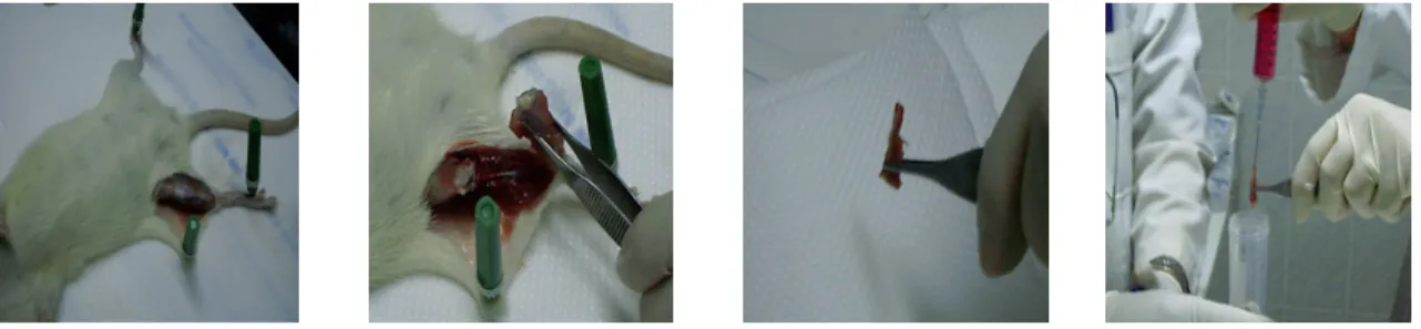
After the animals were sacrificed by cervical dislocation for the isolation of the bone marrow, the gastrocnemius and biceps regions were shaved with a razor. Then the femur and tibia were removed, and the ends of bones were cut and flushed with DMEM (Biochrom FG 0435) which contains 10% Fetal Bovine Serum (Biochrom-S0115) and 1% penicillin/streptomycin solution (Biochrom-A2213), by using a 5ml syringe (Figure 3.1). The mixed media and bone marrow were centrifuged at 4000 rpm for 3 min in order precipitate the cells of the bone marrow. Then the media was removed and the cells were washed three times with 1X PBS buffer to prepare for tissue culturing.
Figure 3.1 The isolation of the bone marrow of the rats
3.2. Cell Culture
3.2.1. Cell Number Detection with Cell Count
After the heterogenous bone marrow cells was washed and precipitated, they were suspended in 10 ml of MesenCult media and the mixture was micropipetted into a hemocytometer. The number of cells in the chamber was determined by counting under the light microscope. The cell number was calculated according to the following formula:
concentration of cells in original mixture =
(http://en.wikipedia.org/wiki/Hemocytometer)
3.2.2. MSC Culture
MSCs were cultured by MesenCult media (Stem Cell technologies) with supplement (Stem Cell Technologies), and 1% penicillin/streptomycin solution (Biochrom-A2213). Next day, the media of the tissue culture plates were changed and the majority of the non-adherent cells were removed. The media of the cells were changed in every 4-5 days, after washing with sterile 1X PBS prior to the change. The adherent cells on the 14th day of the cell culture were mostly MSCs but on the 9th day there were also adherent cells which were not MSCs particularly.
3.2.3. Estrogen Treatment
For the estrogen treatment of cells, 17 ß-estradiol (Sigma) was used, which will be referred to as “estrogen” in the following sections. The cells which will be treated with estrogen were cultured with MesenCult media at first, without estrogen. At every media change including the first day, estrogen was added to the media of the cells at a physiological concentration of 10-7 M(Zhou et al, 2001).
3.3. Standard Solutions and Buffers
DEPC-Treated ddH2O 1ml DEPC
1lt ddH2O