Is pimecrolimus cream (1%) an appropriate
therapeutic agent for the treatment of external
ear atopic dermatitis?
Güçlü Kaan Beriat
1ABCDEFG, Şefik Halit Akmansu
1AEG, Cem Doğan
1ABFG,
Eren Taştan
2BCEF, Ferda Topal
2BEFG, Bizden Sabuncuoğlu
2BEFG
1Department of Otorhinolaryngology, Ufuk University Medical School, Ankara, Turkey
2ENT Department, Ankara Training and Research Hospital, Ankara, Turkey
Source of support: Departmental sources
Summary
Background:
In recent years, pimecrolimus 1% cream has been demonstrated to reduce symptoms of atopic der-matitis in patients when applied topically.Material/Methods:
In our study we compared the therapeutic effects of local 1% pimecrolimus to 1% hydrocortisone, and to a control group in a mouse model with atopic dermatitis in the external ear canals. Atopic dermatitis was created by application of Dinitrochlorobenzene in the external ear canals of mice. The development of atopic dermatitis was detected by clinical observation score and determina-tion of total serum IgE levels. Pimecrolimus and hydrocortisone cream were topically applied to the external ear canal skin once a day for 14 days.Results:
There was no significant difference between the hydrocortisone and the pimecrolimus therapy groups, while there was a statistically significant difference between these 2 groups and the control group (p<0.05) Assessment of the clinical observation scoring carried out on the 14th day of ther-apy revealed that there was no difference between the hydrocortisone and pimecrolimus groups. Biopsies were taken on the 14th day following treatment. Tissue samples were histologicallyeval-uated; contact dermatitis was observed microscopically in the control group, but in the therapy groups only minimal evidence of contact dermatitis was found.
Conclusions:
The results of our study reveal that the therapeutic efficacy of 1% pimecrolimus was equivalent to 1% hydrocortisone treatment in the artificially developed atopic dermatitis model in external ear canals of mice. These results clearly demonstrate that 1% pimecrolimus cream can be an effective alternative therapeutic agent in cases where steroid treatment proves to be insufficient or in cases where treatment must be discontinued due to its adverse effects.key words:
external auditory canal • pimecrolimus • hydrocortisone • atopic dermatitis • albino mice
Full-text PDF:
http://www.medscimonit.com/abstract/index/idArt/882615
Word count:
3763
Tables:
2
Figures:
5
References:
47
Author’s address:
Güçlü Kaan Beriat, Ufuk University Medical School, Department of Otorhinolaryngology, Turkey,
e-mail: [email protected]
Authors’ Contribution: A Study Design B Data Collection C Statistical Analysis D Data Interpretation E Manuscript Preparation F Literature Search G Funds Collection Received: 2011.06.25 Accepted: 2011.10.14 Published: 2012.04.01 Basic ResearchPMID: 22460087
BR
B
ackgroundAtopic dermatitis (AD) is a complex eczematous, inflammatory, chronically relapsing, non-contagious, and pruritic skin disease [1,2]. The elevated allergen-specific serum Ig E levels are the most important biochemical parameters in many patients [3]. AD may occur due to various genetic and environmental fac-tors; however, its precise cause is unclear [4,5]. Environmental factors, nutrition, and hygiene play a role in AD [6]. AD now affects 10–20% of children and 1–3% of adults, and its preva-lence has increased in recent years in developed countries [7]. The skin of a patient with atopic dermatitis reacts abnor-mally and easily to irritants, food and environmental aller-gens. Itching and skin lesions like rash, discharge, desqua-mation, hyperpigmentation, and sometimes fissures are generally observed in patients with atopic dermatitis. These symptoms significantly impair the patient’s quality of life. Patients with isolated external auditory canal atopic der-matitis (EACAD) are common in otolaryngologic practice. Otolaryngologists may treat EACADs with low density hy-drocortisone creams and ointments, topical acetic acid lutions, or dilute aluminum acetate solutions (Burow’s so-lution). Although the use of these types of medication is efficacious in some patients, there are other patients who relapse or continue to have symptoms.
Long-term use of topical steroids is contraindicated because they are associated with thinning of the epidermis, decreased microvasculature and decreased keratinocyte size [8], especial-ly in the external auditory canal (EAC) and auricular skin be-cause of its very thin nature. Furthermore, higher-potency ste-roid creams must not be used on EAC skin where the skin is naturally thin; usually a lower-potency steroid is prescribed for this area. Due to inadequate therapeutic effects and common adverse effects, new treatment options are being investigated. Pimecrolimus belongs to a new class of topical immune modu-lators and was approved by the Food and Drug Administration (FDA) in 2001. It is a calcineurin inhibitor developed for the topical treatment of atopic dermatitis. Pimecrolimus inhibits T cell activation by inhibiting the synthesis and release of cy-tokines from T cells. It has been observed that pimecrolimus binds to macrophilin-12 and inhibits calcineurin. Therefore, it inhibits T cell activation by blocking the transcription of early cytokines, including that of interleukin IL-2, IL-4, IL-10 and interferon gamma. Pimecrolimus also prevents the re-lease of inflammatory cytokines and mediators from mast cells after stimulation by antigens or Ig E [9,10].
Topical pimecrolimus has been recommended in the treat-ment of atopic dermatitis by various authors [9,11,12]. We wanted to explore the probable success of this drug on EACAD. For this purpose the present study evaluated the therapeutic efficacy of 1% topical pimecrolimus compared with 1% topi-cal hydrocortisone on mice with artificially induced EACAD.
M
aterialandM
ethodAnimals
Gazi University’s Animal Research Ethics Committee ap-proved our experimental animal protocol. All procedures
performed on the mice were in accordance with the guidelines of the Gazi University Animal Research Ethics Committee.
Thirty female albino mice, 25–30 grams in weight, obtained from Gazi University Experimental Researches Animal Laboratory (GUERAL), were used. The mice were divided into 3 groups: hydrocortisone treatment group (Group 1), pimecrolimus treatment group (Group 2) and control group (Group 3), each comprising ten animals.
All mice were kept in a pathogen-free environment. They were housed in an air-conditioned animal room with a tem-perature of 22±2°C and a humidity of 60±5%. Food and tap water were provided to the animals ad libitum.
Sensitization
First, all hair on the backs of auriculae of the mice was shaved with an electric clipper and then removed with de-pilatory cream. 2,4-Dinitroklorobenzene powder (DNCB, Merck Chemical Company, Germany) was used to induce controlled dermatitis. DNCB was dissolved in acetone at a concentration of 10%. We painted the auricula and the EAC with 10% DNCB and acetone/olive oil (1:3) solution repeatedly 3 times a day for 28 days.
Drugs
Elidel cream (1% Pimecrolimus, Novartis), Eurax hydro-cortisone cream (1% hydrohydro-cortisone, Novartis), Ketalar Vial (Ketamine HCl 50 mg/ml, Pfizer), and Xylazine Hydrochloride Vial (2% Xylazine hydrochloride solution, Rompun) were used. No therapeutic agent or vehicle has been administered to the control group.
Pimecrolimus and hydrocortisone creams were used for the EACAD therapy topically on the EAC and auricular skin once a day for 14 days. Twenty-five mg/kg ketamine and 2 mg/kg xylazine were injected intramuscularly into the ceps femoris muscle for general anesthesia before the bi-opsies. A disposable sterile insulin injector (1 ml, AYSET) was used for the intramuscular injection.
Macroscopic observation
Macroscopic observing inflammation scale (MOIS) was used and erythema, edema, and ulceration were graded on a scale of 0 to 5. Clinical skin condition was defined in terms of cutaneous lesions that consisted of edema, erythema, erosion and ulceration. The scoring meth-od used in the present study (MOIS) was transformed through the modification of the method described by Hikita et al. [13].
These evaluation parameters were assessed by determining the total area of lesions on the shaved auricular area. The scoring system was as follows: 0, not detectable; 1, 0–25% of total shaved auricular skin surface; 2, 25–50% of total shaved auricular skin surface; and 3, 50–75% of total au-ricular shaved skin surface; 4, 75–100% of total auau-ricular shaved skin surface; and 5,100% of total shaved auricular skin surface.
Biopsies
Biopsies from each of the 3 groups were performed only once at the end of the treatment. Ketamine and xylazine hydrochloride were administered for general anesthesia, applied intramuscularly in the leg prior to the biopsies. The biopsies were performed with a size 11 scalpel in the shape of a full thickness triangle through 2 intersecting in-cisions at the external ear canal that widened outwards to the free edge from the center of the auricula at the exter-nal auditory caexter-nal. Intraoperative bleeding during the bi-opsies was controlled with a bipolar cautery and covered with sterile strips.
Skin changes formed in atopic dermatitis take place in the area where allergenic materials come into contact with the skin. Hence, tissue changes may not be the same in all ar-eas. In addition, performing repetitive biopsies from the same area may cause additional tissue damage and tissue changes. All these factors result in difficult follow-up of the disease through obtaining biopsy samples from the same area. Consequently, follow-up of the disease in our study by repetitive biopsies was avoided.
Histopathological observation
Specimens were obtained from the skin of the patched area, fixed with 10% neutral formalin and embedded in white paraffin for the histological observation of the skin. Serial paraffin sections, 3 µm thick, were prepared and stained with hematoxylin and eosin. For histological evaluation, the removed tissues were fixed in buffered (pH=7.4) 10% formaldehyde solution for 2 days at room temperature. Specimens were dehydrated in increasing concentrations of ethanol and were immersed in xylene until they had a clear appearance, followed by incubation in 3 changes of liq-uid paraffin at 60°C and were embedded in paraffin blocks. Sections of 3 µm were obtained by Leitz-1512 microtome (Leitz, Germany) and stained with hematoxylin and eosin. Specimens were examined under a Nikon Eclipse E600 (Nikon, Tokyo, Japan) bright field microscope and imag-es were captured using a Nikon Coolpix 5000 (Nikon) dig-ital camera attachment.
The histological evaluation was performed by 2 different histologists who did not know to which group the prepa-rations belonged. The histologists assessed tissue integrity and inflammatory findings during their analyses. They ob-served only inter-group differences whether or not there was any difference between samples.
Determination of total IgE in mouse serum
Blood was collected from the tail vein following DNCB sen-sitization period on days 1, 14 and 28, and during the ther-apy period on days 14 and 28. Serum was obtained by cen-trifugation 500×g for 10 min at 4°C and stored at 20°C until use. Total IgE levels in serum were measured by enzyme im-munoassay. The concentration of total IgE in the sera was measured by a sandwich enzyme-linked immunosorbent assay (ELISA) as previously described [14]. Total IgE lev-els in sera were measured by using an ELISA kit (Yamasa Shoyu Co., Ltd., Chiba, Japan) at the Turkish Atomic Energy Authority, Chemical Engineering Laboratories, Ankara.
Statistical analysis
Statistical analysis of total IgE levels
All the results are presented as means ± standard devia-tions for all groups. Differences were assessed using vari-ance analysis (daily total IgE levels between the groups). Post-hoc comparisons were assessed with Tukey’s test. The differences between the days of each group were assessed using the paired t test. P<0.05 was considered to be statis-tically significant.
Statistical analysis of MOIS scores
All results are presented as means ± standard deviations for all groups. Differences between the groups were as-sessed by the Kruskal Wallis test. Paired comparisons were assessed with the Mann Whitney test. Bonferroni correction was applied in order to take multiple testing into account. Differences between the days of each group were assessed using the Friedman test. Paired comparisons were assessed using the Wilcoxon Signed Ranks test after Bonferroni cor-rection for a separate analysis. The power analysis of the study revealed 100% rate.
r
esultsTotal IgE levels
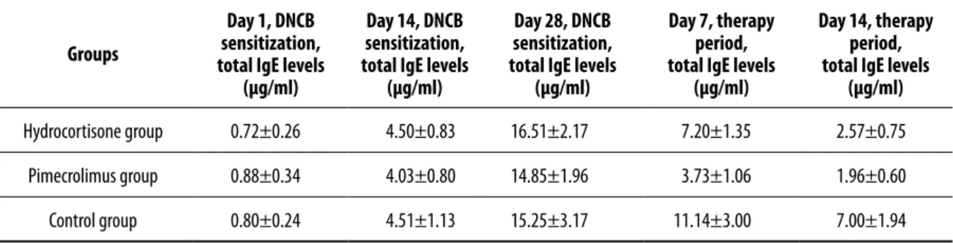
It was observed that the mean levels gradually increased dur-ing the sensitization period in all 3 groups. With the initia-tion of the treatment they started to go down in all groups and that this decrease continued until the last day of the treatment, day 14 (Figure 1, Table 1).
When the total IgE levels were analyzed it was seen that there were no differences among the 3 groups during the DNCB sensitization period.
The assessment carried out on day 7 after treatment revealed that there was a statistically significant difference among the results of the 3 groups (P<0.05). The groups’ mean levels of total IgE were, from the least to the most, Group 2 < Group 1 < Group 3. When the total IgE results were as-sessed on day 14 after treatment, there was no significant difference between Group 1 and Group 2, while there was a statistically significant difference between these 2 groups and Group 3 (P<0.05).
MOIS scores
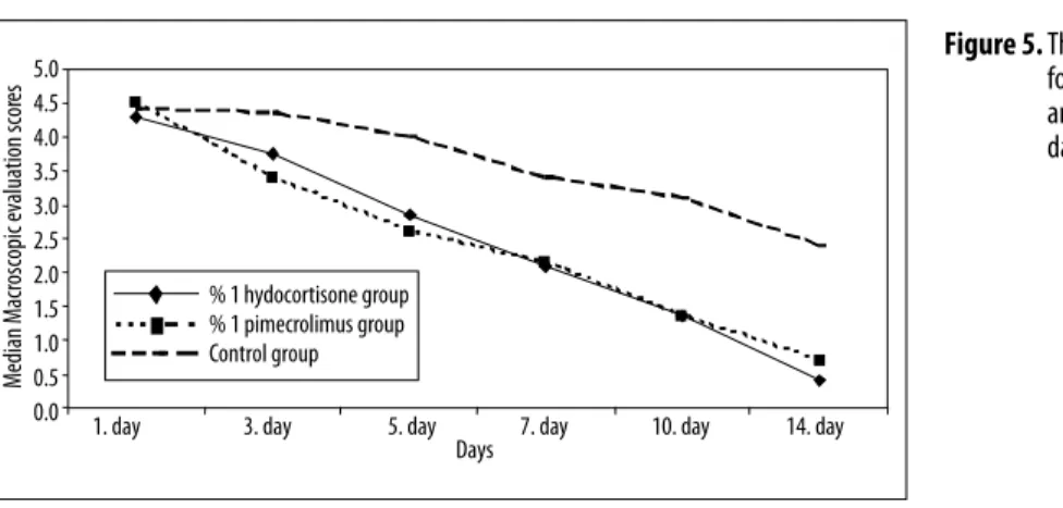
When the MOIS results were assessed, it was seen that there was no difference among the groups for day 1, while there were statistically significant differences for the other days among the groups (P<0.05). The assessments carried out on days 3, 5, 7, 10, and 14 revealed that there was no differ-ence between Group 1 and Group 2, while there were dif-ferences between Groups 1 and 3, and 2 and 3 (P<0.017). Differences were detected among the groups regarding the days (P<0.05). There was a significant difference between Groups 1 and 2 within the framework of binary comparisons for all days (P<0.003). As for Group 3, there was no differ-ence among the days 1–3, 3–5, and 7–10 (P<0.003) (Table 2).
Microscopic evaluation
Tissue samples were taken from all animal groups at the end of the therapy and were evaluated by 2 histologists with light microscopy.
Very few inflammatory cells (mostly macrophages and eo-sinophiles) were detected in the connective tissue under the epithelium. Epithelium and cartilage tissues were also in-tact in the hydrocortisone treatment group (Figures 2A,B). In the pimecrolimus treatment group, inflammation improved. Very few inflammatory cells (mostly eosinophiles) were detect-ed in the connective tissue under the epithelium. The number of eosinophiles in this group was insignificantly higher in com-parison to the hydrocortisone treatment group. Epithelium and cartilage tissues were also intact in this group (Figures 3A,B).
Contact dermatitis was sought microscopically in Group 3. A large number of inflammatory cells (mostly neutrophiles, lymphocytes, macrophages and eosinophiles) were detected in the connective tissue under the epithelium. Epithelium and cartilage tissues were intact (Figures 4A,B). No statisti-cal data on the number of immune cells are shown. Since pre-treatment histopathologic examination was not undertaken, it was not possible to make pre- and treat-ment comparison in this regard. However, although post-treatment abundant and dense eosinophils and inflam-matory cells were seen in the dermis in the control group, eosinophils were scarce in the hydrocortisone and pimecro-limus treatment groups. Apart from this, histologic param-eters such as eosinophilic spongiosis, papillary dermal ede-ma and epiderede-mal thickening, which are all specific for allergic atopic dermatitis, were not observed. This result
Median total IgE lev
els (µg/ml) 18 16 14 12 10 8 6 4 2 0 1. day
before the therapy before the therapy14. day before the therapy28. day after the therapy7. day after the therapy 14. day
Days % 1 hydocortisone group % 1 pimecrolimus group Control group
Figure 1. The mean ± standard deviation (µg/
ml) values for the total IgE values of the
hydrocortisone, pimecrolimus therapies,
and control groups during the DNCB
sensitization period (days 1, 14, and 28)
and the therapy period (days 7 and 14)
are displayed graphically.
Groups
Day 1, DNCB
sensitization,
total IgE levels
(µg/ml)
Day 14, DNCB
sensitization,
total IgE levels
(µg/ml)
Day 28, DNCB
sensitization,
total IgE levels
(µg/ml)
Day 7, therapy
period,
total IgE levels
(µg/ml)
Day 14, therapy
period,
total IgE levels
(µg/ml)
Hydrocortisone group
0.72±0.26
4.50±0.83
16.51±2.17
7.20±1.35
2.57±0.75
Pimecrolimus group
0.88±0.34
4.03±0.80
14.85±1.96
3.73±1.06
1.96±0.60
Control group
0.80±0.24
4.51±1.13
15.25±3.17
11.14±3.00
7.00±1.94
Table 1. The mean ± standard deviation (µg/ml) values for the total IgE values of the hydrocortisone, pimecrolimus therapies, and control groups
during the DNCB sensitization period (days 1, 14, and 28) and the therapy period (days 7 and 14) are displayed.
Groups
Day1
(MOIS, mean
± standard
deviation)
Day 3
(MOIS, mean
± standard
deviation)
Day 5
(MOIS, mean
± standard
deviation)
Day 7
(MOIS, mean
± standard
deviation)
Day 10
(MOIS, mean
± standard
deviation)
Day 14
(MOIS, mean
± standard
deviation)
Hydrocortisone group
4.30±0.57
3.75±0.79
2.85±0.81
2.10±0.72
1.35±0.59
0.40±0.50
Pimecrolimus group
4.50±0.51
3.40±0.50
2.60±0.50
2.15±0.75
1.35±0.67
0.70±0.57
Control group
4.40±0.50
4.35±0.49
4.00±0.56
3.40±0.50
3.10±0.55
2.40±0.50
Table 2. The mean ± standard deviation macroscopic evaluation scores (MOIS) for the hydrocortisone, pimecrolimus, and the control groups during
demonstrates that the disease regressed in accordance with the evaluation parameters in all 3 groups.
d
iscussionThe thin skin of the EAC is normally protected by the nat-ural oils and cerumen produced by the glands in the EAC skin. When the protective layers of the EAC skin are re-moved by the use of cotton-tipped (Q-tip) swabs or by oth-er means, the thin EAC skin is vulnoth-erable to the penetra-tion of haptens.
With multiple sensitizations an allergic dermatitis occurs, which leads to an inflammatory process in the EAC skin. The inflammation in the EAC, in turn, may lead to a reduction or a halt in the secretion of the natural oils and cerumen.
This gives way to a vicious cycle in patients with a lack of ce-rumen, a dry EAC, and continuous contact dermatitis [15]. Allergic contact dermatitis is caused by small molecules that can penetrate the skin, weighing less than 500 Da (haptens). Haptens are weakly allergenic, requiring multiple exposures before they cause sensitization. Allergic contact dermatitis requires the presentation of the antigens to T cells by epi-dermal Langerhans cells. The principal antigen presenting cells of the skin, the Langerhans cells, in turn up-regulate IL-1 and tumor necrosis factor. Keratinocytes are also acti-vated by haptens to secrete immunomodulatory cytokines. The clinical result is itching and irritation of the skin [16]. Preservatives are another large class of molecules that are among the common causes of contact dermatitis and are
Figures 2A,B. Control group samples. A lot of inflammatory cells are seen (*). H.E. Bar 100 µm.
A
B
Figures 3A,B. Hydrocortisone treatment group samples. A few inflammatory cells (arrows) are seen. H.E.Bar 100 µm.
A
B
found in almost all hair care products. Considering the fact that shampoos and conditioners come in contact with the EAC, contact dermatitis can occur in the EAC. Surfactants such as cocamidopropyl betaine in shampoos and its pur-ported allergen amidoamine are among the top 20 most frequent patch test positive allergens. Moreover, fragrances containing protein, lanolin, parabens or formaldehyde are all among the causes of contact dermatitis [17].
Absence or altered pH of the cerumen, high humidity or remaining moisture in the auditory canal, the presence of a foreign material (eg, ear plugs, hearing aid ear molds), local trauma caused while using certain instruments for cleaning the auditory canal, increased susceptibility to con-tact allergens (eg, nickel ear rings, piercing) and second-ary sensitization to ototopic medications, congenital, or ac-quired narrowing of the auditory canal (eg, exostosis), and systemic diseases (eg, diabetes mellitus) provide the basis for the development of chronic eczematous changes of the auricular skin [18].
In the present study, we established an allergic dermatitis model in mice using a contact sensitizer, 2,4-dinitrochloro-benzene (DNCB). DNCB is a contact sensitizer that triggers the Th-1 and Th-2 responses [19–22]. We painted the EAC dermis with DNCB acetone solution repeatedly once a day for 28 days. We selected this skin area because the mouse external ear atopic dermatitis model could be used as an imitated human external ear AD.
We used DNCB (1-Chloro-2,4-Dinitrobenzene) in the for-mation of the disease. DNCB is an agent that was previous-ly used in many studies to form induced allergic dermatitis in mice [23,24]. In our study we successfully formed atop-ic dermatitis through the use of local DNCB in all the trial subjects. Our study revealed that there was an increase in the total IgE levels with the formation and exacerbation of the clinical findings of the disease, and there was a continu-ous increase in the serum IgE levels and the clinical findings with the progression of sensitization (Figure 1). These results
indicate that DNCB is an effective agent in inducing atopic dermatitis as was also put forward by previous studies [25]. The treatment of contact dermatitis depends on avoidance of the allergen and the treatment of the associated inflam-mation in the skin. There are multiple medications used in the treatment of contact dermatitis.
Topical glucocorticoids are the most frequently used first-line medications for this indication and provide an effec-tive treatment choice for AD. They are very beneficial dur-ing acute disease flares, but their prolonged use is generally associated with adverse effects [26].
Since longer durations of corticosteroid exposure can lead to permanent skin atrophy and telangiectasia, its chronic use in EAC is contraindicated [27]. A recent in vivo study has shown that 1 week of glucocorticoid application increased the scratching triggered by an irritant chemical [28]. In recent years pimecrolimus has been introduced for the treatment of AD. Pimecrolimus is an ascomycin macrolac-tam derivative. Pimecrolimus acts through the inhibition of T cell activation by the calcineurin pathway and inhibition of the release of different inflammatory cytokines from T cells and mast cells [29].
Pimecrolimus 1% cream has been approved by the FDA for the treatment of AD. It has been proven to be effective in various inflammatory skin diseases such as seborrheic der-matitis [30], cutaneous lupus erythematosus [31], oral li-chen planus [32], vitiligo [33] and psoriasis [34,35]. It has lower permeation to the skin than topical steroids or topical tacrolimus. Their skin penetration and absorption are 70 to 110 times lower than corticosteroids [36,37]. Furthermore, in contrast to topical steroids, pimecrolimus does not pro-duce skin atrophy [38]. These topical macrolide immuno-suppressive agents can be used in areas with thin skin for prolonged periods of time without the adverse effects of corticosteroids [39].
Figures 4A,B. Pimecrolimus treatment group samples. A few inflammatory cells especially eosinophils (arrows) are seen H.E. Bar 100 µm.
Due to the low adverse effects profile of these agents com-pared to corticosteroids, we examined and comcom-pared the therapeutic effects of pimecrolimus and dexamethasone on the mouse auricular atopic dermatitis model.
The drugs were applied topically to the auricular skin le-sions once a day for 14 days. During the treatment period, the effectiveness of both therapies was evaluated by the total IgE levels and MOIS scores. A perpetual decrease in serum IgE levels, which was initiated by the treatment, was seen in Groups 1 and 2, which received both treatment methods. This result shows that both types of medication are effective in the treatment of atopic dermatitis. This is also in agree-ment with the results of other studies [40–42].
The total IgE measurements carried out on day 7 following treatment revealed that there was a significant difference among the results of the 3 groups (P<0.05). The groups’ mean values of total IgE were: Group 2 < Group 1 < Group 3. This result suggests that the pimecrolimus therapeutic ac-tivity during the first 7 days was more characteristic than the hydrocortisone and the control groups. This result sup-ports the other result derived from the total IgE results in favor of pimecrolimus. When the total IgE results of day 14 were evaluated, we saw that there was no significant differ-ence between the results of Groups 1 and 2, but there was a statistically significant difference between these 2 groups and Group 3 (P<0.05). In other words, both medications have therapeutic activity, but none is superior to the other. In MOIS scoring results based on clinical observation, how-ever, there was no significant difference between Groups 1 and 2 while there were differences between the Groups 1 and 3, and 2 and 3 (P<0.05). The macroscopic evaluation done on days 3, 5, 7, 10, and 14 revealed no difference be-tween Groups 1 and 2, while there were differences bebe-tween Groups 1 and 3, and 2 and 3 (p<0.017) (Figure 5). These results also implied that there was no difference between the therapeutic activities of pimecrolimus and hydrocorti-sone during the therapy period. This result supports the thesis that both medications have therapeutic activity, but their effectiveness is the same.
There was a significant difference between Groups 1 and 2 according to the results of the binary comparison among the days (P<0.003). This result showed that both medica-tions have a gradually increasing therapeutic activity. As for Group 3, there were no differences among the days 1–3, 3–7,
and 7–10, while there was a difference in binary compari-sons among the days. This result reveals the fact that MOIS scores also decreased within the control group that received no treatment with the halting of DNCB sensitization, but this took place much more slowly than in the medicated groups. The histological evaluation of the mice after sacrificing them at the end of the treatment revealed minimal inflammation results for Groups 1 (Figure 2A,B) and 2 (Figure 3A,B) in correlation, while it also showed a continuous inflammation histologically for the control group (Figure 4A,B) that re-ceived no treatment. Histological evaluation of all groups displayed intact cartilage tissue and intact epithelial struc-ture overlying the cartilage. We found that all the inflamma-tory cells located in the subepithelial area and in the con-nective tissue were intensive in the control group (Group 3) (Figure 2). The tissue analysis of the hydrocortisone treated subjects (Group 1) revealed that macrophages and eosino-philes were especially dominant, whereas microscopic assess-ment of the pimecrolimus treated subjects (Group 2) showed that eosinophiles were dominant. This result supports the idea that both medications have an inflammation suppres-sive effect in atopic dermatitis. The fact that we found few-er macrophages, which are cellular immunity elements, in the pimecrolimus treated group as compared to the hydro-cortisone treated group supports the suppressive effect of pimecrolimus on the known cellular immune system [9,10]. There was no difference in the improvement rates between the medications. Treatment was achieved in all animals at the end of therapy, and no complications were observed in any of them. Treatment activity with both medications was satisfactory in the short-term.
When all these findings are assessed together, we see that the clinical, total IgE, and histological findings support one another. The results indicate that local pimecrolimus 1% treatment is as effective as local hydrocortisone 1% treat-ment in the artificially induced mouse auricular atopic der-matitis model with DNCB. It is seen as a good alternative, especially for patients who cannot receive adequate benefit from hydrocortisone use or for those who had to quit the medication because of its adverse effects.
c
onclusionsTopical pimecrolimus can be used only as a short-term, sec-ond-line agent and as an intermittent treatment of atopic
Median Macr
oscopic evaluation scor
es 5.04.5 4.0 3.5 3.0 2.5 2.0 1.5 1.0 0.5
0.0 1. day 3. day 5. day 7. day 10. day 14. day
Days % 1 hydocortisone group % 1 pimecrolimus group Control group
Figure 5. The mean macroscopic evaluation scores
for the hydrocortisone, pimecrolimus,
and the control groups during the 14
dermatitis in patients unresponsive to other treatments. On the other hand, the FDA has issued a warning about topi-cal pimecrolimus being linked with a possible risk of can-cer found in studies associated with high doses and pro-longed use.
The use of these medications are to be avoided in children younger than 2 years and in immune-compromised patients, especially in consideration of a comprehensive review of all the animal studies related to risk of cancer with topical cal-cineurin inhibitors [43]. The cancers and malign lympho-blastic disorders were found in humans treated with a high-er dose of the drug than is routinely used [43–47]. According to our results, local pimecrolimus may be an al-ternative therapeutic choice for EACAD. However, we be-lieve that topical pimecrolimus should only be used as a short-term, second-line agent in EACAD.
r
eferences:
1. Allen BR, Lakhanpaul M, Morris A et al: Systemic exposure, tolerabili-ty and efficacy of pimecrolimus cream 1% in atopic dermatitis patients. Arch Dis Child, 2003; 88(11): 969–73
2. Meingassner JG, Kowalsky E, Schwendinger H et al: Pimecrolimus does not affect Langerhans cells in murine epidermis. Br. J. Dermatol, 2003; 149(4): 853–57
3. Billich A, Aschauer H, Aszódi A, Stuetz A: Percutaneous absorption of drugs used in atopic eczema: pimecrolimus permeates less through skin than corticosteroids and tacrolimus. Int J Pharm, 2004; 269(1): 29–35 4. Gupta AK, Chow M: Pimecrolimus: a review. J Eur Acad Dermatol
Venereol, 2003; 17(5): 493–503
5. Firooz A, Solhpour A, Gorouhi F et al: Pimecrolimus cream 1% vs. hy-drocortisone acetate cream 1% in the treatment of facial seborrheic der-matitis: a randomized investigator blind clinical trial. Arch Dermatol, 2006; 142(8): 1066–67
6. Gorouhi F, Solhpour A, Beitollahi JM et al: Randomized trial of pimecro-limus cream versus triamcinolone acetonide paste in the treatment of oral lichen planus. J Am Acad Dermatol, 2007; 57(5): 806–13 7. Kreuter A, Gambichler T, Breuckmann F et al: Pimecrolimus 1% cream
for cutaneous lupus erythematosus. J Am Acad Dermatol, 2004; 51(3): 407–10
8. Kolbe L, Kligman AM, Schreiner V et al: Corticosteroid induced atro-phy and barrier impairment measured by noninvasive methods in hu-man skin. Skin Res Technol, 2001; 7: 73–77
9. Amrol D, Keitel D, Hagaman D et al: Topical pimecrolimus in the treatment of human allergic contact dermatitis. Ann. Allergy Asthma Immunol, 2003; 91: 563–66
10. Allen BR, Lakhanpaul M, Morris A et al: Systemic exposure, tolerabili-ty, and efficacy of pimecrolimus cream 1% in atopic dermatitis patients. Arch Dis Child, 2003; 88(11): 969–73
11. Meurer M, Fartasch M, Albrecht G et al: Long-term efficacy and safety of pimecrolimus cream 1% in adults with moderate atopic dermatitis. Dermatology, 2004; 208: 365–72
12. Paul C, Cork M, Rossi AB et al: Safety and tolerability of 1% pimecro-limus cream among infants: experience with 1133 patients treated for up to 2 years. Pediatrics, 2006; 117(1): 118–28
13. Hikita I, Yoshioka T, Mizoguchi T et al: Characterization of dermatitis arising spontaneously in DS-Nh mice maintained under conventional conditions: another possible model for atopic dermatitis. J Dermatol Sci, 2002; 30(2): 142–53
14. Yamashita H, Michibata Y, Mizukami H et al: Dermal mast cells play a central role in the incidence of scratching behavior in mice induced by multiple application of the hapten, 2,4,6-trinitrochlorobenzene. Exp Dermatol, 2005; 14(6): 438–44
15. Seftel DM: Itching in the ear canal due to hypocerumenosis. An effec-tive method of treatment. Calif Med, 1963; 98: 151–52
16. Li LY, Cruz PD Jr: Allergic contact dermatitis: pathophysiology applied to future therapy. Dermatol Ther, 2004; 17: 219–23
17. Cohen DE: Contact dermatitis: a quarter century perspective. J Am Acad Dermatol, 2004; 51: 60–63
18. Osguthorpe JD, Nielsen DR: Otitis externa: review and clinical update. Am Fam Physician, 2006; 74: 1510–16
19. Bellinghausen I, Brand U, Enk AH et al: Signals involved in the early TH1/TH2 polarization of an immune response depending on the type of antigen. J Allergy Clin Immunol, 1999; 103: 298–306
20. Kitagaki H, Ono N, Hayakawa K et al: Repeated elicitation of contact hypersensitivity induces a shift in cutaneous cytokine milieu from a T helper cell type 1 to a T helper cell type 2 profile. J Immunol, 1997; 159: 2484–91
21. Nagai H, Matsuo A, Hiyama H et al: Immunoglobulin E production in mice by means of contact sensitization with a simple chemical, hapten. J Allergy Clin Immunol, 1997; 100: 39–44
22. Yokozeki H, Ghoreishi M, Takagawa S et al: Signal transducer and ac-tivator of transcription 6 is essential in the induction of contact hyper-sensitivity. J Exp Med, 2000; 191: 995–1004
23. Pokharel YR, Lim SC, Kim SC et al: Sopungyangjae-Tang Inhibits Development of Dermatitis in Nc/Nga Mice. Evid Based Complement Alternat Med, 2008; 5(2): 173–80
24. Inoue Y, Isobe M, Hayashi H: The combined effect of topical CX-659S, a novel diaminouracil derivative, with topical corticosteroid on the three types of allergic responses in mice or guinea pigs. J Pharmacol Sci, 2003; 91(1): 71–78
25. Inoue Y, Isobe M, Shiohara T et al: Protective and curative effects of top-ically applied CX-659S, a novel diaminouracil derivative, on chronic pic-ryl chloride-induced contact hypersensitivity responses. Br J Dermatol, 2002; 147(4): 675–82
26. Hengge UR, Ruzicka T, Schwartz RA, Cork MJ: Adverse effects of topi-cal glucocorticosteroids. J Am Acad Dermatol, 2006; 54(1): 1–15 27. Li LY, Cruz PD Jr: Allergic contact dermatitis: pathophysiology applied
to future therapy. Dermatol Ther, 2004; 17: 219–23
28. Fujii Y, Sengoku T, Takakura S: Repeated topical application of gluco-corticoids augments irritant chemical-triggered scratching in mice. Arch Dermatol Res, 2010; 302(9): 645–52
29. Amrol D, Keitel D, Hagaman D et al: Topical pimecrolimus in the treatment of human allergic contact dermatitis. Ann Allergy Asthma Immunol, 2003; 91: 563–66
30. Esparza-Gordillo J, Weidinger S, Fölster-Holst R et al: A common variant on chromosome 11q13 is associated with atopic dermatitis. Nat Genet, 2009; 41(5): 596–601
31. Saito H: Much atopy about the skin: genome-wide molecular analysis of atopic eczema. Int Arch Allergy Immunol, 2005; 137(4): 319–25 32. Schreiber S, Rosenstiel P, Albrecht M et al: Genetics of Crohn disease,
an archetypal inflammatory barrier disease. Nat Rev Genet, 2005; 6(5): 376–88
33. Boone B, Ongenae K, Van Geel N et al: Topical pimecrolimus in the treatment of vitiligo. Eur J Dermatol, 2007; 17(1): 55–61
34. Kreuter A, Sommer A, Hyun J et al: 1% pimecrolimus, 0.005% calcipot-riol and 0.1% betamethasone in the treatment of intertriginous psoria-sis: a double-blind, randomized controlled study. Arch Dermatol, 2006; 142(9): 1138–43
35. Jacobi A, Braeutigam M, Mahler V et al: Pimecrolimus 1% cream in the treatment of facial psoriasis: a 16-week open-label study. Dermatology, 2008; 216(2): 133–36
36. Ohameje NU, Loveless JW, Saini SS: Atopic dermatitis or hyper-IgE syn-drome? Allergy Asthma Proc, 2006; 27: 289–91
37. Billich A, Aschauer H, Aszodi A et al: Percutaneous absorption of drugs used in atopic eczema: pimecrolimus permeates less through skin than corticosteroids and tacrolimus. Int J Pharm, 2004; 9(6): 29–35 38. Pastar Z, Lipozencic J, Ljubojevic S: Etiopathogenesis of atopic
derma-titis: an overview. Acta Dermatovenerol. Croat, 2005; 13: 54–62 39. Meurer M, Fartasch M, Albrecht G et al: Long-term efficacy and safety
of pimecrolimus cream 1% in adults with moderate atopic dermatitis. Dermatology, 2004; 208: 365–72
40. Yun MY, Yang JH, Kim DK et al: Therapeutic effects of Baicalein on atop-ic dermatitis-like skin lesions of NC/Nga matop-ice induced by dermatopha-goides pteronyssinus. Int Immunopharmacol, 2010; 10(9): 1142–48 41. Ohbuchi T, Sakaino M, Takahashi T et al: Oral administration of acidic
xylooligosaccharides prevents the development of atopic dermatitis-like skin lesions in NC/Nga mice. J Nutr Sci Vitaminol, 2010; 56(1): 54–59 42. Watanabe T, Hamada K, Tategaki A et al: Oral administration of lac-tic acid bacteria isolated from traditional South Asian fermented milk ‘dahi’ inhibits the development of atopic dermatitis in NC/Nga mice. J Nutr Sci Vitaminol, 2009; 55(3): 271–78
43. Ring J, Barker J, Behrendt H et al: Review of the potential photo-cocar-cinogenicity of topical calcineurin inhibitors: position statement of the European Dermatology Forum. J. Eur. Acad. Dermatol Venereol, 2005; 19: 663–71
44. Wang Y, Chen J: Bronchogenic carcinoma after lung transplantation: a case report and literature review]. Zhongguo Fei Ai Za Zhi, 2011; 14(1): 75–78
45. Chinnakotla S, Davis GL, Vasani S et al: Impact of sirolimus on the re-currence ofhepatocellular carcinoma after liver transplantation. Liver Transpl, 2009; 15(12): 1834–42
46. Suthanthiran M, Hojo M, Maluccio M et al: Post-transplantation ma-lignancy: a cell autonomous mechanism with implications for therapy. Trans Am Clin Climatol Assoc, 2009; 120: 369–88
47. Pulsipher MA, Wall DA, Grimley M et al: A phase I/II study of the safe-ty and efficacy of the addition ofsirolimus to tacrolimus/methotrexate graft versus host disease prophylaxis after allogeneic haematopoietic cell transplantation in paediatric acute lymphoblastic leukaemia (ALL). Br J Haematol, 2009; 147(5): 691–99