ContentslistsavailableatScienceDirect
Data
in
Brief
journalhomepage:www.elsevier.com/locate/dib
Data
Article
Experimental
data
of
labeling
the
heart
and
cardiac
cultures
with
a
retrograde
tracer
in
vitro
and
in
vivo
Tuba
Akgul
Caglar
a
,
b
,
Mehmet
Yalcin
Gunal
c
,
Mehmet
Ugurcan
Turhan
a
,
d
,
Gurkan
Ozturk
a
,
b
,
e
,
Esra
Cagavi
a
,
f
,
g
,
∗
a Regenerative and Restorative Medicine Research Center (REMER), Research Institute for Health Sciences and Technologies (SABITA), Istanbul Medipol University, Istanbul, Turkey
b Neuroscience Program, Institute of Health Sciences, Istanbul Medipol University, Istanbul, Turkey c Department of Physiology, School of Medicine, Alanya Alaaddin Keykubat University, Antalya, Turkey d School of Medicine, Istanbul Medipol University, Istanbul, Turkey
e Department of Physiology, School of Medicine, Istanbul Medipol University, Istanbul, Turkey
f Medical Biology and Genetics Program, Institute of Health Sciences, Istanbul Medipol University, Istanbul, Turkey g Department of Medical Biology, School of Medicine, Istanbul Medipol University, Istanbul, Turkey
a
r
t
i
c
l
e
i
n
f
o
Article history:
Received 7 November 2020 Revised 23 January 2021 Accepted 1 February 2021 Available online 4 February 2021 Keywords: In vivo labeling Di-8-ANEPPQ Cardiac afferents Retrograde tracers In vitro labeling Sensory system Cardiac system
a
b
s
t
r
a
c
t
Retrogradedyes areoftenusedinbasicresearchto investi-gateneuronalinnervationsofanorgan.Thisarticledescribes theexperimentaldataontheapplicationofretrogradedyes onthemouseheartin vivoand onthecardiacorneuronal culturesinvitro.Byprovidingthisinformation,cardiacor in-neinnervationscanbeevaluatedinvivo.Therefore,unknown cellularandmolecularmechanismsandsystemicinteractions inthe bodycanbe investigated.Inparticular, weprovided practicaltips tolower mortalityrisks followingthe cardiac surgeryand evaluatedthestainingcapacityand fluorescent characteristics of the Di-8-ANEPPQ dye in the cardiac tis-sue and cell cultures.First, primarycultures ofmouse no-dose ganglia (NG) neurons and mouse neonatal cardiomy-ocytes were stained with Di-8-ANEPPQ. The Di-8-ANEPPQ signalfromlivecultureswerevisualizedusingspinningdisk confocal microscopytoverify thelipophilicand fluorescent
DOI of original article: 10.1016/j.brainres.2020.147201
∗ Corresponding author at: Regenerative and Restorative Medical Research Center (REMER), Research Institute for Health Sciences and Technologies (SABITA), Istanbul Medipol University, Istanbul, Turkey.
E-mail addresses: [email protected] , [email protected] (E. Cagavi). https://doi.org/10.1016/j.dib.2021.106834
2352-3409/© 2021 The Authors. Published by Elsevier Inc. This is an open access article under the CC BY-NC-ND license ( http://creativecommons.org/licenses/by-nc-nd/4.0/ )
labeling capacity ofDi-8-ANEPPQ. Next,the excitation and emission data of Di-8-ANEPPQ were collected between 415nm and690nm usingpower spectrummoduleof con-focalmicroscopy.Thisspectrumanalysiscouldbeusefulfor the researcherswhoplanto useDi-8-ANEPPQin combina-tionwithotherfluorescentdyestoeliminateanyflorescent overlap. Inorder tolabelthe heart tissuewith tracerdyes Di-8-ANEPPQorDiIinvivo,the heartwasexposedwithout damaging lungs orother tissues following anesthetization, then the retrograde dyewas applied as apaste for DiI or injected totheapex oftheheart forDi-8-ANEPPQ andthe operationareawassutured.Thesurgicalprocedurerequired intubationtocontroltherespiratoryreflexwithouttheneed toperformatracheotomyandyieldedhighviability. Follow-inglabelingtheheart invivo,theheart wasdissected,and images of injectionarea werecaptured using confocal mi-croscopy.AllfluorescentimagesofDi-8-ANEPPQlabeledcells wereanalyzedbyusingtheFijisoftware.Overall,thesedata provide applicable data to other investigators to trace the sensoryneuronsinnervatingnotonlytheheartbutalsoother organs usingDi-8-ANEPPQ. Thesedatasupporttheoriginal researcharticletitled“Evaluationofbilateralcardiacafferent distributionatthespinalandvagalgangliabyretrograde la-beling” thatwas acceptedforpublicationinBrainResearch Journal[1] .
© 2021TheAuthors.PublishedbyElsevierInc. ThisisanopenaccessarticleundertheCCBY-NC-ND license(http://creativecommons.org/licenses/by-nc-nd/4.0/ )
Specifications
Table
Subject Neuroscience: Sensory Systems
Specific subject area Application of Di-8-ANEPPQ or other retrograde tracers to the primary neurons or cardiomyocytes in vitro and to the murine heart in vivo , that could be used to investigate cardiac afferents
Type of data Image
Figure
How data were acquired Stereomicroscopy, Fluorescent Microscopy Instruments: Fiji software (2.0.0-rc-69/1.52i)
Data format Raw
Analyzed Filtered
Parameters or data collection Adult BALB/c mice at 8-12 weeks were housed in the animal facility of MEDITAM of Istanbul Medipol University. The animal care was given according to Ethical community approval of HADHEK. The experimental protocol was approved by the Institutional Animal Experimentation Ethical Committee with the approval number 38328770-30. The in vitro staining capacity of Di-8-ANEPPQ was explored in primary neonatal mouse cardiomyocyte cultures and primary mouse NG neurons. Di-8-ANEPPQ dye has a lipophilic nature [2 , 3] and binds to the plasma membrane. The incubation of Di-8-ANEPPQ dye with the cell cultures was kept at RT to allow the staining of only the cell membrane. Furthermore, the images of live D ˙I-8-ANEPPQ labeled primary cardiomyocyte cultures were captured following the incubation of 10-15 minutes at 37 °C for the cells to regain their basal activity. To obtain emission and excitation properties of Di-8-ANEPPQ dye, the labeled primary
cardiomyocyte culture was excited by not only 488 nm but also 561 nm and 633 nm using the power spectrum module of confocal microscopy. In addition to the application of Di-8-ANEPPQ on the live cultured cells for further
labeling evaluations, the mice weighing at least 25 g were chosen to visualize the epiglottis and to insert the intubation tube without tissue damage. First, ketamine (50 mg/kg, Pfizer) and xylazine (5 mg/kg, Bayer) of anesthesia was administrated into the animal, checked vital signs and sedation parameters, another dosage was given if it was necessary. The intubation was verified by checking the synchronization of the animal chest movement. Next, the animal was transferred to an operation area equipped with a heating pad to maintain hemostasis and prevent hypothermic shock. To expose the heart for delivery of the Di-8-ANEPPQ, the third and fourth intercostal muscles were separated using retractors to prevent lung and diagram damage. For the retrograde labeling, 1 μl of 10 mg/ml freshly thawed Di-8-ANEPPQ was used and warmed up to RT before administration. Following the Di-8-ANEPPQ injection into the heart, negative pressure was applied to the lung before suturing the muscle and skin.
Description of data collection For evaluation of in vitro labeling capacity of Di-8-ANEPPQ, primary cultures of NG neurons and cardiomyocytes were stained with 20 μM Di-8-ANEPPQ at RT. After incubation, live images of cultured cells were taken using spinning disk confocal microscopy (Zeiss). The excitation and emission properties of the Di-8-ANEPPQ was obtained using the power spectrum module of confocal microscopy (LSM 780, Zeiss). The images of the in vivo heart operation were captured using the stereomicroscope (Discovery 8, Zeiss). The Di-8-ANEPPQ, in 1 μl containing 10 mg/ml, was injected into the apex of the heart. Following 2-3 hrs of the surgery, the heart was dissected out and the injection area was imaged using confocal microscopy (LSM 780, Zeiss). All images were analyzed and prepared using Fiji software.
Data source location Institution: Istanbul Medipol University City/Town/Region: Istanbul
Country: Turkey
Data accessibility The corresponding data were provided within this article
Related research article Akgul Caglar T., Durdu Z. B., Turhan M. U., Gunal M. Y., Aydın M. ¸S ., Ozturk G., Cagavi E., “Evaluation of bilateral cardiac afferent distribution at the spinal and vagal ganglia by retrograde labeling” Brain Research.
https://doi.org/10.1016/j.brainres.2020.147201 .
Value
of
the
Data
•
Our
data
described
a
reproducible
approach
to
label
the
primary
cardiac
and
neural
cells
in
vitro
and
the
murine
heart
in
vivo
.
Our
data
provided
important
tips
for
other
researchers
to
enhance
labeling
efficiency
and
the
recovery
after
cardiac
surgery,
in
which
mortality
rates
are
often
high.
The
evaluation
of
the
fluorescent
characteristics
of
the
Di-8-ANEPPQ
dye
has
not
been
reported
elsewhere
in
such
detail
that
has
applicable
value.
•
The
data
would
be
an
important
reference
for
the
scientists
at
different
disciplines
who
are
interested
in
tracing
neurons
in
vivo
not
only
at
the
cardiac
system
but
other
organs
as
well.
These
data
would
be
of
great
interest
to
researchers
who
plan
to
conduct
experiments
with
Di-8-ANEPPQ
or
similar
dyes
for
fluorescently
staining
and
tracing
cells
in
vitro
.
•
The
data
might
be
used
for
tracing
neurons
not
only
innervating
the
heart
but
also
other
visceral
organs
using
Di-8-ANEPPQ.
Researchers
who
study
cardiac
physiology,
regeneration
or
nervous
system
interactions
would
be
expected
to
benefit
and
use
the
provided
data.
•
The
power
spectrum
data
of
Di-8-ANEPPQ
dye
would
guide
scientist
to
select
the
best
ex-perimental
and
imaging
settings
in
combination
to
other
staining
methodologies.
1.
Data
Description
The
data
in
this
article
described
the
experimental
steps
for
the
application
of
retrograde
dyes
on
primary
cardiac
and
neuron
cultures
in
vitro
and
the
mouse
heart
in
vivo.
To
determine
the
efficiency
of
Di-8-ANEPPQ
in
vitro;
primary
NG
neurons
and
neonatal
cardiomyocytes
were
stained
with
20
μM
Di-8-ANEPPQ.
The
live
images
of
cells
labeled
with
Di-8-ANEPPQ
were
cap-tured
using
spinning
disk
confocal
microscopy
and
the
green
fluorescence
signal
of
Di-8-ANEPPQ
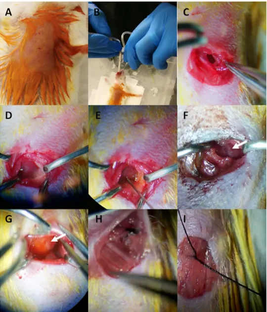
Fig. 1. Imaging data of Di-8-ANEPPQ labeled primary neuronal and cardiac cells in vitro The Di-8-ANEPPQ dye label- ing efficiency was determined by flourescent imaging of the primary NG neurons (A) or primary neonatal cardiomyocytes
(B) imaged by floresence microscopy. Di-8-ANEPPQ: green, scale bar: 100 μm.
Fig. 2. Emission spectrum analysis of Di-8-ANEPPQ labeled primary cell culture. Di-8-ANEPPQ (gray) labeled cardiac cells were excited (A) 488 nm, (B) 561 nm, and (C) 633 nm. Emission spectrum data between 415 nm and 690 nm were captured and displayed. Scale bars: 100 μm.
was
detected
at
the
plasma
membrane
(
Fig.
1
).
Next,
the
emission
and
excitation
properties
of
Di-8-ANEPPQ
labeled
cardiomyocytes
were
analyzed
by
collecting
emissions
between
415
nm
and
690
nm
using
the
power
spectrum
module
of
confocal
microscopy
(
Fig.
2
).
Interestingly,
we
observed
that
Di-8-ANEPPQ
dye
could
be
excited
by
both
488
nm
(
Fig.
2
A)
and
561
nm
(
Fig.
2
B),
but
not
excited
by
633
nm
(
Fig.
2
C)
wavelength
expanding
the
previous
knowledge
[4]
.
More-over,
the
emission
spectrum
of
Di-8-ANEPPQ
was
found
to
be
at
a
broad
range
between
571
and
690
nm
(
Fig.
2
).
In
addition
to
the
application
of
Di-8-ANEPPQ
on
the
live
primary
cultures,
10
mg/ml
Di-8-ANEPPQ
or
DiI
paste
were
applied
to
the
heart
apex
in
vivo
(
Fig.
3
).
Experimen-tal
steps
for
application
of
a
retrograde
dye
to
the
heart
in
vivo
were
illustrated
in
Fig.
3
.
First,
the
anesthetized
mouse
was
shaved
in
the
operation
area,
and
the
skin
was
aseptically
cleaned
(
Fig.
3
A).
Then,
the
mouse
was
intubated
using
a
cannula
and
connected
to
the
ventilation
ap-paratus
(
Fig.
3
B).
The
incisions
were
made
at
the
left
side
of
the
thorax
above
the
xiphoid,
and
muscles
were
moved
gently
to
expose
the
heart
(
Fig.
3
C).
The
retractors
were
placed
to
separate
the
third
and
fourth
ribs
(
Fig.
3
D).
Following
stabilization
of
the
vital
signs
of
the
mouse
and
clear
visualization
of
the
heart,
the
Di-8-ANEPPQ
dye
was
injected
into
the
apex
(
Fig.
3
E)
or
DiI
paste
was
applied
onto
the
heart
(
Fig.
3
F).
The
application
area
was
observed
by
the
spread-ing
of
the
retrograde
dye
in
the
cardiac
tissue
(
Fig.
3
G).
The
intercostal
muscles
(
Fig.
3
H)
and
Fig. 3. Experimental steps for application of a retrograde dye to the heart in vivo. (A) The anesthetized mouse was shaved at the operation area and the skin was aseptically cleaned. (B) The mouse was intubated using a cannula and connected to the ventilation apparatus. (C) The incisions were made at the left side of the thorax above the xiphoid, and muscles were moved gently to expose the heart. (D) The retractors were placed to separate the third and fourth ribs.
(E) T he Di-8-ANEPPQ was injected into apex or (F) DiI paste was applied on the heart. Arrow showed the tissue after DiI paste was applied. (G) The application area observed by the spread of the retrograde dye in the cardiac tissue. The injection site is shown by the arrow. (H) The intercostal muscles and (I) the skin were sutured.
the
skin
were
sutured
before
the
mouse
was
awakened
from
the
anesthesia
(
Fig.
3
I).
To
verify
the
proper
injection
of
Di-8-ANEPPQ
dye,
an
unlabeled
heart
which
was
dissected
for
determin-ing
the
fluorescence
background
(
Fig.
4
A),
and
the
heart
in
which
Di-8-ANEPPQ
was
adminis-trated
in
vivo
were
imaged
by
confocal
microscopy
(
Fig.
4
B).
The
green
fluorescence
signal
of
Di-8-ANEPPQ
dye
was
detected
at
the
injection
site
of
the
heart
tissue.
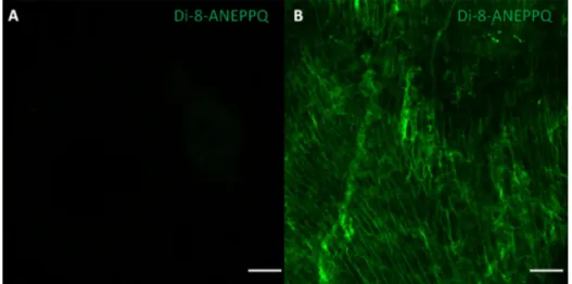
Fig. 4. Imaging data of Di-8-ANEPPQ from in vivo labeled cardiac tissue . Fluorescence imaging was performed on unlabeled control heart tissue for determining (A) the background or (B) following the Di-8-ANEPPQ administration to the murine heart in vivo by confocal microcopy. Di-8-ANEPPQ: green. Scale bar: 100 μm.
2.
Experimental
Design,
Materials
and
Methods
2.1.
Preparation
of
the
primary
murine
cardiomyocyte
and
neuron
cultures
for
in
vitro
labeling
The
primary
neonatal
cardiomyocytes
or
adult
nodose
ganglia
(NG)
neurons
were
prepared
and
cultured
as
previously
described
[5
,
6]
.
Briefly,
the
heart
of
the
BALB/c
neonatal
mice
at
1-3
days
after
birth
was
dissected
out
and
put
in
the
ice-cold
Trypsin
solution
(T4174,
Sigma)
for
pre-digestion
overnight
at
4
°C.
The
next
day,
the
heart
tissue
was
collected
and
incubated
with
450
U/ml
collagenase
II
(17101-015,
Gibco)
enzyme
at
a
37
°C
water
bath
for
one
hour
with
gen-tle
agitation.
The
cell
suspension
was
spun
at
120
g
for
5
min,
and
the
pellet
was
resuspended
with
cardiomyocyte
medium
containing
Na-pyruvate
(11360-070,
Gibco),
MEM
NEAA
(11140-050,
Gibco),
fetal
calf
serum
(10270-106,
Gibco),
horse
serum
(16050-130,
Gibco)
and
newborn
calf
serum
(16010167,
Gibco).
The
neonatal
cardiomyocytes
were
plated
on
culture
dishes
coated
with
1
mg/ml
Fibronectin
(F1141,
Sigma).
NG
neuron
cultures
were
prepared
by
dissecting
the
NG
from
an
adult
BALB/c
mouse
at
8-12
weeks
as
previously
described
[5]
.
The
mouse
was
euthanized
with
cervical
dislocation
and
bilateral
NG
tissues
were
dissected
out
by
trimming
the
ganglia
from
the
neural
fibers
and
extra
connective
tissue.
Next,
nerve
bodies
were
enzymatically
dissociated
using
collagenase
XI
(C7657,
Sigma),
then
trypsin
(25300-054,
Gibco)
at
the
37
°C
incubator.
To
dissociate
single
cells,
the
NGs
were
triturated
by
pipetting
and
DNAse
(D4513,
Sigma)
was
added
to
the
cell
suspen-sion
to
inhibit
free
DNA
fragments.
Next,
the
cell
suspension
was
spun
at
120g
for
3
min
and
re-suspended
using
fetal
calf
serum
and
trypsin
inhibitor
(T6522,
Sigma).
Then,
the
cell
suspension
was
collected
and
resuspended
with
Neural
basal
medium
(10888-022,
Gibco)
supplemented
with
B27
(17504-044,
Gibco).
The
collected
neurons
were
plated
on
the
culture
dishes
coated
with
1mg/ml
Laminin
(L2020,
Sigma).
2.2.
The
labeling
of
in
vitro
primary
cultures
with
Di-8-ANEPPQ
The
cultured
cells
were
stained
with
Di-8-ANEPPQ
to
evaluate
the
staining
efficiency
of
Di-8-ANEPPQ
in
vitro
bef
ore
perf
orming
in
vivo
applications.
Di-8-
ANEPPQ
dye
was
reported
to
have
a
lipophilic
nature
binding
to
the
plasma
membrane
[2
,
3]
.
In
the
second
or
third
day
of
the
primary
cultures,
the
neurons
or
cardiomyocytes
were
stained
with
Di-8-ANEPPQ
to
test
the
lipophilic
staining
capacity
of
Di-8-ANEPPQ
for
live
cells.
Di-8-ANEPPQ
(61014,
Biotium)
at
a
stock
solution
of
10
mM
was
prepared
to
add
697
μl
DMSO
into
the
5
mg
dye
powder,
then
vortexed
and
stored
at
−20
°C
in
small
aliquots.
It
is
better
not
to
freeze
an
aliquot
once
thawed
and
use
fresh
aliquots
at
each
time.
Di-8-ANEPPQ
dye
was
warmed
up
to
25
°C
for
10-15
min
before
staining
the
cells.
The
NG
neuron
or
cardiomyocyte
cultures
were
incubated
with
20
μM
Di-8-ANEPPQ
in
culture
media
for
20-30
mins
at
room
temperature
to
allow
the
staining
of
only
the
cell
membrane.
The
dye
solution
was
removed
and
or
the
cells
to
regain
their
basal
activity,
cultures
were
incubated
for
10-15
minutes
at
37
°C.
The
images
of
live
Di-8-ANEPPQ
labeled
primary
cultures
were
captured
as
detailed
below.
2.3.
In
vivo
labeling
of
the
mouse
heart
For
the
application
of
the
tracer
dyes
to
the
murine
heart,
adult
BALB/c
mice
at
8-12
weeks
of
age
were
used.
Before
the
operation,
the
mice
were
weighed,
and
25
grams
or
heavier
mice
were
selected
for
the
operation.
This
was
empirically
determined
since
the
visualization
of
the
epiglottis
with
mice
smaller
than
25
grams
was
difficult
during
intubation,
and
the
mortality
rates
were
higher
due
to
repeated
attempts
and
tissue
damage.
Before
the
operation,
mice
were
anesthetized
by
an
intraperitoneal
injection
of
a
minimum
dose
of
ketamine
(50
mg/kg,
Pfizer)
and
xylazine
(5
mg/kg,
Bayer)
to
prevent
the
possible
side
effects
in
difficulty
in
breathing
and
sudden
death
[7
,
8]
.
Vital
signs
and
sedation
parameters
such
as
pedal
reflex
and
eye
movement
were
checked,
and
another
dose
was
given
if
it
was
necessary.
After
confirming
sedation
by
monitoring
the
sedation
parameters,
the
chest
of
mice
was
cleaned
aseptically
with
povidone-iodine
solution
and
shaved.
The
anesthetized
mice
were
positioned
into
the
intubation
platform
having
60-80
degrees
to
visualize
the
epiglottis
[9]
.
After
stabilizing
the
mouth
of
the
mouse,
the
tongue
was
lifted
by
the
help
of
forceps,
and
a
fiber
optic
cable
connected
to
the
light
source
was
placed
to
the
throat
to
visualize
the
movement
of
the
epiglottis.
When
the
epiglottis
was
open,
the
mouse
was
endotracheally
intubated
with
an
intubation
cannula
with
a
Y
adapter
(1.2
mm
od,
27
mm
length;
732844,
Harvard
apparatus),
and
then
the
adapter
was
connected
to
the
ventilation
apparatus
(Harvard
Apparatus
Minivent
Type
845).
In
the
meantime,
stroke
volume
and
stroke
rate
were
adjusted
according
to
the
parameters
indicated
in
the
manufac-turer’s
manual
for
the
ventilation
apparatus
[10]
.
To
make
sure
the
air
was
moving
to
both
lungs,
the
chest’s
movement
was
monitorized.
Next,
the
mouse
was
transferred
to
the
operation
area
equipped
with
a
heating
pad
(WPI)
in
order
to
maintain
hemostasis
and
prevent
hypothermic
shock.
In
addition,
sterile
saline
drops
were
applied
to
the
eyes
in
order
to
prevent
dryness.
After
the
mouse
was
positioned
for
the
surgery
by
taping
the
extremities
and
the
tail,
all
operational
steps
were
performed
under
the
stereo
microscopy
(Discovery
8,
Zeiss).
To
expose
the
intercostal
space,
the
incisions
were
made
at
the
left
side
of
the
thorax
above
the
xiphoid,
and
muscles
were
dissected.
The
ribcage
under
the
intercostal
area
was
observed
and
the
incision
between
the
third
and
fourth
intercostal
muscle
was
made
using
fine
scissors
(FM010R,
Aesculap)
and
forceps
(BD329R,
Aesculap).
To
expose
the
heart
for
delivery
of
the
Di-8-ANEPPQ
dye,
the
third
and
fourth
intercostal
muscles
were
separated
using
retractors
to
pre-vent
any
damage
to
the
lung
or
the
diaphragm.
The
light
was
spotted
on
the
operational
area
to
visualize
the
apex.
The
thin
layer
of
epicardium
was
removed
using
fine
forceps
(FD048R,
Aesculap).
Next,
1
μl
Di-8-ANEPPQ
(10
mg/ml)
was
injected
into
the
apex
of
the
heart
to
label
cardiac
afferent
retrogradely
using
30
G
Hamilton
injector
(5221002,
Hamilton).
For
each
dye
administration,
frozen
Di-8-ANEPPQ
aliquot
was
thawed
and
warmed
up
at
room
temperature
because
storage
of
Di-8-ANEPPQ
at
4
°C
could
reduce
labeling
efficieny.
Alternatively,
another
lipophilic
retrograde
dye
DiI
Tissue-Labeling
Paste
(N22880,
Thermofisher)
was
applied
on
the
heart
surface
using
a
pipet
tip.
Following
the
administration
of
the
retrograde
dye
to
the
apex,
negative
pressure
was
applied
to
the
lungs
before
the
muscle
and
skin
were
sutured.
The
dye
application
was
verified
by
the
spreading
of
the
Di-8-ANEPPQ
over
the
heart.
Next,
intercostal
muscles
and
skin
were
closed
using
a
6-0
silk
suture
(Do
˘gsan)
and
the
povidone-iodine
solution
was
applied
at
the
incision
area.
The
mouse
was
removed
from
the
intubation
apparatus
and
the
chest
movement
with
the
rhythmic
inhalation
was
monitorized.
Occasionally
the
breathing
was
observed
to
be
stopped,
then
a
25-gauge
sterile
needle
was
carefully
administered
between
the
operated
ribs
to
remove
excess
air.
While
pulling
up
the
plunger
to
remove
air,
the
needle
was
constantly
checked
to
be
clear
from
blood
to
prevent
damaging
other
tissue.
Alternatively,
a
gentle
heart
message
could
be
performed
to
restart
the
breathing.
For
post-operational
care,
the
10
0-20
0
μl
saline
was
administered
subcutaneously.
At
the
end
of
the
operation,
the
mouse
was
placed
into
the
recovery
cage
equipped
with
a
warm
bath
and
was
observed
until
the
mouse
was
awaken
from
the
anesthesia.
The
food
and
the
water
were
put
into
the
reachable
area
of
the
cage.
The
cage
was
placed
into
post-operative
room
and
the
mouse
was
monitored
daily.
2.4.
Imaging
data
collection
Following
two
or
three
hours
of
Di-8-ANEPPQ
administration
in
vivo
,
the
mouse
was
eutha-nized
with
cervical
dislocation,
then
the
heart
was
dissected
out
and
put
on
the
ice-cold
Roswell
Park
Memorial
Institute
(RPMI)
1640
Medium
(R0883,
Sigma).
Live
images
from
the
heart
tis-sue
was
captured
using
laser
scanning
confocal
microscope
(Carl
Zeiss,
LSM
780)
with
488
nm
excitation
laser,
green
fluorescence
emission
detected
between
493-630
nm
with
GaAsP
photo-multiplier
tube,
plan-apochromat
10x/0.45
Ph1
objective,
1024
× 1024
resolution,
1.58
μ
s
pixel
dwell
time
and
20
μ
m
total
Z
stack
divided
by
five
slices.
In
addition,
the
primary
neuron
or
neonatal
cardiomyocyte
cultured
cells
were
stained
with
20
μM
Di-8-ANEPPQ.
Live
images
from
the
primary
cells
were
taken
under
cell
observer
SD
spinning
disk
time-lapse
microscope
(Carl
Zeiss)
with
488
nm
excitation
laser
with
38
HE
green
flurosecent
prot
reflector,
500-550
nm
fil-ters
for
emisson
detection,
C-Apochromat
40x/1.2
water
korr
objective,
522.4
ms
exposure
time
and
1388
× 1040
resolution.
To
detect
the
excitation
and
emission
spectrum
of
Di-8-ANEPPQ,
car-diomyocyte
cells
were
stained
with
Di-8-ANEPPQ
and
imaged
using
the
power
spectrum
module
of
LSM
780
confocal
microscopy
(Zeiss).
For
excitation
of
the
Di-8-ANEPPQ
dye,
488
nm,
561
nm,
and
633
nm
laser
settings
were
all
kept
at
the
pinhole
diameter
of
90
μ
m
,
gain
at
800
and
the
digital
gain
at
1.
Laser
power
for
633
nm
was
used
at
14%,
for
561
nm
laser
2%
and
for
the
488nm
laser
2%.
The
emission
was
captured
between
410
nm
and
695
nm.
Ethics
Statement
The
experiments
comply
with
international
guidelines
and
in
accordance
with
HADHEK.
Our
experimental
protocols
were
approved
by
the
Institutional
Animal
Experimentation
Ethical
Com-mittee
with
the
approval
number
38328770-30.
In
this
study,
two
adult
BALB/c
independent
of
gender
mice
and
one
BALB/c
neonatal
mouse
were
used.
Declaration
of
Competing
Interest
The
authors
declare
that
they
have
no
known
competing
financial
interests
or
personal
rela-tionships
which
have
or
could
be
perceived
to
have
influenced
the
work
reported
in
this
article.
Acknowledgments
This
work
was
supported
by
Scientific
and
Technological
Research
Council
of
Turkey
(TUBITAK)
under
1001
Scientific
and
Technological
Research
Projects
Funding
Program
by
project
number
115S381
and
by
Istanbul
Medipol
University
under
project
number
BAP
2018/19.
We
thank
REMER-SABITA
personnel
for
their
technical
help
and
our
animal
facility
MEDITAM
per-sonnel
for
their
kind
assistance.
References
[1] Akgul Caglar T., Durdu Z. B., Turhan M. U., Gunal M. Y., Aydın M. ¸S ., Ozturk G., Evaluation of the bilateral cardiac afferent distribution at the spinal and vagal ganglia by retrograde labeling, Brain Res. doi: 10.1016/j.brainres.2020. 147201 .
[2] P. Wenner, Y. Tsau, L.B. Cohen, M.J. O’Donovan, Y. Dan, Voltage-sensitive dye recording using retrogradely trans- ported dye in the chicken spinal cord: Staining and signal characteristics, J. Neurosci. Methods. (1996), doi: 10.1016/ S0165-0270(96)00108-2 .
[3] Y. Tsau, P. Wenner, M.J. O’Donovan, L.B. Cohen, L.M. Loew, J.P. Wuskell, Dye screening and signal-to-noise ratio for retrogradely transported voltage-sensitive dyes, J. Neurosci. Methods. (1996), doi: 10.1016/S0165-0270(96)00109-4 . [4] L.M. Loew, Potentiometric dyes: Imaging electrical activity of cell membranes, Pure Appl. Chem. (2007), doi: 10.1351/
pac199668071405 .
[5] N. Cengiz, G. Öztürk, E. Erdo ˘gan, A. Him, E.K. O ˘guz, Consequences of neurite transection in vitro, J. Neurotrauma. (2010), doi: 10.1089/neu.2009.0947 .
[6] M.Y. Lee, B. Sun, S. Schliffke, Z. Yue, M. Ye, J. Paavola, E.C. Bozkulak, P.J. Amos, Y. Ren, R. Ju, Y.W. Jung, X. Ge, L. Yue, B.E. Ehrlich, Y. Qyang, Derivation of functional ventricular cardiomyocytes using endogenous promoter sequence from murine embryonic stem cells, Stem Cell Res. (2012), doi: 10.1016/j.scr.2011.08.004 .
[7] H.R. Amouzadeh, C.W. Qualls, J.H. Wyckoff, G.K. Dzata, S. Sangiah, A. Mauromoustakos, L.E. Stein, Biochemi- cal and morphological alterations in xylazine-induced pulmonary edema, Toxicol. Pathol. (1993), doi: 10.1177/ 019262339302100607 .
[8] S. Buitrago , T.E. Martin , J. Tetens-Woodring , A. Belicha-Villanueva , G.E. Wilding , Safety and efficacy of various com- binations of injectable anesthetics in BALB/c mice, J. Am. Assoc. Lab. Anim. Sci. (2008) .
[9] T.C. Vandivort, D. An, W.C. Parks, An improved method for rapid intubation of the trachea in mice, J. Vis. Exp. (2016), doi: 10.3791/53771 .
[10] HUGO SACHS ELEKTRONIKHARVARD, MiniVent Ventilator (Type 845) User’s Manual, (2012). https://www. harvardapparatus.com/media/manuals/ProductManuals/Mini _ Micro _ MidiVentManual.pdf .