O
RIGINAL PAPEREtiological factors and management in priapism patients
and attitude of emergency physicians
Mehmet Giray Sönmez1, Leyla Öztürk Sönmez2, Hakkı Hakan Taşkapu1, Cengiz Kara3,
Zerrin Defne Dündar2, Yunus Emre Gög˘er1, Togay Evrin4, Ahmet Öztürk1
1 Department of Urology, Meram Medical Faculty, Necmettin Erbakan University, Konya, Turkey;
2 Department of Emergency Medicine, Meram Medical Faculty, Necmettin Erbakan University, Konya, Turkey; 3 Department of Urology, Medical Park Ankara Hospital, Ankara, Turkey;
4 Department of Emergency Medicine, Ufuk University Medical Faculty, Ankara, Turkey.
Objective: To present the underlying etiolog-ical factors in patients referring with pri-apism, sharing how they are managed according to etiology and priapism type together with our experiences, creating awareness so that urologists and emergency physicians may play a more active role together in priapism management. Materials and methods: Patients referring to emergency service with priapism were examined. Penile Doppler ultrasonography (PDU) and/or corporeal aspiration and blood gas analysis were made in order to determine priapism type after anamnesis and physical examination. The most appropriate treatment option was chosen and applied on the patients considering priapism type, underlying etiological factors and priapism time. Presence of a statistical difference between etiological factors causing priapism, priapism type and applied treatment methods was calculated using Chi square (χ2) test.
Results: A total of 51 patients referring to emergency service with priapism attacks for 53 times were included in the evalua-tion.When compared to other etiological factors, number of pri-apism cases developing secondary to papaverine after PDU was found statistically significantly high (p < 0.001). Ischemic pri-apism ratio was detected statistically higher compared to other groups (p < 0.001). Aspiration and/or irrigation treatment were the most common method used for treatment at a statistically significant level (p < 0.001). All patients (100%) were hospital-ized in urology service without applying any treatment in emer-gency service and had treatment and intervention under the control of the urologist.
Conclusions: Application of non-invasive treatments in suitable priapism patients would protect patients from invasive painful interventions. We believe that emergency physicians should be more effective in priapism phase management and at least non-invasive treatment phase.
KEY WORDS: Priapism; Prolonged erection; Emergency.
Submitted 14 June 2017; Accepted 15 July 2017
Summary
result in permanent erectile dysfunction unless treatedquickly. Even though the incidence is rare (0.3-1.5/100.000), it is more common in males than females. It is frequent in 20-50 age group of males (3, 4). Although the possible causes of priapism differ accord-ing to priapism types, it is observed that they are mostly related to idiopathic and iatrogenic causes. Alcohol, medicine, drug use (21%), perineal trauma (12%) and sickle cell nephropathy (5%) are other possible etiologi-cal causes in order (4). Among iatrogenic priapism caus-es, penile papaverine application made for penile Doppler ultrasonography (PDU) used most commonly for erectile dysfunction diagnosis and use of phosphodi-esterase 5 enzyme (PDE5) inhibitors used for erectile dysfunction treatment are responsible (5, 6). In order to be able to start priapism treatment, it is required to pres-ent priapism etiology primarily.
There are three different types of priapism: ischemic (veno-occlusive, low flow), non-ischemic (arterial, high flow) and stuttering (recurrent) priapism.
Pathophysiological causes and treatment methods of every priapism type are different (3).
Although priapism patients routinely refer to emergency service, patient management is frequently made by urol-ogists. It is observed that generally emergency physicians demand urology consultation before intervening these patients.
The aim of this study is to present underlying etiological factors causing pathology in patients referring with pri-apism and sharing how these patients are managed according to the etiology and priapism type together with our experiences. At the same time, our aim was to create awareness so that emergency physicians may also actively participate in priapism management together with urologists.
MATERIALS AND METHODS
Without any relief through orgasm and ejaculation, erec-tion state lasting longer than 4 hours was defined as pri-apism. Patients referring to emergency services between October 2006 and November 2016 were examined. A total of 51 patients referring to emergency service in two centers with a total of 53 priapism attacks were included
DOI: 10.4081/aiua.2017.3.203
INTRODUCTION
Priapism takes its name from god Priapus who is the symbol of virility and fertility in ancient Greek culture and is constantly in the state of erection (1). Priapism is a painful erection condition of penis or clitoris lasting more than four hours without sexual desire. Glans and corpus spongiosum do not participate in this period (2). Although observed rarely, it is one of the urological emergency pathologies. It is an urgency which may
in the evaluation. In the anamnesis of these patients, erection duration, previous priapism story, medicine, drug, alcohol use, phosphodi-esterase type 5 inhibitor use (PDE5 inh), penile Doppler ultrasonography (PDU) story, penile papaverine and intracavernosal medicine appli-cation during or apart from PDU, presence of sickle cell anemia, trauma history, previous penile surgery, urinary system surgery and ver-tebra surgery story were questioned. All patients were examined physically after anamnesis. Continuation/discontinuation of erection, accompanying pain, presence of rigid erection, color of the penis, color and tissue changes for considering permanent circulation disorder in penis were evaluated. Penile Doppler ultra-sonography and/or corporeal aspiration and blood gas analysis were made in order to deter-mine priapism type. Blood count, prothrombin time and activated partial thromboplastin time were studied in order to evaluate possible hema-tological parameters in patients. The most appropriate treatment option was chosen and applied on the patients considering priapism type, underlying etiological factors and priapism time. Statistical analysis
Statistical analysis was performed with SPSS 15.0 for Windows version 15.0 (SPSS Inc., Chicago, IL, USA). Presence of a statistical difference between etiological factors causing priapism, priapism type and applied treatment methods was calculated using Chi square test (X2). P < 0.05 was used as a threshold for statistical sig-nificance.
RESULTS
A total of 51 patients referring to emergency service in two centers with a total of 53 priapism attacks between October 2006 and November 2016 were included in the evaluation. Mean age of the patients was measured as 47.2 (10-69). Mean priapism duration was detected as 17.68 (5-104) hours. In relation to etiology, priapism was observed after Doppler ultrasonography (secondary to intracavernosal 60 mg papaverine application) in 31 patients (60.7%), after PDE5 inhibitor use in nine patients (17.6%), secondary to urethral intervention in two patients (3.9%), after pelvic trauma in two patients (3.9%), pelvic mass related in one patient (1.88%), antipsychotic drug use related in one patient (1.88%) and related to unexplainable idiopathic causes since no cause was found in five patients (9.8%). When compared to other etiological factors, number of priapism cases devel-oping secondary to papaverine after PDU was found sta-tistically significantly more frequent (p < 0.001). According to the penile Doppler ultrasonography and/or corporeal aspiration with blood gas analysis and the patient's clinic, four out of 53 priapism attacks (7.5%) were stutter (two attacks each for two patients), three (5.6%) were non-ischemic and 46 were ischemic priapism (86.7%). Ischemic priapism ratio was detected statistically more frequently compared to other groups (p < 0.001).
In the etiology of non-ischemic type of patients, two patients had pelvic trauma and one patient had urethral intervention. Among two stutter type of patients, one had overdose PDE5 use and one had idiopathic etiology. General information for priapism patients are available in Table 1.
All patients (100%) had urology consultation, were hos-pitalized in urology service without applying any treat-ment in emergency service and had treattreat-ment and inter-vention under the control of the urologist.
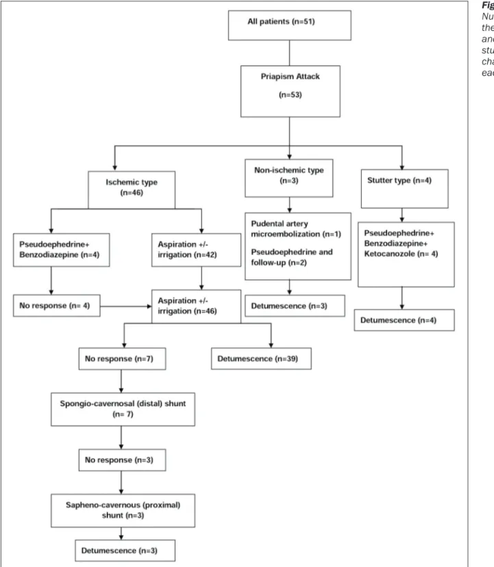
During the treatment of 53 priapism attacks, one non-ischemic priapism patient with pelvic trauma etiology was treated with pudendal artery micro-embolization by inter-ventional radiology, two patients with 4 stutter attacks were orally given pseudoephedrine 60 mg+diazepam 5 mg+ketoconazole 200 mg and two non-ischemic priapism patients were orally given pseudoephedrine 60 mg. Four ischemic priapism patients were given pseudoephedrine 60 mg+ diazepam 5 mg medical treatment. But since de-tumescence was not provided, corporeal aspiration and/or phenylephrine irrigation were applied to a total of 46 patients including these four patients (200 mcg/ml, maxi-mum: 1 mg). For corporeal aspiration and irrigation, a transcavernosal 18 Gauge needle was placed percuta-neously into the lateral aspect of the proximal penile shaft. Aspiration and evacuation of blood from the corpora cav-ernosa were performed with irrigation of normal saline followed by irrigation with phenylephrine (200 ug/mL) in saline and administered intermittently as 1.0 mL, every 3-5 min to a maximum dosage of 1 mg. Detumescence was provided in 39 patients through aspiration and/or irriga-tion. Spongiocavenous (distal) shunt was applied to seven patients unresponsive to aspiration and irrigation. Winter shunt and T shunt were applied as spongiocavernous shunt. Safenocavernous (proximal) shunt was applied to three patients not benefitting from this approach. Aspiration and/or irrigation treatment were the most com-mon method used for treatment at a statistically significant
Table 1.
General information of patients.
P value*
Number of patients 51
Number of priapism attacks 53 Mean age (years) 47.2 (10-69) Mean priapism duration (hours) 17.68 (5-104) Mean hospitalization duration (hours)
19.2 (4-215) Etiology (n = 51) PDU: 31 (60.7%) < 0.001 PDE 5 inh.: 9 (17.6%) Idiopathic: 5 (9.8%) Urethral intervention: 2 (3.9%) Pelvic trauma: 2 (3.9%) Pelvic mass: 1 (1.88%) Antipsychotic drugs: 1 (1.88%) Priapism type (n = 53) Ischemic: 46 (86.7%)
Non-ischemic: 3 (5.6%)
Stutter: 4 (7.5%) < 0.001
PDU: Penile Doppler ultrasonography;
level (p < 0.001). Mean hospitalization time of the patients was measured as 19.2 hours (4-215). Number of the patients and the study flow chart in each step are demon-strated in Figure 1.
DISCUSSION
Priapism was published in 1845 for the first time in modern medicine literature (7). Recurrent priapism was defined in 1980 together with sickle cell anemia cases (8). After that it became a pathology covering a wide range of the studies in literature.
During the first referral of priapism patient, priapism duration, possible etiological factors, previous priapism
story, presence of accompanying pain should be ques-tioned and penile Doppler ultrasonography and/or cor-poreal aspiration with blood gas analysis should defi-nitely be made in order to determine priapism type (2, 3). There are three different types of priapism: ischemic (veno-occlusive, low flow), non-ischemic (arte-rial, high flow) and stuttering (recurrent) priapism. Pathophysiological causes and treatment methods of every priapism type are different. There is no or very low arterial flow in corpus cavernosum in ischemic priapism (IP). In non-ischemic priapism, cavernosal flow can be normal, high or irregular and arteriosinusoidal fistule or pseudoaneurism may be observed. Stuttering priapism has repetitive, spaced erections (1-4). Ischemic priapism
Figure 1. Number of the patients and the study flow chart in each step.
is the most common type among priapism types. Ischemic priapism constitute 95% of all priapism cases. It is characterized by painful erection accompanied by significant permanent hardness in corpus cavernosum due to venous blood exit disorder. Occurrence of hypox-ia, hypercapnia and acidosis is similar to penile com-partment syndrome and this situation may cause tissue damage. In ischemic priapism, the ultrastructural changes in cavernosal flat muscle are observed 12 hours later, focal necrosis 24 hours later and finally necrosis and transformation of wide necrosis and fibroblast-like cells are observed 48 hours later.
Thus emergency inspection and management are required and delayed treatment may cause total erectile dysfunction (ED) (2-4). In the patient group in this study, ischemic priapism ratio was detected as 86.7%. There is an increase in priapism cases recently due to the frequent use of phosphodiesterase 5 enzyme (PDE5) inhibitors in ED treatment and to penile Doppler ultra-sonography (PDU) generally accompanied by intracaver-nosal papaverine injection used for erectile dysfunction (ED) diagnosis. Prolonged erections after intracavornos-al injection may be seen with a ratio of 5-35% (9). In our priapism patient group, intracavernosal injection related priapism accounted for 60.7% and priapism developing after PDE 5 use for 17.6% of cases. All patients were using papaverine in priapism occurring due to intracav-ernosal injections. Idiopathic causes with a ratio of 9.8%, urethral intervention and pelvic trauma with 7.5%, antipsychotic drug use with 1.96% and pelvic mass with 1.96% played a role in etiology in this order. One of the most common causes of priapism is sickle cell disease (10). Interestingly, sickle cell disease was not detected as etiology in any of the patients in our group. This may be due to the fact that these patients were generally fol-lowed up by hematology department and were referred to their own hematologists or to child emergency service since they had their first attack between the ages 15 and 18 generally.
Suggestions of European Association of Urology guidelines and American Urological Association guidelines were used for the treatment of all patients (2, 11). Spontaneous res-olution ratio is 62% with follow-up in non-ischemic pri-apism treatment and selective artery embolization can be made on demand (12). We followed up one patient with selective artery embolization and two patients with pseu-doephedrine treatment in this group.
A complete response was obtained in all three patients. It is recommended to manage stutter priapism like pri-apism (13).
Medical agents such as pseudoephedrine, ketoconazole, GnRh agonists and 5-alpha reductase inhibitors were used for priapism attack and for preventing the attack (2, 12, 14).
We applied pseudoephedrine+benzodiazepine+keto-conazole medical treatment for our stutter priapism patients and we were successful in the treatment of four attacks. Time is important in ischemic priapism since serious complications may occur. So aspiration and/or irrigation should be the primary treatment. Primarily dis-tal and then proximal shunt treatments should be used in irresponsive cases. Penile prosthesis may provide an
effective treatment to preserve penile length in ischemic priapism patients not responding to conventional treat-ment (15). Penile prosthesis application was not required in any of the patients in this study.
Non-invasive treatment approaches are recommended for ischemic priapism in current studies. Habous et al. provided detumescence in 34% of priapism patients in 60 minutes with salbutamol 4 mg oral treatment which is a β2 adrenergic agonist (9). Lowe and Jarow compared terbutaline which is a X2 adrenergic agonist and pseu-doephedrine which is β- and β2-adrenergic agonist in priapism patients and with a respond ratio of 38% with terbutaline and 28% with pseudoephedrine (16). Due to their antiandrogenic effect, gonadotropin-releasing hor-mone agonists, estrogens, anti- androgens and 5α-reduc-tase inhibitors can also be used as non-invasive medical treatment of priapism (2, 3). We recommend the use of these medicines in early-period priapism and stutter type priapism especially. This is due to the fact that delay in ischemic priapism treatment especially may result in per-manent erectile dysfunction in the patient.
Especially in case of the patient being responsive to the application of symptomatic medicines such as terbu-taline, salbutamol and pseudoephedrine during the first intervention in emergency service, the patient would be saved from an invasive intervention.
Emergency physicians generally leave priapism inter-vention to urologists in Turkey. In our study, treatment of all patients were managed by urologists. Since patients primarily refer to emergency service for pri-apism which is among the important urological emer-gencies, we believe that emergency physicians should have a good mastery of non-invasive and invasive treat-ment protocols for priapism and have equiptreat-ment to provide required intervention on this.
CONCLUSIONS
Underlying etiological factors and priapism type should definitely be determined before starting priapism man-agement. Application of non-invasive treatments in suit-able patients would protect patients from invasive painful interventions. We believe that emergency physi-cians be more effective in priapism first phase manage-ment and at least in non-invasive treatmanage-ment. Deficiencies during this phase may be made up through common education programs in urology and emergency medicine clinics.
REFERENCES
1. Kadıoglu A, Sanlı Ö, Ersay A, et al. Practical Management of Priapism. Turkish J Urol. 2006; 32:182-192.
2. Salonia A, Eardley I, Giuliano F, et al. European Association of Urology guidelines on priapism. Eur Urol. 2014; 65:480-9. 3. Shigehara K, Namiki M. Clinical Management of Priapism: A Review. World J Mens Health. 2016; 34:1-8.
4. Song PH, Moon KH. Priapism: current updates in clinical man-agement. Korean J Urol. 2013; 54:816-23.
an intracavernosal injection therapy programme. BJU Int. 2012; 110:1787-91.
6. Broderick GA, Kadioglu A, Bivalacqua TJ, et al. Priapism: patho-genesis, epidemiology, and management. J Sex Med. 2010; 7:476-500. 7. Tripe JW. Case of continued priapism. Lancet 1845; 2:8. 8. Emond AM. Holman R, Hayes RJ, Serjeant GR. Priapism and impotence in homozygous sickle cell disease. Arch Intern Med. 1980; 58:113-8.
9. Habous M, Elkhouly M, Abdelwahab O, et al. Noninvasive treat-ments for iatrogenic priapism: Do they really work? A prospective multicenter study. Urol Ann. 2016; 8:193-6.
10. Cita KC, Brureau L, Lemonne N, et al. Men with sickle cell ane-mia and priapism exhibit increased hemolytic rate, decreased red blood cell deformability and increased red blood cell aggregate strength. PLoS One. 2016; 11:e0154866.
11. Montague DK, Jarow J, Broderick GA, et al. Members of the
Erectile Dysfunction Guideline Update Panel; Americal Urological Association. American Urological Association guideline on the man-agement of priapism. J Urol. 2003; 170:1318-24
12. Muneer A, Ralph D. Guideline of Guidelines Priapism. BJU Int. 2017; 119:204-208.
13. Muneer A, Garaffa G, Minhas S, Ralph DJ. The management of stuttering priapism within a specialist unit: a 25 years experience. British Journal of Medical and Surgical Urology. 2009; 2:11-16 14. Levey HR, Kutlu O, Bivalacqua TJ. Medical management of ischemic stuttering priapism: a contemporary review of the litera-ture. Asian J Androl. 2012; 14:156-63.
15. Zacharakis E, Raheem AA, Freeman A, et al. Early insertion of a malleable penile prosthesis in ischaemic priapism allows later upsizing of the cylinders Scan J Urol. 2015; 26:1-4.
16. Lowe FC, Jarow JP. Placebo controlled study of oral terbutaline and pseudoephedrine in management of prostaglandin E1 induced prolonged erections. Urology. 1993; 42:51-3.
Correspondence
Mehmet Giray Sönmez, MD Assistant Prof (Corresponding Author) [email protected]
Hakkı Hakan Taskapu, MD Assistant Prof Yunus Emre Gög˘er, MD Assistant Prof Ahmet Öztürk, MD Prof
Department of Urology, Meram Medical Faculty, Necmettin Erbakan University, Yunus Emre quarter, 42080 Konya, Turkey
Leyla Öztürk Sönmez, MD
Zerrin Defne Dündar, MD Associate Prof
Department of Emergency Medicine, Meram Medical Faculty, Necmettin Erbakan University, Konya, Turkey
Cengiz Kara, MD Associate Prof
Department of Urology, Medical Park Ankara Hospital, Ankara, Turkey Togay Evrin, MD Assistant Prof
Department of Emergency Medicine, Ufuk University Medical Faculty, Ankara, Turkey