Celiac artery stenosis (CAS) is a common finding and oc-curs in up to 24% of the general population (1,2). It is usually asymptomatic. The collateral arterial blood supply prevents ischemia in the pancreas via the superior mesenteric artery. The pancreaticoduodenal arcades between the gastroduodenal artery (GDA) and inferior pancreaticoduodenal artery (PDA) are the major collateral pathways from the superior mesenteric artery to the celiac truncus.
The median arcuate ligament (MAL), a fibrous band that originates from diaphragmatic crura, is one of the major rea-sons for stenosis. The MAL may cause stenosis either as a result of inferior positioning of the diaphragm or a high origin of the celiac truncus (3).
In a small group of these patients, CAS causes symptoms. Because of the collaterals, CAS is not hemodynamically
significant in most cases. During pancreaticoduodenectomy (PD), intraoperative division of the GDA, which is the main vessel for the collateral blood, is necessary (4). In order to pre-vent postoperative hepatic and biliary ischemic complications the celiac artery should be patent.
In this paper, we present a patient with a mass in the gastric bulbus who was diagnosed with celiac artery stenosis during PD.
CASE PRESENTATION
A 60-year-old woman was referred to our hospital with a mass in the gastric bulbus that was detected with gastroscopy. The physical examination and laboratory findings were
un-Background: Celiac artery stenosis (CAS) is a not a
rare finding in the general population. The median ar-cuate ligament (MAL) is a fibrous arch that unites the diaphragmatic crura on either side of the aortic hiatus and, while it normally passes cranial to the origin of the celiac truncus, a low-lying ligament may lead to compression of the celiac artery and is the one of the major causes of CAS.
Case Report: In this paper, we present a patient with
a mass in the gastric bulbus who was diagnosed with celiac artery stenosis during the pancreaticoduodenec-tomy (PD). MAL was the cause of the celiac artery ste-nosis, determined based on the findings of preoperative computed tomography (CT).
Conclusion: Although CAS is usually asymptomatic
due to the collateral blood supply, it may be associated with potentially disastrous results due to ischemia of the upper abdominal organs as a result of disruption of the collateral pathways. It is especially important to recognize the presence of CAS and its etiology before interventional procedures. With the increasing use of multidetector computed tomography (MDCT), it be-comes essential for radiologists to be aware of this en-tity and the cross-sectional findings.
Keywords: Celiac artery, median arcuate ligament,
mul-tidetector computed tomography, pancreaticoduodenec-tomy
Address for Correspondence: Dr. Özdil Başkan, Department of Radiology, İstanbul Medipol University, İstanbul, Turkey Phone: +90 533 437 26 87 e-mail: [email protected]
Received: 31.01.2014 Accepted: 05.11.2014 • DOI: 10.5152/balkanmedj.2015.15082 Available at www.balkanmedicaljournal.org
Cite this article as:
Başkan Ö, Özdenkaya Y, Erol C, Dolay K. Problems with the Median Arcuate Ligament Should Be Recognized Before Surgery; its Importance in Pancreaticoduodenectomy. Balkan Med J 2015;32:312-5.
Copyright 2015 © Trakya University Faculty of Medicine Balkan Med J 2015;32:312-5 Case Report | 312
Problems with the Median Arcuate Ligament Should Be Recognized
before Surgery; Its Importance in Pancreaticoduodenectomy
1Department of Radiology, İstanbul Medipol University, İstanbul, Turkey 2Department of Surgery, İstanbul Medipol University, İstanbul, Turkey
remarkable. The patient underwent abdominal multiphase computed tomography (CT, Brilliance iCT, Phillips Health-care, Best, the Netherlands) for evaluation of the mass. The CT showed a 6 cm mass extending between the bulbus and the Papilla Vateri. It was decided that the lesion was not suitable for endoscopic or transduodenal local excision because of its location, size and broad-based configuration. Informed con-sent form was obtained. After further assessment the patient was organized to undergo PD.
A potential pitfall in PD is misidentification of a replacing common hepatic artery (CHA), or even a replacing right he-patic artery as the GDA. A technique to avoid this mistake is the GDA occlusion test; placing a vascular clamp across the presumed GDA and checking for pulsations at the porta hepa-tis before this vessel is divided. After clamping the GDA, the adequacy of the blood supply was assessed by digital palpa-tion on the CHA. In our case, it was noted there was a reduced pulsation in the CHA following the clamping of the GDA.
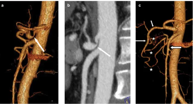
When the preoperative CT examination was evaluated ret-rospectively during the operation, she was found to have a stenosis at the origin of the celiac artery with patent branches. The CT demonstrated acute angulation and narrowing of the proximal celiac artery. There was post-stenotic dilatation and a classical hooked appearance of the proximal celiac artery that is characteristic of MAL compression (Figure 1). Well-developed collateral pathways between the SMA and GDA
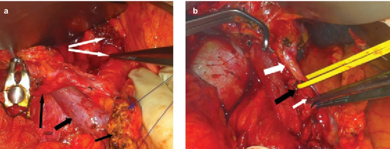
were detected (Figure 1). We decided that MAL was the cause of the celiac artery stenosis based on the CT findings. The MAL was divided and the origin of the celiac truncus was completely cleared to remove all MAL fibers. This led to an immediate release of the compressed celiac artery. A pulsatile flow was achieved in the lumen of the CHA despite clamping of the GDA (Figure 2). The patient underwent elective PD. Postoperatively, there were no signs of hepatic or anastomotic ischemia. The patient was discharged on the 7th postoperative
day without any complication. Histopathological diagnosis of the lesion was villous adenoma with in situ carcinoma and adjuvant therapy was not planned.
DISCUSSION
The MAL is a fibrous band that originates from a combi-nation of the left and right portions of diaphragmatic crura. The ligament crosses the aortic hiatus at the level of the L1 vertebral body proximal to the origin of the celiac truncus. In 10–24% people the MAL may cause compression and down-ward angulation of the celiac artery (5). Median arcuate liga-ment syndrome (MALS) is defined as ischemia in the gastro-intestinal system due to compression of the celiac truncus by the MAL, and was first described in 1963 (6). The incidence of MALS is 1.76–4% (7,8).
FIG. 1. a-c. Sagittal 3D VR image (a), Sagittal MIP image (b), CT angiogram demonstrating acute angulation and narrowing of the proximal celiac artery. There is post-stenotic dilatation and a classical hooked appearance (long white arrow) of the proximal celiac artery that is characteristic of MAL compression. Sagital oblique 3D VR image (c), CT angiogram shows prominent collateral vessels (stars) and dilatation of the GDA (long arrow) that is supplying the common hepatic artery (short arrow) off the superior mesenteric artery (thick arrow). This configuration is a common collateral pathway seen in patients with celiac axis stenosis.
a b c
Balkan Med J, Vol. 32, No. 3, 2015
313 Başkan et al. Median Arcuate Ligament
The celiac artery arises from the aorta and supplies blood to the liver, spleen, stomach, and duodenum. The causes of CAS can be classified as extrinsic stenosis (MAL), intrinsic stenosis (arteriosclerosis) and various other causes, including congenital, neoplastic etc. (7,9). The MAL is the most com-mon cause of CAS in Eastern countries (7), in contrast to Western countries where arteriosclerosis is the most common cause (9). The incidence of celiac artery stenosis is 7.6% (10) in the population undergoing PD. MAL compression is the major cause of stenosis in this group, which is detected in up to 5% of patients (11-13).
Hepatic and biliary system infarction is a well-known com-plication of PD (14). In most cases, there is an underlying pa-thology in the arterial system, especially at the origin of the celiac artery. Awareness of these conditions during preopera-tive examinations is important and may prevent postoperapreopera-tive complications.
In the presence of CAS, the arterial blood supply to the hepatobiliary system, pancreas, stomach, and spleen is sup-ported by the collaterals between the celiac artery and SMA. Because of these collateral pathways (15) CAS usually does not cause clinical symptoms. During PD, many of these col-lateral vessels are ligated. In the presence of CAS, ligation of the GDA can cause ischemia of the liver, biliary system, stomach and spleen. Ischemia of these organs can complicate early and late results of PD and hepatic function (14). Celiac artery stenosis due to MAL can be identified intraoperatively by clamping the GDA. The absence of pulsatile flow in the CHA should lead one to suspect stenosis of the celiac artery and the presence of MAL. Removing the MAL will solve the problem. Otherwise in these cases ischemia might cause seri-ous complications, including death.
Therefore, it is important to maintain the blood supply to the upper abdominal organs in these patients based on a detailed preoperative evaluation. MAL compression of the celiac truncus is conventionally diagnosed by catheter an-giography; however, with recent developments, MDCT has become a valuable tool for examination of vasculature sys-tems. In patients with celiac artery compression by the MAL, the findings include focal narrowing of the proximal celiac artery with a characteristic hooked appearance, hypertrophy of the diaphragmatic crura, a prominent GDA, and the pres-ence of prominent collateral arteries (12,16). The three-di-mensional MDCT images allow detailed visualization of the complex vascular anatomy with multiplanar reformatting, maximum intensity projection (MIP) and volume rendering (VR) techniques. This provides the diagnosis and the possi-bility of treating compression of MAL preoperatively. Endo-vascular celiac artery stenting, laparoscopic median arcuate ligament release, and intraoperative revascularization are the treatment options.
In conclusion, prior to PD, preoperative evaluation of the celiac artery, pancreatic and liver arterial supply should be performed in order to exclude celiac artery stenosis and pre-vent disastrous outcomes, include significant liver necrosis and death. MDCT is a non- invasive and useful method for diagnosing compression of the MAL. Careful preoperative preparation and awareness of vascular anatomical variations for vascular reconstruction will make PD a safer procedure.
Ethics Committee Approval: N/A.
Informed Consent: Written informed consent was obtained from the patient who participated in this study.
FIG. 2. a,b. Intraoperative images before (a) and after (b) release of the MAL. After clamping the GDA, the GDA and CHA were seen to lose their luminal tension (a). MAL (long white arrows), GDA (long black arrow), CHA (short white arrow), Portal vein (short thick black arrow), pancreatic remnant (short thin black arrow). After the MAL was divided and the origin of the celiac trunk was completely cleared to remove all MAL fibers, as expected, the lumen of the CHA and hepatic propria were filled (b). Hepatic propria (thick white arrow), CHA (thin white arrow), stump of GDA (black arrow)
a b
Balkan Med J, Vol. 32, No. 3, 2015
Peer-review: Externally peer-reviewed.
Author contributions: Concept - Ö.B., C.E; Design - ÖB.; Su-pervision - C.E., K.D.; Resource - Ö.B, Y.Ö., C.E., K.D.; Materials - Ö.B., Y.Ö., K.D.; Data Collection &/or Processing - Ö.B., Y.Ö., K.D.; Analysis &/or Interpretation - Ö.B., Y.Ö., C.E., K.D.; Litera-ture Search - Ö.B., K.D.; Writing - Ö.B., K.D.; Critical Reviews - Ö.B., Y.Ö., C.E., K.D.
Conflict of Interest: No conflict of interest was declared by the authors.
Financial Disclosure: The authors declared that this study has re-ceived no financial support.
REFERENCES
1. Bron KM, Redman HC. Splanchnic artery stenosis and occlu-sion. Radiology 1969;92:323-8. [CrossRef]
2. Kwon JW, Chung JW, Song SY, Lim HG, Myung JS, Choi YH, et al. Transcatheter arterial chemoembolization for hepatocel-lular carcinomas in patients with celiac axis occlusion. J Vasc
Interv Radiol 2002;13:689-94. [CrossRef]
3. Gloviczki P, Duncan AA. Treatment of celiac artery compres-sion syndrome: does it really exist? Perspect Vasc Surg
Endo-vasc Ther 2007;19:259-63. [CrossRef]
4. Bull DA, Hunter GC, Crabtree TG, Bernhard VM, Putnam CW. Hepatic ischemia, caused by celiac axis compression, compli-cating pancreaticoduodenectomy. Ann Surg 1993; 217:244-7.
[CrossRef]
5. Levin D C, Baltaxe H A. High incidence of celiac axis narrow-ing in asymptomatic individuals. Am J Roentgenol Radium Ther
Nucl Med 1972;116:426-9. [CrossRef]
6. Harjola PT. A rare obstruction of the coeliac artery: report of a case. Ann Chir Gynaecol Fenn 1963;52:547-50.
7. Park CM, Chung JW, Kim HB, Shin SJ, Park JH. Celiac axis stenosis: incidence and etiologies in asymptomatic individuals.
Korean J Radiol 2001;2:8-13. [CrossRef]
8. Ilica AT, Kocaoglu M, Bilici A, Ors F, Bukte Y, Senol A, et al. Median arcuate ligament syndrome: multidetector computed to-mography findings. J Comput Assist Tomogr 2007;31:728-31. 9. Berney T, Pretre R, Chassot G, Morel P. The role of
revascular-ization in celiac occlusion and pancreatoduodenectomy. Am J
Surg 1998;176:352-6. [CrossRef]
10. Kurosaki I, Hatakeyama K, Nihei KE, Oyamatsu M. Celiac axis stenosis in pancreaticoduodenectomy. J Hepatobiliary Pancreat
Surg 2004;11:119-24. [CrossRef]
11. Kohler TR, Debas H, Crames M, Strandness DE Jr. Pancreatico-duodenectomy and the celiac artery compression sndrome. Ann
Vasc Surg 1990;4:77-80. [CrossRef]
12. Farma JM, Hoffman JP. Nonneoplastic celiac axis occlusion in patients undergoing pancreaticoduodenectomy. Am J Surg 2007;193:341-4. [CrossRef]
13. Sugae T, Fujii T, Kodera Y, Kanzaki A, Yamamura K, Yamada S, et al. Classification of the celiac axis stenosis owing to median arcuate ligament compression, based on severity of the stenosis with subsequent proposals for management during pancreatico-duodenectomy. Surgery 2012;151:543-9. [CrossRef]
14. Raman SP, Horton KM, Cameron JL, Fishman EK. CT after pan-creaticoduodenectomy: spectrum of normal findings and compli-cations. AJR Am J Roentgenol 2013;201:2-13. [CrossRef]
15. Song SY, Chung JW, Kwon JW, Joh JH, Shin SJ, Kim HB, et al. Collateral pathways in patients with celiac axis stenosis: angio-graphic-spiral CT correlation. Radiographics 2002;22:881-93.
[CrossRef]
16. Horton KM, Talamini MA, Fishman EK. Median arcuate liga-ment syndrome: evaluation with CT angiography.
Radiograph-ics 2005;25:1177-82.[CrossRef]
Balkan Med J, Vol. 32, No. 3, 2015
315 Başkan et al. Median Arcuate Ligament