Department of Anesthesiology and Reanimation, Dumlupinar University, Kutahya, Turkey
Submitted (Başvuru tarihi) 03.04.2018 Accepted after revision (Düzeltme sonrası kabul tarihi) 28.05.2018 Available online date (Online yayımlanma tarihi) 28.06.2018
Correspondence: Dr. Lokman Demir. Department of Anesthesiology and Reanimation, Dumlupinar University, Kutahya, Turkey. Phone: +90 - 505 - 153 71 13 e-mail: [email protected]
© 2018 Turkish Society of Algology
150 JULY 2018
Ultrasound-guided distal adductor canal catheter placement for
continuous postoperative analgesia in lower leg surgery
Alt ekstremite cerrahisinde postoperatif ağrı kontrolü için ultrason eşliğinde distal
adduktor kanala kateter yerleştirilmesi
Onur BALABAN, Tayfun AYDIN, Lokman DEMIR Agri 2018;30(3):150-151
doi: 10.5505/agri.2018.54037
L E T T E R T O T H E E D I T O R
PAINA RI
To the Editor,
Distal adductor canal block is performed under ul-trasound guidance by administering high volume of a local anesthetic into the adductor canal, close to the hiatus.[1] Local anesthetic injection into the
ad-ductor canal leads to its distribution, thus reaching the sciatic nerve in the popliteal fossa.[2] Single
injec-tion into the adductor canal at the distal hiatus level blocks both the sciatic and saphenous nerves and provides analgesia at the lower leg, ankle, or foot.[3, 4]
We would like to present a case of ultrasound-guided catheter placement to the adductor canal for contin-uous postoperative analgesia in lower leg surgery. Written informed consent was obtained for the pub-lication of this report. The patient was a 23-year-old male who was operated for second, third, and fourth metatarsal fractures. The operation started under spinal anesthesia; however, spinal block was worn off due to elongation of the surgery, and the local anes-thesia was converted to general. The surgery contin-ued for 3.5 h. After the surgery, the patient suffered from pain with an intensity of 8/10, evaluated by the numerical rating score (NRS) system. We performed ultrasound-guided single injection distal adductor canal block at post anesthesia unit using 30 ml of 0.25% bupivacaine. Pain intensity decreased to 0/10 in 20 min, and the patient was shifted to the surgery ward. After 4 h, the patient reported pain, which was rated as 6/10. We decided to place a catheter to the
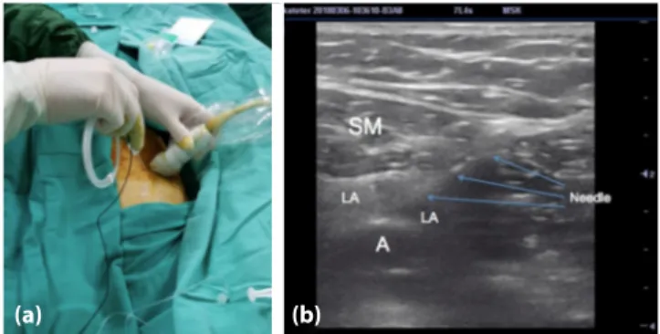
distal adductor canal under ultrasound guidance for prolonged analgesia. The patient was in prone po-sition during the procedure. After providing sterile conditions, a high frequency linear probe was placed transversally at the medial side of 1/3 distal thigh, and a cross-sectional image of the femoral artery was obtained. The needle was inserted in the anterior-to-posterior direction using the in-plane technique at the distal hiatus level of the adductor canal (Fig. 1a). When the tip of the needle reached the posterior of the sartorius muscle, 30 ml of 0.25% bupivacaine was administered. Distribution of the local anes-thetic solution was observed, in real time, posteriorly to the sartorius muscle and around the artery (Fig. 1b). A peripheral nerve catheter (Techniplex, Vygon, Ecouen, France) was placed through the needle into the area, which was enlarged due to the local anes-thetic solution. After 20 min, the intensity of pain
re-(a) (b)
Figure 1. (a) Position of the ultrasound probe and needle during
catheterization. (b) Ultrasound image showing the needle in the adductor canal and spread of the local anesthetic solution. A: artery; SM: sartorius muscle; LA: local anesthetic.
duced to 0/10. The patient needed a second dose 3.5 h after the catheter placement, third after 7 h, and fourth after 11 h, with the administration of 10 ml of 0.25 ml bupivacaine. The catheter was withdrawn af-ter 36 h, and the patient was discharged afaf-ter 2 days of stay in the hospital without any complications. No motor block was observed in the patient during the treatment. Distal adductor canal blocks are usually performed for analgesia at the lower leg, and low concentrations of the local anesthesia are usually administered. In previous anecdotal reports, no mo-tor block involving the sciatic nerve was reported.[1, 4]
Also, because the needle was not directly advanced around the sciatic nerve, high accumulation of drugs around the nerve leading to motor block was not ex-pected.
Continuous and sufficient analgesia was achieved at the lower leg with adductor canal catheter
place-ment in our case. Distal adductor canal block has the advantage of blocking both sciatic and saphenous nerves with a single injection in the prone position, and continuous postoperative analgesia is possible with catheter placement.
References
1. Morozumi K, Takahashi H, Suzuki T. Distal adductor canal block for administering postoperative analgesia in lower limb surgery. J Clin Anesth 2018;44:44. [CrossRef]
2. Andersen HL, Andersen SL, Tranum-Jensen J. The spread of injectate during saphenous nerve block at the ad-ductor canal: a cadaver study. Acta Anaesthesiol Scand 2015;59(2):238–45. [CrossRef]
3. Marian AA, Ranganath Y, Bayman EO, Senasu J, Brennan TJ. A Comparison of 2 Ultrasound-Guided Approaches to the Saphenous Nerve Block: Adductor Canal Versus Distal Transsartorial: A Prospective, Randomized, Blinded, Nonin-feriority Trial. Reg Anesth Pain Med 2015;40(5):623–30. 4. Tulgar S, Selvi O. Ultrasound guided distal adductor canal
block provides effective postoperative analgesia in lower leg surgery. J Clin Anesth 2018;45:51. [CrossRef]
JULY 2018 151