Evidence for the Control of Aggrecanases by Insulin and
Glucose in Alzheimer’s Disease
Sumeyya Akyol1, Veli Ugurcu2, Ozlem Cakmak3, Aynur Altuntas4, Yunus Yukselten5, Omer Akyol6, Asuman Sunguroglu5, Kadir Demircan1
ÖZET:
Alzheimer hastalığında insülin ve glukoz ile agrekanazların kontrolü üzerine kanıtlar Amaç: Alzheimer hastalığı (AH) kişinin hafıza ve bilişsel becerilerini, hatta en sonunda en basit görevleri yapma becerisini bile yavaş yavaş bozabilecek geri dönüşümsüz ve ilerleyici bir beyin hastalığıdır. AH’nın başlaması ve ilerlemesi, az anlaşılmış kompleks bir süreçtir. Bu çalışmada AH’da diyabetle ilişkili olduğu düşünülen, beyin fonksiyonlarının bozulması açısından risk teşkil eden muhtemel bazı mekanizmalar araştırılacaktır.
Yöntem: U87 (insan primer glioblastoma) hücrelerinin Dulbecco’s modifiye Eagle’s ortamında kültürü yapıldı. Hücreler 48 saat süre ile insülin (10 μg/ml), düşük miktar glukoz (11 mM, 2 mg/ml) ve yüksek miktar glukoza (55 mM, 10 mg/ml) maruz bırakıldı. Daha sonra harvest işlemi yapılarak protein izolasyonu gerçekleştirildi. Western blot membranları ve özel bir program ile primer anti-ADAMTS5, anti-IL-33, anti-NFκB, ve anti-GAPDH antikorları kullanılarak ilgili proteinlerin bantlarının tayini yapıldı ve bunların dansiteleri ölçülerek gruplar birbiri ile karşılaştırıldı. Bulgular: Western blot sonuçları insülin uygulanan U87 hücrelerinde ADAMTS5 proteininin azaldığını gösterdi. Düşük miktar glukoz uygulanması hücrelerde ADAMTS5 düzeylerinde orta derecede bir artışa yol açarken yüksek miktar glukoz uygulanması belirgin bir artışa yol açtı. Yüksek miktar glukoz uygulanan hücrelerde IL-33 protein düzeyinde açık bir indüksiyon tespit edilirken düşük miktar glukoz uygulanan grupta orta düzeyde bir azalma bulundu. İnsülin uygulanması ise IL-33 düzeyinde azalmaya neden oldu. NFκB ile immün reaksiyon veren antikorla yapılan muamelede insülin uygulanan hücrelerde NFκB protein düzeyinde keskin bir düşüş bulunurken düşük miktar ve yüksek miktar glukoz verilen gruplarda orta düzeyde bir azalma tespit edildi. Sonuç: Bu çalışma, U87 hücrelerinde aynı deneysel düzenek ile hem agrekanazlar hem de inflamasyon medyatörlerinin birlikte çalışıldığı ve sonuçlarının AH patofizyolojisiyle diyabet ve hipergliseminin birlikte yorumlanmaya çalışıldığı ilk çalışmadır. Çalışmanın sonuçları, insülin ve glukozun henüz bilinmeyen mekanizma(lar) ile ADAMTS, IL-33 ve NFκB sentezinde önemli fonksiyonları olabileceğini göstermektedir. Bütün agrekanazların ve diğer sınıf ADAMTS enzimlerinin yukarıdaki parametrelerle birlikte çalışılıp yorumlanacağı, ayrıca moleküler biyoloji, genetik, immunoloji ve diğer ilgili bilim dallarının ışığında AH’da ADAMTS enzimleri ve inflamasyon medyatörlerinin patofizyolojiye katkılarının değerlendirileceği ileri çalışmalara ihtiyaç vardır.
Anahtar sözcükler: ADAMTS5, IL-33, NFκB, diyabet, hiperglisemi, Alzheimer hastalığı, matriks yıkımı
Kli nik Psikofarmakoloji Bulteni 2014;24(4):323-32
ABS TRACT:
Evidence for the control of aggrecanases by insulin and glucose in Alzheimer’s disease Objective: Alzheimer’s disease (AD) is a progressive and irreversible central nervous system disease, which slowly destroys cognitive skills and memory, and eventually even the ability to handle the simplest tasks. The initiation and progression of AD is a poorly understood complex process. Here, we have investigated possible biological mechanisms that could be responsible for the increased risk for diminished brain function associated with diabetes in AD. Method: The U87 cell line (human primary glioblastoma cell line) was cultured in Dulbecco’s modified Eagle’s medium. Cells were incubated with insulin (10 μg/ml), low glucose (11 mM, 2 mg/ml) and high glucose (55 mM, 10 mg/ml) for 48 hours. Cells were harvested and protein isolations were performed. Primary ADAMTS5, anti-IL-33, anti-NFκB, and anti-GAPDH antibodies were used to detect corresponding proteins and to measure band densities in Western membranes using a specific program. Results: Western blot analysis showed ADAMTS5 protein decreases in insulin-applied U87 cells. High glucose application led to a notable increase in ADAMTS5 levels in cells, while low glucose application caused a moderate increase in ADAMTS5 levels. An apparent induction of IL-33 protein was observed in high glucose-applied cells, while a moderate decrease was noted in the low-glucose applied group. Insulin administration led to a decrease in IL-33 levels. Immunoreaction of NFκB with corresponding antibody was found to be sharply decreased in insulin-applied cells while low and high glucose application led to a moderate decrease in NFκB.
Conclusion: This is the first reported study that has investigated both aggrecanases and inflammation mediators in the same experimental setup with U87 cells and interpreted the results in the various aspects of AD pathophysiology related to diabetes and hyperglycemia. Our findings suggest that insulin and glucose may have important functions in the synthesis of ADAMTS, IL-33, and NFκB through undefined mechanism(s). Further investigations dealing with all aggrecanases and other class of ADAMTS enzymes should be carried out together with the above-mentioned parameters with the collaboration of molecular biology, genetics, immunology, and other related disciplines in order to elaborate the pathophysiological importance of ADAMTS enzymes and inflammation mediators in AD.
Keywords: ADAMTS5, IL-33, NFκB, diabetes, hyperglycemia, Alzheimer’s disease, matrix degradation
Bulletin of Clinical Psychopharmacology 2014;24(4):323-32
1Turgut Özal University, Faculty of
Medicine, Department of Medical Biology, Ankara - Turkey
2Dumlupınar University, Faculty of
Medicine, Department of Medical Biochemistry, Kütahya - Turkey
3Gazi University, Faculty of Education,
Department of Biology Education, Ankara - Turkey
4Ankara Regional Office of Council of
Forensic Medicine, Department of Chemistry, Ankara - Turkey
5Ankara University, Faculty of Medicine,
Department of Medical Biology, Ankara - Turkey
6Hacettepe University, Medical School,
Department of Medical Biochemistry, Ankara - Turkey
Corresponding author:
Sumeyya Akyol, PhD
Turgut Ozal University, Faculty of Medicine, Department of Medical Biology, Camlica Mh. Anadolu Bulvari No: 16/A, Gimat, Yenimahalle, Ankara - Turkey
Phone: +90-312-397-7400 Fax: +90-312-397-7448 E-ma il add ress:
[email protected] Date of submission: July 25, 2014 Date of acceptance: September 05, 2014 Declaration of interest:
S.A., V.U., O.C., A.A., Y.Y., O.A., A.S., K.D.: The authors reported no conflict of interest related to this article.
INTRODUCTION
A l z h e i m e r ’ s d i s e a s e ( A D ) i s a neurodegenerative disease and the most common cause of dementia that affects approximately 36 million individuals worldwide. The best known hallmarks of AD are the accumulation of extracellular amyloid beta peptide (Aβ)-rich senile plaques which stem from the cleavage of amyloid precursor protein (APP), the gathering of intracellular neurofibrillary tangles (NFTs) which are built up by the aggregated form of hyperphosphorylated Tau, and synapse loss. The exact etiology of AD remains unknown and there is no definite treatment yet available.1,2 The four genes encoding apolipoprotein E (APOE), APP, presenilin-1 (PSEN1), and presenilin-2 (PSEN2) are believed to be responsible for genetic as well as sporadic cases of AD.3
Healthy individuals exhibit a strict control between glucose metabolism and insulin. However, the close interaction between glucose metabolism and insulin function is not well controlled in diabetes mellitus (DM). The early phases of type 2 DM mainly include insulin resistance and hyperinsulinemia.4 Insulin and insulin-like growth factor type 1 (IGF-I) receptors are present in the central nervous system (CNS) and they are involved in growth and metabolic functions. Insulin and IGF-I are neurotropic substances because they increase neuronal growth and differentiation. They also promote neuronal protein synthesis, neuronal migration and synapse formation.5-7 The medial temporal lobe is involved in the pathophysiology of AD8; for this reason, identification of its structures may provide useful knowledge concerning the effect of insulin on memory and AD pathophysiology.
AD patients have been reported to have higher levels of plasma glucose.9 Additionally, non-diabetic patients with AD or vascular dementia have significantly higher insulin and glucose blood levels compared to those of control subjects.10 As a pathophysiological mechanism, insulin may interact with the metabolism of proteins related with amyloid plaques and NFTs; namely Aβ and tau.11
Several proteins like tau, APP, neurofilaments, and synaptosomal proteins are involved in the pathophysiology of the AD. In normal brain, these proteins are GlcNAcylated but in AD, the brain may reduce the normal GlcNAcylation of tau due to defective glucose metabolism. This may induce tangle formation and finally neuronal death. AD and type 2 DM represent two metabolic disorders, in which dysfunctional protein GlcNAcylation/ phosphorylation might be important for disease pathology.12
An additional potential factor that may contribute to the onset of AD is interleukins. IL-33, a member of the interleukin-1 (IL-1β) family, may have an important function in AD.13 IL-33 acts as a proinflammatory cytokine as well as an intra-cellular nuclear factor with transcriptional regulatory properties. It may be specifically proinflammatory in the CNS.13 Microglia and astrocytes express IL-33 receptors. The glial cells respond by proliferating and releasing tumor necrosis factor-alpha (TNF-α), IL-1β, and nitric oxide (NO).14 It is known that both IL-1β and TNF-α increase in the brains of AD patients compared to age-matched control subjects.15 IL-33 release in excess above the physiological level may induce proinflammatory reactions and may act as a prominent inflammatory mediator of neuroinflammation in the CNS. Further, IL-33 is known to activate inflammatory cells, including glial cells.16,17 NFκB controls many genes that are involved in inflammation and immune responses, as well as cell division and apoptosis. In the murine diabetes model, intranasal insulin has been found to decrease NFκB activation.18 Studies of postmortem brains from AD have shown increased NFκB activity in cells involved in the n e u r o d e g e n e r a t i v e p r o c e s s .1 9 Immunohistochemical studies have demonstrated that NFκB activity was increased in cholinergic neurons in the AD patients; therefore dysfunction of cholinergic neurons is thought to contribute to the cognitive loss in AD.20
A disintegrin-like and metalloproteinase with thrombospondin motifs (ADAMTSs) are a family of new proteinases belonging to the
metzincin-superfamily of metalloendopeptidases. This proteinase enzyme group is primarily located in the extracellular matrix (ECM). It is a member of the Matrix Metalloproteinase (MMP) family in the ECM that breaks down substrates such as agrecan, versican, brevican, nidogen and procollagen. It has 19 members and different functions have been identified for each member. They are involved in many physiological processes and also many pathological processes such as arthritis and cancer. ADAMTS1, 4, 5, 8, 9 and 15 have aggrecanase activity whereas other members do not.
Biomarkers for insulin resistance and inflammation and the risk for dementia and AD have attracted scientists’ attention.21 Insulin resistance and inflammation are important hallmarks of type 2 DM and could provide a potential mechanism explaining the association between type 2 DM and AD. Although a central role for insulin in the pathology of AD has been suggested11,22, and intervention trials using insulin-sensitizing drugs to slow down cognitive decline are ongoing23, there remains uncertainty regarding the role of insulin and glucose tolerance. The deterioration in the insulin/glucose relationship in AD patients might indicate the possible role of hyperglycemia and insulin level changes in the initiation and prognosis of AD. It is crucial to investigate whether these changes affect the ECM or not, because the molecules produced/ degraded in the ECM might play a role in accumulation in the brain in AD. To our knowledge, no prior studies have simultaneously related measure of glucose homeostasis and markers of inflammation to the risk for AD patients. We have hypothesized that factors that underlie type 2 DM (insulin signaling and inflammation) may also contribute to the development of AD. To scrutinize this hypothesis, we aimed to investigate the difference in the levels of ADAMSTS5, an aggrecanase in glial cells, and the levels of IL-33 and NFκB, as inflammation mediators, in relation to glucose and insulin changes. Observing difference in levels of ADAMTS5, IL-33, and NFκB in relation to glucose and insulin changes will contribute to the
understanding of AD pathophysiology by determining the role of certain ECM and inflammation mediators.
METHODS
Cell culture and treatment: The U87 cell line (human primary glioblastoma cell line) was cultured in Dulbecco’s modified Eagle’s medium (Lonza) supplemented with 10% fetal bovine serum, 1% L-glutamine, and antibiotics (100 U/ mL penicillin, 100 U/mL streptomycin). The U87 cell line was maintained at 37°C in a 5% CO2-humidified atmosphere. Cells (initial cell density: 1.0 × 10⁵/ml) were incubated with insulin (10 μg/ ml), low glucose (11mM, 2mg/ml) and high glucose (55mM, 10 mg/ml) for 48 h. Powdered insulin was dissolved within 0.01N HCl solution. The stock solution had a 2 mg/mL concentration in 0.01N HCl and the concentration of the working solution was 10 μg/mL in the medium. After the experiment, cells were harvested and protein isolations were performed.
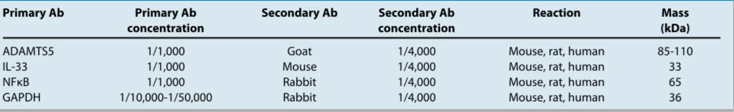
Protein Extraction, Western Blot Analysis, and Antibodies: Anti-ADAMTS5, anti-IL-33, anti-NFκB, and anti-GAPDH antibodies were purchased from Santa Cruz Biotechnology, Inc. (Santa Cruz, CA, USA) and Abcam (Cambridge, UK) and used at different dilutions. Before the study, cross-reactivity was confirmed as stated in the manufacturer’s data sheet. After the stimulation of the cells, they were washed once with phosphate buffered saline (PBS) and then scraped from the plates. The cells were solubilized with CelLytic TMM (200 μL, by Sigma) and protease inhibitor. The cells were centrifuged after incubating at 4 °C for 15 min, and then the supernatant was collected. The protein concentration was determined with standard bovine serum albumin and a protein-assay kit (Thermo Scientific Bradford Assay Kit, Rockford, IL, USA).
The samples were treated with Laemli Sample buffer and β-mercaptoethanol for 8 min, at 95 °C. A 5 μL aliquot of protein marker (BioRad Precision Plus Protein Western C Standard, Cat#161-0376) was added to the protein samples (15 μg) for
Western blot analysis. A 15 μL portion of each protein sample was loaded onto Western blot gel (BioRad Mini-PROTEAN TGX Stain-Free Gels, 4–15%, 12-well comb, 15 μL, Cat#456-8096). A BioRad 1X Tris/Glycerine/SDS running buffer was used and the samples were run at 250 V for 20 min. Later, proteins were moved to a polyvinylidene difluoride membrane (BioRad Trans-Blot Turbo Transfer Pack, 0.22 μM PVDF, Cat#170-4156) by using a transfer system (BioRad Trans-Blot Turbo Transfer System, Singapore) after the electrophoresis. Membranes were blocked for 1 h in 2.5% nonfat dried skim milk in Tris-buffered saline (TBS) with 0.05% of Tween 20 (TBS-T). That was incubated overnight with the primary anti-GAPDH antibodies, anti-ADAMTS5, anti-NFκB, and anti-IL-33 (Table 1) diluted in blocking buffer. After stringent washing with TBS-T three times for 8 min each at room temperature, the membranes were incubated approximately 1 hour with the suitable secondary antibodies (Table 1). Following three washes in TBS-T, immunoreactive bands were visualized for 90 seconds with the chemiluminescence system (BioRad Immun-Star Western C kit, Cat#170-5070). Signals were analyzed with BioRad ChemiDoc MP Imaging System (Singapore), and the density was measured using Image J software (W. Rasband, Research Services Branch, NIMH, NIH, Bethesda, MD) and normalized with the signal from GAPDH as standard control in every experiment. The standard curve by the GAPDH was used for the quantification of the samples.
RESULTS
Overall, one of the aggrecanase family members, ADAMTS5, as well as two of the
inflammation mediators, IL-33 and NFκB, were found to be expressed in U87 cells. Western blot analysis showed that ADAMTS5 protein was decreased in insulin-applied U87 cells up for 48 h (Figure 1) while high glucose application led to a notable increase in ADAMTS5 levels and low glucose led to a moderate increase in ADAMTS5 levels in cells. The ratios of ADAMTS5 protein levels in insulin-, low- and high-glucose-applied U87 cells compared to control cells were 0.708, 1.361, and 1.559, respectively. Immunoreactive proteins with molecular mass of 75 kDa for ADAMTS5 were detected. In accordance with the Tab le 1: Primary and secondary antibodies (Ab) used for Western blot techniques
Primary Ab Primary Ab Secondary Ab Secondary Ab Reaction Mass
concentration concentration (kDa)
ADAMTS5 1/1,000 Goat 1/4,000 Mouse, rat, human 85-110 IL-33 1/1,000 Mouse 1/4,000 Mouse, rat, human 33 NFκB 1/1,000 Rabbit 1/4,000 Mouse, rat, human 65 GAPDH 1/10,000-1/50,000 Rabbit 1/4,000 Mouse, rat, human 36
Figure 1: Western blot analysis of ADAMTS5. The graphic was obtained by calculating the intensity of protein bands of both ADAMTS5 and GAPDH and dividing them by each other
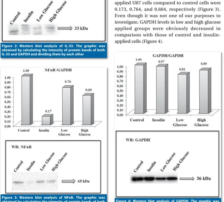
ADAMTS5 increase, an obvious induction of IL-33 protein (Figure 2) was seen in high glucose-applied cells while a moderate decrease was seen in the low-glucose applied group. Insulin led to a decrease in IL-33 levels just similar to that of low glucose. Immunoreactive proteins with molecular mass of 33 kDa for IL-33 were detected. A sharp decrease in NFκB levels was observed in cells treated with insulin (Figure 3). On the other hand, there were moderate decreases in NFκB levels in the low and high dose glucose groups of cells compared to control cells. The ratios of NFκB protein levels in insulin-, low-, and high-glucose-applied U87 cells compared to control cells were 0.173, 0.764, and 0.604, respectively (Figure 3). Even though it was not one of our purposes to investigate, GAPDH levels in low and high glucose applied groups were obviously decreased in comparison with those of control and insulin-applied cells (Figure 4).
Figure 2: Western blot analysis of IL-33. The graphic was obtained by calculating the intensity of protein bands of both IL-33 and GAPDH and dividing them by each other
Figure 3: Western blot analysis of NFκB. The graphic was obtained by calculating the intensity of protein bands of both
DISCUSSION
Recent evidence has indicated that type 2 DM increases the risk of development of AD. There are lots of studies that aimed to investigate the potential common processes that could explain this relationship. Determination of the interaction between DM and inflammation is of high importance in AD patients. In order to assess the involvement of the ADAMTS5, IL-33, and NFκB as pathophysiological factors in AD, we analyzed the immunoreactive proteins of these three important parameters, one of which is a member of the aggrecanases and the other two of which are inflammation mediators. High glucose led to up-regulation of both ADAMTS5 and IL-33 while insulin led to down-regulation of both proteins. Otherwise, insulin led to sharp down-regulation of NFκB while low- and high-glucose concentration lead to moderate down-regulation of NFκB.
Some epidemiological studies suggest the association between AD and insulin-resistant pathologic conditions, such as type 2 DM and hyperinsulinemia. In the Honolulu–Asia Aging Study, Type 2 DM was associated with an increased risk for dementia and AD.24 In the Rotterdam and the Mayo studies, type 2 DM increased the risk for AD, regardless of vascular dementia.25,26 Luchsinger et al. have reported that hyperinsulinemia is a risk factor for AD and general memory decrease in the elderly.27 As a conclusion, these reports suggest that AD may be related to insulin resistance.28 Hoyer has indicated that desensitization of the neuronal insulin receptor contributes to AD pathophysiology.29 In his model, decreasing CNS insulin levels lead to reduced acetylcholine levels and cerebral blood flow. Additionally, these conditions are associated with chronic deficits in brain oxidative metabolism, increased intracellular acidosis in the endoplasmic reticulum and Golgi and interference with Aβ protein, which is a peptide strongly related with AD. Supporting these findings, AD patients show reduced insulin receptor density and tyrosine kinase activity markers in their CNS.30
Inflammation
Pathologic conditions related to insulin resistance and hyperinsulinemia are associated with increased inflammation and increase the risk for AD.26,27 Insulin may contribute to CNS inflammation partly by its effect on Aβ. The Aβ interacts with inflammatory factors and increases pro-inflammatory cytokines.31 The inflammatory process starts with the activation of the precursors of proinflammatory caspases, that degrade the precursor forms of IL-1β, IL-18 and IL-33 into their active forms. The secretion of these substances leads to a potent inflammatory response, and influences the release of toxins from glial and endothelial cells. The production of inflammatory cytokines by other cells in the CNS contributes to the total inflammatory response as this also affects the function of the blood brain barrier (BBB).32 Studies demonstrate that many inflammatory substances like prostaglandins, chemokines, and cytokines are increased in the CSF and in neuroinflammatory conditions as well as neurodegenerative diseases such as multiple sclerosis, Parkinson’s disease, and AD. These studies suggest that inflammation is a key pathogenic factor for the development of AD.33 Increased levels of IL-6 and E2 isoprostane have been reported in the CSF of patients with AD.34 In
vitro and in vivo studies suggest that inflammation
interacts with processing and deposition of Aβ.35 Insulin modulates many characteristics of the inflammatory network in the periphery. Low doses of insulin exert anti-inflammatory effects;36 however, during chronic hyperinsulinemia, insulin may exacerbate oxidative stress markers and inflammatory response.37 Although the concepts of cerebral insulin resistance and insulin-induced amyloid pathology are attractive explanations for some of the effects of type 2 DM on the brain, there are still many unclear points to be clarified. A previous study has shown decreased IL-33 in the brains of AD patients compared to control brains by mRNA and immunohistochemical examination.38 The authors reported that IL-33 is restricted to vascular capillaries in the brain and
suggested IL-33 as a potential genetic determinant of AD.38 They suggested that there is clear evidence of an association between the risk of developing AD and the IL-33 gene. Our results revealed that insulin led to a decrease of IL-33 levels contrary to the decreased levels in AD, mentioned above. Increased expression of IL-33 and its receptor ST2 have been shown in the vicinity of amyloid plaques and NFTs in the affected entorhinal cortex of AD brains, as previously reported for IL-18 in AD brain.39 We first confirmed that IL-33 expression was increased after high glucose application compared with that of controls in glial cells showing the potential problem in brains of AD cases. Therefore, it can be suggested that there is clear evidence of an association between the risk of developing AD and the IL-33 gene.
Hormone Regulation
Insulin is not a major regulator of glucose metabolism in CNS, in contrast to its main function in peripheral organs like liver, muscle and fat.40 However, insulin receptors are widely present in the brain, especially in certain parts, like the hypothalamus and the hippocampus.41 The relation between insulin and Aβ/tau proteins is getting more attention.42 Aβ is converted from APP. It can aggregate with other proteins and form senile plaques just after its secretion into the extracellular area. Alternatively, excessive amounts of Aβ can be degraded via endocytosis, or direct extracellular cleavage.43 Insulin-degrading enzyme (IDE) is involved in the latter process.44 Insulin stimulates production of Aβ, and inhibits the extracellular cleavage of Aβ by competing with IDE.45 In a recent study AD patients have been reported to have a marked reduction in IDE levels in the hippocampus, compared to that of the controls.46 Interestingly, this only happened in patients carrying the Apolipoprotein E (APOE) ε4 allele. The APOE ε4 genotype has also been associated with increased risk for AD in patients with DM24. This finding has also been supported by some other neuropathological studies.24
The mechanism of acceleration of Aβ
degradation by insulin is not clear. Obtained findings suggest that insulin indirectly decreases the degradation of aggrecanase by decreasing ADAMTS activity. Therefore, it prevents the process of inflammation, accelerating plaque formation. Hormone levels were detected in mild AD patients relevant to glucoregulation before and during a hyperglycemic clamp.47 Before and during the hyperglycemic clamp, levels of insulin were higher in very mild AD patients, on the other hand that was lower for mild AD than those of controls. Stages of AD seem to have a role in the insulin response to the hyperglycemic clamp; the patients were tested again 1 to 1.5 years later. Patients progressing from very mild to mild AD have been reported to have higher levels of insulin before and during the hyperglycemic clamp than those of controls or patients who were in later stages of AD. These studies suggest that changes in the hormones involved in glucose regulation due to hyperglycemia might be associated with AD progression. Additionally, higher insulin levels are related with the early stage of the disease and these levels later decrease with the progression of the disease. The importance of these findings in the pathophysiology of AD is still not identified clearly. However, there may be an immediate clinical relevance to the phenomenon of improved memory under hyperglycemia at certain stages of AD.48 Impaired Aβ processing may have a potential to explain APOE modulation of the association between type 2 DM and AD. Aβ is the main component of the neuritic plaques. Literature data suggests that insulin may modulate Aβ levels by increasing intracellular Aβ release49 and by interfering with Aβ cleavage via insulin degrading enzyme that is highly present in the brain.50 Independent investigators with different studies have suggested that IDE may play a critical role in Aβ degradation in CNS.51
The effect of insulin resistance has also been examined in the rest of the body on Aβ levels in the brain. Experiments in animal models of AD showed that inducing insulin resistance and hyperinsulinemia increased the burden of Aβ in the brain. In humans, it was found through an
experimental diet intervention study that 4 weeks of a diet high in saturated fat and with a high glycemic index not only had a negative impact on metabolism, increasing insulin resistance and LDL cholesterol, but also significantly increasing markers of AD pathology (including Aβ) and oxidative injury detectable in the CSF, a representation of brain metabolism.25,34
Hyperglycemia
Literature data suggest that the toxic effects of hyperglycemia are involved in the brain damage caused by DM.52 Hyperglycemic rodents are reported to have some functional/structural changes in the CNS.52 Toxic effects of hyperglycemia are related to increased glucose transport through the hexosamine and polyol pathways, impairments of intracellular second messenger pathways, and an impairment of ROS.53 These steps affect brain tissue directly and cause m i c r ov a s c u l a r c h a n g e s.5 2 , 5 4 C h r o n i c a l l y, hyperglycemia in DM may cause changes in the cerebral capillaries, like thickening of the basal membrane.55 These microvascular changes may also cause chronic brain ischemia, which contributes to the development of subcortical white-matter pathologies.
Hyperglycemia is one of the factors that induce tau cleavage in diabetes. Tau cleavage in diabetes may be a key link for the increased incidence of AD in diabetic patients. Here we can speculate that increased ADAMTS enzymes might modify the extracellular matrix of brain cells and lead to more tau cleavage in diabetic conditions.
Mechanisms of Aβ clearance
There is a balance in the metabolism of Aβ in normal conditions. Recent studies have
demonstrated that AD is associated with impairment of BBB transport of Aβ and its enzyme-dependent degradation.56 Aβ is mainly degraded by two zinc metalloendopeptidases namely n e p h r i l y s i n a n d I D E .5 6 O t h e r metalloendopeptidases, like angiotensin-converting enzyme (ACE) and endothelin-converting enzyme (ECE) also cleave Aβ. ECE-2 can also hydrolyze Aβ in CNS.57 Epidemiological studies show the relationship between ACE and AD.58 Similarly, ACE has been recently shown to degrade Aβ.59 These pathways of Aβ elimination may become a potential target for the treatment of AD.56 H i g h l e v e l s o f i n s u l i n o r c h r o n i c hyperinsulinemia increase the inflammatory process. AD is a chronic neuroinflammatory pathologic condition; therefore, insulin abnormalities might affect AD progress via its effects on the neuroinflammatory pathways. Recent studies focus on preventive and therapeutic effects of anti-inflammatory drugs on AD. Accumulating findings recommend that insulin abnormalities have a role in the progress of neurodegenerative pathologies and DM. For this reason, management of insulin resistance may have some impact on diseases like AD and type 2 DM.
As a conclusion, there is a strong need for more research conducted with the collaboration of molecular biology, genetics, immunology and other related disciplines in order to show the pathophysiological importance of ADAMTS enzymes and inflammation mediators in AD. Further studies of all ADAMTS enzymes, NFκB and IL-33 for their function, signal transduction pathways involved in AD disease are also required. Results achieved through these future studies will provide new ideas for the treatment of chronic diseases such as AD, DM, and other similar diseases.
References:
1. Carter MD, Simms GA, Weaver DF. The development of new therapeutics for Alzheimer’s disease. Clin Pharmacol Ther 2010;88(4):475-86. [CrossRef]
2. Korczyn AD. Why have we failed to cure Alzheimer’s disease? J Alzheimers Dis 2012;29(2):275-82.
3. Kaminsky YG, Marlatt MW, Smith MA, Kosenko EA. Subcellular and metabolic examination of amyloid-beta peptides in Alzheimer disease pathogenesis: evidence for Abeta (25-35). Exp Neurol 2010;221(1):26-37. [CrossRef]
4. Ramlo-Halsted BA, Edelman SV. The natural history of type 2 diabetes. Implications for clinical practice. Prim Care 1999;26(4):771-89. [CrossRef]
5. D’Ercole AJ, Ye P, O’Kusky JR. Mutant mouse models of insulin-like growth factor actions in the central nervous system. Neuropeptides 2002;36(2-3):209-20. [CrossRef]
6. Popken GJ, Hodge RD, Ye P, Zhang J, Nq W, O’Kusky JR, et al. In vivo effects of insulin-like growth factor-I (IGF-I) on prenatal and early postnatal development of the central nervous system. Eur J Neurosci 2004;19(8):2056-68.
[CrossRef]
7. O’Kusky JR, Ye P, D’Ercole AJ. Insulin-like growth factor-I promotes neurogenesis and synaptogenesis in the hippocampal dentate gyrus during postnatal development. J Neurosci 2000;20(22):8435-42.
8. Squire LR. Memory and the hippocampus: a synthesis from findings with rats, monkeys, and humans. Psychol Rev 1992;99(2):195-231. [CrossRef]
9. Meneilly GS, Hill A. Alterations in glucose metabolism in patients with Alzheimer’s disease. J Am Geriatr Soc 1993;41(7):710-4.
10. Carantoni M, Zuliani G, Munari MR, D’Elia K, Palmieri E, Fellin R. Alzheimer disease and vascular dementia: relationships with fasting glucose and insulin levels. Dement Geriatr Cogn Disord 2000;11(3):176-80. [CrossRef]
11. Biessels GJ, Kappelle LJ. Increased risk of Alzheimer’s disease in Type II diabetes: insulin resistance of the brain or insulin-induced amyloid pathology? Biochem Soc Trans 2005;33(Pt 5):1041-4.
12. Dias WB, Hart GW. O-GlcNAc modification in diabetes and Alzheimer’s disease. Mol Biosyst 2007;3(11):766-72.
[CrossRef]
13. Van Eldik LJ, Thompson WL, Ralay Ranaivo H, Behanna HA, Martin Watterson D. Glia proinflammatory cytokine upregulation as a therapeutic target for neurodegenerative diseases: function-based and target-based discovery approaches. Int Rev Neurobiol 2007;82:277-96. [CrossRef]
14. Yasuoka S, Kawanokuchi J, Parajuli B, Jin S, Doi Y, Noda M, et al. Production and functions of IL-33 in the central nervous system. Brain Res 2011;1385:8-17. [CrossRef]
15. Llano DA, Li J, Waring JF, Ellis T, Devanarayan V, Wittle DG, et al. Cerebrospinal fluid cytokine dynamics differ between Alzheimer disease patients and elderly controls. Alzheimer Dis Assoc Disord 2012;26(4):322-8. [CrossRef]
16. Espinassous Q, Garcia-de-Paco E, Garcia-Verdugo I, Synquelakis M, von Aulock S, Sallenave JM, et al. IL-33 enhances lipopolysaccharide-induced inflammatory cytokine production from mouse macrophages by regulating lipopolysaccharide receptor complex. J Immunol 2009;183(2):1446-55. [CrossRef]
17. Theoharides TC, Zhang B, Kempuraj D, Tagen M, Vasiadi M, Angelidou A, et al. IL-33 augments substance P-induced VEGF secretion from human mast cells and is increased in psoriatic skin. Proc Natl Acad Sci U S A 2010;107(9):4448-53.
[CrossRef]
18. Francis GJ, Martinez JA, Liu WQ, Xu K, Ayer A, Fine J, et al. Intranasal insulin prevents cognitive decline, cerebral atrophy and white matter changes in murine type I diabetic encephalopathy. Brain 2008;131(Pt 12):3311-34.
19. Mattson MP, Camandola S. NF-kappaB in neuronal plasticity and neurodegenerative disorders. J Clin Invest 2001;107(3):247-54. [CrossRef]
20. Boissiere F, Hunot S, Faucheux B, Duyckaerts C, Hauw JJ, Aqid Y, et al. Nuclear translocation of NF-kappaB in cholinergic neurons of patients with Alzheimer’s disease. Neuroreport 1997;8(13):2849-52. [CrossRef]
21. van Himbergen TM, Beiser AS, Ai M, Seshadri S, Otokozawa S, Au R, et al. Biomarkers for insulin resistance and inflammation and the risk for all-cause dementia and alzheimer disease: results from the Framingham Heart Study. Arch Neurol 2012;69(5):594-600. [CrossRef]
22. Watson GS, Craft S. Insulin resistance, inflammation, and cognition in Alzheimer’s Disease: lessons for multiple sclerosis. J Neurol Sci 2006;245(1-2):21-33. [CrossRef]
23. Luchsinger JA, Gustafson DR. Adiposity, type 2 diabetes, and Alzheimer’s disease. J Alzheimers Dis 2009;16(4):693-704. 24. Peila R, Rodriguez BL, Launer LJ. Type 2 diabetes, APOE
gene, and the risk for dementia and related pathologies: The Honolulu-Asia Aging Study. Diabetes 2002;51(4):1256-62.
[CrossRef]
25. Leibson CL, Rocca WA, Hanson VA, Cha R, Kokmen E, O’Brien PC, et al. The risk of dementia among persons with diabetes mellitus: a population-based cohort study. Ann N Y Acad Sci 1997;826:422-7. [CrossRef]
26. Ott A, Stolk RP, van Harskamp F, Pols HA, Hofman A, Breteler MM. Diabetes mellitus and the risk of dementia: The Rotterdam Study. Neurology 1999;53(9):1937-42. [CrossRef]
27. Luchsinger JA, Tang MX, Shea S, Mayeux R. Hyperinsulinemia and risk of Alzheimer disease. Neurology 2004;63(7):1187-92.
[CrossRef]
28. Messier C. Diabetes, Alzheimer’s disease and apolipoprotein genotype. Exp Gerontol 2003;38(9):941-6. [CrossRef]
29. Hoyer S. The aging brain. Changes in the neuronal insulin/ insulin receptor signal transduction cascade trigger late-onset sporadic Alzheimer disease (SAD). A mini-review. J Neural Transm 2002;109(7-8):991-1002. [CrossRef]
30. Frolich L, Blum-Degen D, Bernstein HG, Engelsberger S, Humrich J, Laufer S, et al. Brain insulin and insulin receptors in aging and sporadic Alzheimer’s disease. J Neural Transm 1998;105(4-5):423-38. [CrossRef]
31. Mrak RE, Griffin WS. Interleukin-1, neuroinflammation, and Alzheimer’s disease. Neurobiol Aging 2001;22(6):903-8.
[CrossRef]
32. Chakraborty S, Kaushik DK, Gupta M, Basu A. Inflammasome signaling at the heart of central nervous system pathology. J Neurosci Res 2010;88(8):1615-31.
33. Akiyama H, Barger S, Barnum S, Bradt B, Bauer J, Cole GM, et al. Inflammation and Alzheimer’s disease. Neurobiol Aging 2000;21(3):383-421. [CrossRef]
34. Montine TJ, Kaye JA, Montine KS, McFarland L, Morrow JD, Quinn JF. Cerebrospinal fluid abeta42, tau, and f2-isoprostane concentrations in patients with Alzheimer disease, other dementias, and in age-matched controls. Arch Pathol Lab Med 2001;125(4):510-2.
35. Sheng JG, Bora SH, Xu G, Borchelt DR, Price DL, Koliatsos VE. Lipopolysaccharide-induced-neuroinflammation increases intracellular accumulation of amyloid precursor protein and amyloid beta peptide in APPswe transgenic mice. Neurobiol Dis 2003;14(1):133-45. [CrossRef]
36. Dandona P. Endothelium, inflammation, and diabetes. Curr Diab Rep 2002;2(4):311-5. [CrossRef]
37. Krogh-Madsen R, Plomgaard P, Keller P, Keller C, Pedersen BK. Insulin stimulates interleukin-6 and tumor necrosis factor-alpha gene expression in human subcutaneous adipose tissue. Am J Physiol Endocrinol Metab 2004;286(2):E234-8. [CrossRef]
38. Chapuis J, Hot D, Hansmannel F, Kerdraon O, Ferreira S, Hubans C, et al. Transcriptomic and genetic studies identify IL-33 as a candidate gene for Alzheimer’s disease. Mol Psychiatry 2009;14(11):1004-16. [CrossRef]
39. Sutinen EM, Pirttila T, Anderson G, Salminen A, Ojala JO. Pro-inflammatory interleukin-18 increases Alzheimer’s disease-associated amyloid-beta production in human neuron-like cells. J Neuroinflammation 2012;9:199. [CrossRef]
40. Schwartz MW, Porte D, Jr. Diabetes, obesity, and the brain. Science 2005;307(5708):375-9. [CrossRef]
41. Zhao WQ, Chen H, Quon MJ, Alkon DL. Insulin and the insulin receptor in experimental models of learning and memory. Eur J Pharmacol 2004;490(1-3):71-81. [CrossRef]
42. de la Monte SM, Wands JR. Review of insulin and insulin-like growth factor expression, signaling, and malfunction in the central nervous system: relevance to Alzheimer’s disease. J Alzheimers Dis 2005;7(1):45-61.
43. Ling Y, Morgan K, Kalsheker N. Amyloid precursor protein (APP) and the biology of proteolytic processing: relevance to Alzheimer’s disease. Int J Biochem Cell Biol 2003;35(11):1505-35. [CrossRef]
44. Farris W, Mansourian S, Chang Y, Lindsley L, Eckman EA, Frosch MP, et al. Insulin-degrading enzyme regulates the levels of insulin, amyloid beta-protein, and the beta-amyloid precursor protein intracellular domain in vivo. Proc Natl Acad Sci U S A 2003;100(7):4162-7. [CrossRef]
45. Gasparini L, Xu H. Potential roles of insulin and IGF-1 in Alzheimer’s disease. Trends Neurosci 2003;26(8):404-6.
[CrossRef]
46. Cook DG, Leverenz JB, McMillan PJ, Kulstad JJ, Ericksen S, Roth RA, et al. Reduced hippocampal insulin-degrading enzyme in late-onset Alzheimer’s disease is associated with the apolipoprotein E-epsilon4 allele. Am J Pathol 2003;162(1):313-9. [CrossRef]
47. Craft S, Dagogo-Jack SE, Wiethop BV, Murphy C, Nevins RT, Fleischman S, et al. Effects of hyperglycemia on memory and hormone levels in dementia of the Alzheimer type: a longitudinal study. Behav Neurosci 1993;107(6):926-40.
[CrossRef]
48. Messier C, Gagnon M. Glucose regulation and cognitive functions: relation to Alzheimer’s disease and diabetes. Behav Brain Res 1996;75(1-2):1-11. [CrossRef]
49. Gasparini L, Gouras GK, Wang R, Gross RS, Beal MF, Greengard P, et al. Stimulation of beta-amyloid precursor protein trafficking by insulin reduces intraneuronal beta-amyloid and requires mitogen-activated protein kinase signaling. J Neurosci 2001;21(8):2561-70.
50. Authier F, Posner BI, Bergeron JJ. Insulin-degrading enzyme. Clin Invest Med 1996;19(3):149-60.
51. Qiu WQ, Walsh DM, Ye Z, Vekrellis K, Zhang J, Podlisny MB, et al. Insulin-degrading enzyme regulates extracellular levels of amyloid beta-protein by degradation. J Biol Chem 1998;273(49):32730-8. [CrossRef]
52. Gispen WH, Biessels GJ. Cognition and synaptic plasticity in diabetes mellitus. Trends Neurosci 2000;23(11):542-9.
[CrossRef]
53. Brownlee M. Biochemistry and molecular cell biology of diabetic complications. Nature 2001;414(6865):813-20.
[CrossRef]
54. Biessels GJ, van der Heide LP, Kamal A, Bleys RL, Gispen WH. Ageing and diabetes: implications for brain function. Eur J Pharmacol 2002;441(1-2):1-14. [CrossRef]
55. Mankovsky BN, Metzger BE, Molitch ME, Biller J. Cerebrovascular disorders in patients with diabetes mellitus. J Diabetes Complications 1996;10(4):228-42. [CrossRef]
56. Wang YJ, Zhou HD, Zhou XF. Clearance of amyloid-beta in Alzheimer’s disease: progress, problems and perspectives. Drug Discov Today 2006;11(19-20):931-8. [CrossRef]
57. Eckman EA, Reed DK, Eckman CB. Degradation of the Alzheimer’s amyloid beta peptide by endothelin-converting enzyme. J Biol Chem 2001;276(27):24540-8. [CrossRef]
58. Elkins JS, Douglas VC, Johnston SC. Alzheimer disease risk and genetic variation in ACE: a meta-analysis. Neurology 2004;62(3):363-8. [CrossRef]
59. Hemming ML, Selkoe DJ. Amyloid beta-protein is degraded by cellular angiotensin-converting enzyme (ACE) and elevated by an ACE inhibitor. J Biol Chem 2005;280(45):37644-50. [CrossRef]