KADIR HAS UNIVERSITY
GRADUATE SCHOOL OF SCIENCE AND ENGINEERING
IN SILICO DESIGN OF NOVEL AND HIGHLY SELECTIVE CYCLOOXYGENASE-2 INHIBITORS
TUĞBA MEHMETOĞLU
Tuğba Mehme toğl u M.S c. The sis 2014 S tudent‘ s F ull Na me P h.D. ( or M.S . or M.A .) The sis 2011
IN SILICO DESIGN OF NOVEL AND HIGHLY SELECTIVE CYCLOOXYGENASE-2 INHIBITORS
TUĞBA MEHMETOĞLU
Submitted to the Graduate School of Science and Engineering in partial fulfillment of the requirements for the degree of
Master of Science in
Computational Biology and Bioinformatics
KADIR HAS UNIVERSITY
January, 2014
iii
Abstract
IN SILICO DESIGN OF NOVEL AND HIGHLY SELECTIVE CYCLOOXYGENASE-2 INHIBITORS
TUĞBA MEHMETOĞLU
Master of Science in Computational Biology and Bioinformatics Advisor: Prof. Dr. Kemal Yelekçi
January, 2014
For many years, prevention of inflammation is achieved by inhibition of both cyclooxygenase (COX) enzymes; the eventual outcome is gastrointestinal toxicity. Selective inhibitor design for COX-2 initialized just after discovery of two distinct types of COX enzymes. Both isoforms of COX show great similarities at the active sites. It is still essential to find more potent, more selective and reversible COX-2 inhibitors.
Crystallographic structures of COX-1 (pdb code: 1Q4G; Ovis aries COX-1 crystallized with Alpha-Methyl-4-Biphenylacetic, resolution 2.00 Å) and COX-2 (pdb code: 3NT1; Mus musculus COX-2 crystallized with naproxen, resolution 1.73 Å) isozymes have paved the way for computational modeling.
In the present work, from receptor cavities of enzyme, suitable scaffolds for both isozyme are generated by using ZINCv12 fragment library. Accelrys 3.1‘s Discovery Studio Protocols and de novo design module were assigned in the derivation process of the scaffolds via link library to produce 1129 analogs. GOLD and AutoDock 4 are used to scan and define poses in catalytic sites of both COX isozymes. Known inhibitors were taken as a reference for verification of modeling studies. The best resultant inhibitors are subjected to ADMET test and validity is confirmed.
iv
Özet
BĠLGĠSAYAR DESTEKLĠ ĠLAÇ TASARIMI KULLANARAK SEÇĠCĠ SĠKLOOKSĠJENAZ-2 ĠNHĠBĠTÖRÜ TASARIMI
TUĞBA MEHMETOĞLU
Hesaplamalı Biyoloji ve Biyoinformatik, Yüksek Lisans DanıĢman: Prof. Dr. Kemal Yelekçi
Ocak, 2014
Yıllarca, vücutta oluĢan enflamasyonu engellemek için her iki COX enzim inhibisyonunun sağlanması gerektiği düĢünülmüĢ ve sonuçta gastrointestinal zehirlenmeler ortaya çıkmıĢtır. Seçimli COX-2 inhibitör tasarımı, iki ayrı COX enzimi bulunmasından hemen sonra baĢlamıĢtır. Her iki enzim de aktif bölgelerinde yüksek benzerlik gösterir. Daha etkili, seçimli ve tersinir COX-2 inhibitör tasarımı çok önemlidir.
COX-1 (pdb kodu: 1Q4G; Alfa-Metil-4-Bifenilasetik asit ile koyun COX-1 enzimi, çözünürlük 2.00 Å) ve COX-2 (pdb kodu: 3NT1; naproksen ile fare COX-2 enzimi, çözünürlük 1.73 Å) kristalleri in silico olarak hesaplamalı modellemenin yolunu açmıĢtır.
Bu çalıĢmada reseptör oyuklarından her iki enzim içinde ZINCv12 parçacık kütüphanesi yardımı ile uygun iskelet yapılar oluĢturulmuĢ, küçük grupları Accelrys 3.1 Discovery Studio protokolleri ve de novo dizayn modülü ile farklı pozisyonlarda kullanılarak 1129 analog elde edilmiĢtir. GOLD ve Autodock 4 kullanılarak tarama gerçekleĢtirilmiĢ ve bağlanma pozları belirlenmiĢtir. Piyasada bulunan bilinen inhibitörler çalıĢmada temel referans olarak alınmıĢtır. En iyi çıkan inhibitörler ADMET testine tabi tutulmuĢ ve geçerli sayılmıĢtır.
Anahtar Kelimeler: Siklooksijenaz-2 inhibitörü, yapı odaklı ilaç tasarımı, docking, modelleme
v
Acknowledgements
I would like to express my deepest gratitude for my thesis supervisor Prof. Dr. Kemal Yelekçi for accepting me into study group, making research concept straightforward with his immense knowledge and patience and also for expressing his trust in me in many occasions.
Above and beyond my advisor, I would like to express my acknowledge to Prof. Dr. Safiye Sağ Erdem, Dr. Hatice Bahar ġahin for participating in thesis jury and also for insightful comments. I also appreciate Prof. Dr. Taylan Akdoğan, Dr. Ebru Demet Akten Akdoğan, and Dr. Tuğba Arzu Özal for their encouragement and perceptive notes.
Mr. Serkan AltuntaĢ for supporting and motivating me during the entire period of my master study as being the topmost colleague in academia.
Mr. Okan Gürbüz for his never-ending patience, passion, inspiration, support...and beyond all enduring love, moral in my life and also for assistance in technical issues throughout writing thesis all in all for being in my life.
Before I finish, I would love to express thanks to my family especially Hilmiye Mehmetoğlu for her endless support during writing of this thesis.
vi
Table of Contents
Abstract ... ii Özet ... iv Acknowledgements ... v Table of Contents ... viList of Figures ... vii
List of Tables... ix
1. Chapter 1: Properties of Cyclooxygenases ... 1
1.1 Introduction ... 1
1.2 Mechanism of COX enzymes ... 2
1.3 COX isoenzymes ... 3
1.4 Structure and Physiochemical Properties of COX enzymes ... 4
1.5 COX enzymology ... 6
1.6 Crystallographic and structural properties of COX enzymes ... 8
2. Chapter 2: COX inhibition ... 10
2.1 Mechanisms of COX Inhibition ... 11
2.2 Classification of COX inhibitors ... 12
3. Chapter 3: Methods and Procedures Used in Molecular Modeling ... 14
3.1 Introduction ... 14
3.2 Preparation of Enzymes and Ligands ... 18
3.3 Selecting a Docking Method for Virtual Screening ... 20
3.4 Docking Based Virtual Screening with CHEMPLP ... 20
3.5 Validating CHEMPLP Results with Other Docking Tools ... 22
3.6. ADMET ... 23
4. Chapter 4: Results and Discussion... 24
4.1 Docking Results ... 24
4.3 ADMET results ... 63
5. CONCLUSION ... 65
Curriculum Vitae ... 68
vii
List of Figures
Figure 1. 1: Schematic view of Prostanoids Mechanism. ... 2
Figure 1. 2: 3-D view of COX-1 and COX-2... 4
Figure 1. 3: Scheme of binding site of COX enzymes. ... 5
Figure 1. 4: Schematic view of selectivity feature of COX-2againist COX-1... 6
Figure 1. 5: Equation showing reduce state of Fe. ... 7
Figure 1. 6:Interacting residue of COX and HOX ... 7
Figure 1. 7:General view and binding site of COX-enzyme. ... 9
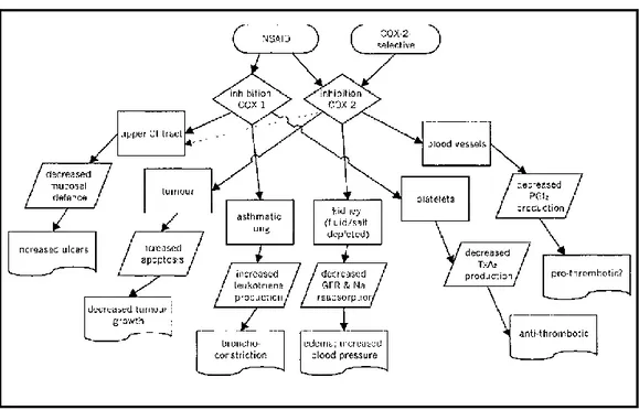
Figure 1. 8: Outcomes of COX-1 and/or COX-2 inhibition. ... 10
Figure 1. 9: Classification of COX-2 specific drugs. ... 12
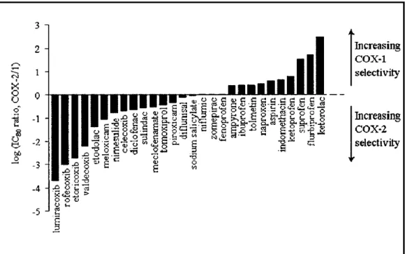
Figure 1. 10: Graph representing COX2/COX1 ... 13
Figure 4. 1: 2-D Molecule structures of highly selective ligands ... 37
Figure 4. 2: 3-D molecular view of TM_01 ... 38
Figure 4. 3: 3-D molecular view of TM_02 ... 38
Figure 4. 4: 3-D molecular view of TM_04 ... 39
Figure 4. 5: 3-D molecular view of TM_07 ... 39
Figure 4. 6: 3-D molecular view of TM_09 ... 40
Figure 4. 7: 3-D molecular view of TM_12 ... 40
Figure 4. 8: 3-D molecular view of TM_16 ... 41
Figure 4. 9: 3-D molecular view of TM_18 ... 41
Figure 4. 10: 3-D molecular view of TM_24 ... 42
Figure 4. 11: 3-D molecular view of TM_27 ... 42
Figure 4. 12: 3-D molecular view of TM_28 ... 43
Figure 4. 13: 3-D molecular view of TM_31 ... 43
Figure 4. 14: 3-D molecular view of TM_34 ... 44
Figure 4. 15: 3-D molecular view of TM_v_20 ... 44
Figure 4. 16: 2-D interaction diagram of ligand TM_01 with COX-2... 45
Figure 4. 17: COX-2 enzyme surface around ligand TM_01 ... 45
Figure 4. 18: Interacting residues of COX-2 with TM_01... 46
Figure 4. 19: 2-D interaction diagram of ligand TM_02 with COX-2... 46
Figure 4. 20: COX-2 enzyme surface around ligand TM_02 ... 47
Figure 4. 21: 2-D interaction diagram of ligand TM_04 with COX-2... 47
Figure 4. 22: COX-2 enzyme surface around ligand TM_04 ... 48
Figure 4. 23: : Interacting residues of COX-2 with TM_04 ... 48
Figure 4. 24: 2-D interaction diagram of ligand TM_07 with COX-2... 49
Figure 4. 25: COX-2 enzyme surface around ligand TM_07 ... 49
Figure 4. 26: : Interacting residues of COX-2 with TM_07 ... 50
Figure 4. 27: 2-D interaction diagram of ligand TM_09 with COX-2... 50
viii
Figure 4. 29: : Interacting residues of COX-2 with TM_09 ... 51
Figure 4. 30: 2-D interaction diagram of ligand TM_12 with COX-2... 52
Figure 4. 31: COX-2 enzyme surface around ligand TM_12 ... 52
Figure 4. 32: : Interacting residues of COX-2 with TM_12 ... 53
Figure 4. 33: 2-D interaction diagram of ligand TM_16 with COX-2... 53
Figure 4. 34: COX-2 enzyme surface around ligand TM_16 ... 54
Figure 4. 35: : Interacting residues of COX-2 with TM_16 ... 54
Figure 4. 36: 2-D interaction diagram of ligand TM_18 with COX-2... 55
Figure 4. 37: COX-2 enzyme surface around ligand TM_18 ... 55
Figure 4. 38: 2-D interaction diagram of ligand TM_24 with COX-2... 56
Figure 4. 39: COX-2 enzyme surface around ligand TM_24 ... 56
Figure 4. 40: 2-D interaction diagram of ligand TM_27 with COX-2... 57
Figure 4. 41: COX-2 enzyme surface around ligand TM_27 ... 57
Figure 4. 42: 2-D interaction diagram of ligand TM_28 with COX-2... 58
Figure 4. 43: COX-2 enzyme surface around ligand TM_28 ... 58
Figure 4. 44: : Interacting residues of COX-2 with TM_28 ... 59
Figure 4. 45: 2-D interaction diagram of ligand TM_31 with COX-2... 59
Figure 4. 46: COX-2 enzyme surface around ligand TM_31 ... 60
Figure 4. 47: : Interacting residues of COX-2 with TM_31 ... 60
Figure 4. 48: 2-D interaction diagram of ligand TM_34 with COX-2... 61
Figure 4. 49: COX-2 enzyme surface around ligand TM_34 ... 61
Figure 4. 50: 2-D interaction diagram of ligand TM_v_20 with COX-2... 62
Figure 4. 51: COX-2 enzyme surface around ligand TM_v_20 ... 62
ix
List of Tables
Table 4. 1: Docking scores of each candidate ... 25
Table 4. 2: Scores of known COX-1 or COX-2 inhibitors ... 27
Table 4. 3: Folds of ratiosof COX-2/1 inhibitor ... 27
Table 4. 4: COX-2 based designed drug‘s folds ... 29
Table 4. 5: COX-1 based de novo designed drug scores ... 30
Table 4. 6:COX-1 based designed drug‘s folds ... 31
Table 4. 7: Fold of Known Drugs ... 31
1
1. Chapter 1: Properties of Cyclooxygenases
1.1 Introduction
In the 1990‘s a noteworthy breakthrough was emerged from sophisticated molecular and cellular biological studies that; two cyclooxygenase (COX) enzyme systems present and taking part in steps of the generating of prostanoids; COX-1 products regulates biological functions on the other hand COX-2 products regulate generation of prostaglandins taking part in inflammation, aching and fever. Specifically inhibiting COX-2 products have been the principal aim for remedying rheumatoid and osteo-arthritis and other arthritic diseases, dental and surgical pain in post-operative states, dysmenorrhoea, and acute injuries. According to World Health Organization (WHO) population statistics; 10-50% of individuals suffer from musculoskeletal disorders and the majority suffer from pain. Approximately all will require Non-Steroidal Anti-Inflammatory Drugs (NSAIDs) and other analgesics for their pain management1.
The focus of this thesis is defining the necessary inhibitors for the management of COX associated infections. Due to high homology between the active sites of two isoenzymes, selectivity of the resultant drug is the most important target. Searching for new, reversible and highly selective COX-2 inhibitors via in silico drug design methods is the main objective of this study.
Synthesizing new drugs, even with rational drug design methods, takes ages to find effective solutions. In addition, needed effort and project budget could be unpredictable. These kinds of studies may end up without successful result. Computational modeling studies are more economic and faster alternative to start with best possible pathway. This generates an opportunity to start searching
2
candidate drug via computational technique rather than following the traditional method to find better solutions2.
1.1 Mechanism of COX enzymes
Both a broad array of stimuli in the cell and mobilization of calcium activate phospholipase A2. It is widely known that vast majority of biologically active lipids are originated from esterified arachidonic acid (AA) by the engagement of oxidative enzymes. AA biotransformation (Figure-1.1) is catalyzed by phospholipase A2, to eventually produce unoccupied arachidonate, which is the preliminary rate-limiting step throughout formation of sequential eicosanoids including prostanoids (prostaglandins E2, D2, F2α, I2, and thromboxane A2). Crucial enzyme group; prostaglandin endoperoxide synthases (also known as COX) and hydroperoxidase (HOX) catalyze the first assigned stage in the transformation of AA into the prostanoid associated metabolites3. COX enzymes maintain two distinctive catalytic actions: (1) a cyclooxygenase that biotransforms AA and two molecules of molecular oxygen to generate PGG2 and (2) a peroxidase (HOX) that reduces PGG2 to PGH2. Both actions necessitate heme groups that exist one per enzyme subunit4.
3 1.2 COX isoenzymes
At the moment, there are three types of COX enzymes known; namely, COX-1, COX-2 and COX-3. Starting with COX-1; it yields products, which largely provide ‗housekeeping‘ functions, such as gastric cyto-protection as well as homeostasis. By contrast, expression of COX-2 is strictly controlled by cytokines and mitogens, and considered to be essential for stimulating the inflammatory response in prostaglandin formation, mostly taking place in inflammation and cancer. Nevertheless, prostaglandins, which are originated through COX-1, can contribute to inflammation6. COX-1 is expressed in several tissues such as brain, liver, lung, spleen, kidney, stomach as well as other gastrointestinal tract tissue; but not in renal medulla. Basically, mRNA for COX-2 was not noticeable in tissues except brain7,8. However, immunocytochemical localization of COX-1 and COX-2 indicated that both isoforms were present in the rat stomach, in the alveolar, peritoneal macrophages of mice and in amniotic epithelium8-10. Also constitutive expression of COX-2 in the brain and kidneys is well documented and expression of COX-2 increased in labor7,11.At cellular level, both COX-1 and COX-2 are positioned on the luminal side of the ER, but COX-2 also seems to exist in the nuclear envelope12. Despite these variations from the simple hypothesis of bearing two distinct roles, the hypothesis has been the touchstone for rationale drug discovery and development of selective COX-2 inhibitors, which mainly focus on the lateral extension of the hydrophobic channel in the isozyme3,13.COX-3 is variant of COX-1 and is only detected in dog brain14.
4
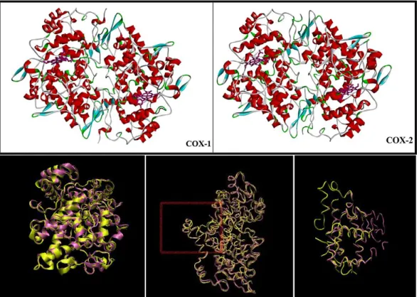
Figure 1. 2: 3-D view of COX-1 and COX-2 retrieved from Accelery‘s Discovery Studio 3.1and
superimpose of them two COX enzymes retrieved from Visual Molecular Dynamics (VMD). COX-1: mauve, COX-2: yellow.
1.3 Structure and Physiochemical Properties of COX enzymes
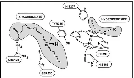
COX-1and COX-2 (EC 1.14.99.1) are both found in integral membrane protein family. Unlike many membrane related receptors, COX enzymes do not contain hydrophobic membrane penetrating arrangements by means of primary structure; instead they seem to have monotopic interaction through three N-terminal mini α-helices, which are amphipathic (Figure-1.2).15 Active site of the cyclooxygenase is confined in 25 Å constricted hydrophobic channel that elongates throughout the membrane binding motif up via the epicenter of the protein terminating at the heme binding site, which locates in cytoplasm, adjoining to the peroxidase active site. At upper end of channel is majorly consisted of Tyr 385 and Ser 530. Residue Arg 120 is located in the middle of the channel, in appropriate position to make interaction
5
with either the arachidonate carboxylate or, as was comprehended in the ovine X-ray structure which is with flubiprofen, the carboxylate of an NSAID (Figure-1.3)16.
Figure 1. 3: Binding site of COX enzymes.
COX-1 enzyme isolated from sheep, mouse and human demonstrates roughly 90% homology at amino acid level. COX-1 and COX-2 have approximately 60% homology by means of amino acid sequence in the same species of sheep, mouse and human17. Excluding N- and C-termini of the COX enzymes, since two proteins are mostly dissimilar in these regions, they are approximately 75% identical. Human COX-1 has 576 amino acid and the predicted molecular weight of subunit is 65kDa, whereas ovine COX-1 has 580 amino acid18,19. Molecular weight increases up to 72kDa by the action of post-translational modification occurring at glycosylation sites found within three high mannose oligosaccharides19. In COX-2, mouse homolog has 587 amino acid and a supplementary glycosylation site exists on the C-terminal with 18-amino acid insert in human 16,20. Fractional glycosylation arises at C-terminal and COX-2 seems as a 72kDa/74kDadouble band on SDS-gel. Both enzymes preserve activity after removal of the sugars, but they become less stable20. The N-terminus includes signal sequences of 25 (COX-1) and 17 (COX-2) amino
6
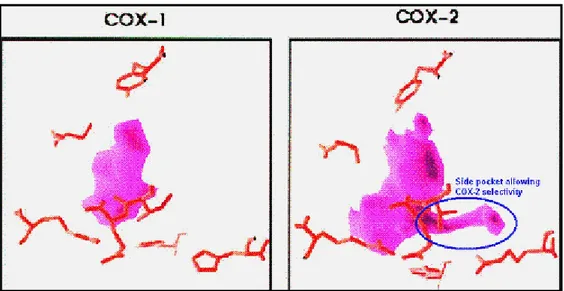
acids that lacks in the processed polypeptide. A remarkable variation among isoforms exists close to the C-terminus of COX-2 as an 18-amino acid insertion. This exceptional sequence has been used to develop COX-2 targeted antibodies in the market and also for rational drug design (Figure 1.4)21.
Figure 1. 4: Schematic view of selectivity feature of COX-2 against COX-1
1.4 COX enzymology
COX-1 was the first isoenzyme isolated from sheep seminal vesicles as a sedentary homodimer. Adding heme group, one per each dimer subunit, is involved for catalytic activities. At the end of peroxidase reaction of resting heme, a ferryl-oxo complex (Fe+4-O) is produced in COX enzyme (the iron is ferric in the inactive enzyme and is thermodynamically unable to oxidize Tyr385). Inactive Fe is represented as a radical cation Fe+4 intermediate that may either take a hydrogen atom away from Tyr385 or go through a two-electron reducing back to the sedentary state of Fe+3enzyme (Figure 1.5). Tyrosine radical is supposed to be initiator of the COX reaction. A study with T385F mutant in COX states that; mutant enzyme loses
7
its cyclooxygenase activity nevertheless retains its peroxidase activity that is compatible with the suggested mechanism 22,23.
Figure 1. 5: Equation showing reduce state of Fe adapted from Marnett et al24.
The heme irons of most HOXs are organized by the four nitrogens of the protoporphyrin ring and at the fifth coordination position by the Nδ atoms of the imidazole group of the proximal histidine. In some cases, the iron is also coordinated at the sixth position with either a small inorganic ion or water. Distal histidine located near the sixth coordination position pulls a proton from the peroxide substrate and this becomes substrate for COX. His 207, Gln 203 and His 388 are important for catalysis and heme coordination (Figure-6)25-27.
8
1.5 Crystallographic and structural properties of COX enzymes
Many crystallization, computational simulation and molecular modeling studies deduce very important data that contribute to design of COX inhibitors. Both COX isoenzymes of ovine and mouse crystallizes as dimer, whereas human COX enzymes crystallizes as four chains16,18,29. COX enzyme has two sites; one is for reducing Fe+4 to Fe+3 by peroxide (HOX), and the other is catalytic site for AA. These 2 sites elongate in opposite direction (Figure-1.7)3.Active site of COX-2 is nearly 20% larger and more accommodating than that of COX-1 .This difference in active site size and shape is due to three amino acid differences between COX-1 and COX-2: Ile523 to Val523 in the first shell of the active site, Ile 434 to Val434 and His513 to Arg513 in the covering second shell. Both COX entrance cavity volume is about 25Å for each monomer of hydrophobic channel that originates at the membrane-binding domain (MBD) that is assembled from residues 111-122 and projects into the core of the globular domain13.
COX-2 has 2090 Å3 cavity volume. A number of amino acids constituting the superior half of the channel have a key role in cyclooxygenase catalysis. Active site of COX-2 is restricted by H-bonding network done by side chains of Arg 120, Glu524, Tyr355 and Arg513. Twenty-four residues reside within the hydrophobic cyclooxygenase active site with only one difference between COX isozymes—Ile at position 523 in COX-1 and Val at position 523 in COX-2.Amino acids exist in the hydrophobic cyclooxygenase active site channel include; Leu117, Arg120, Phe205, Phe209, Val344, Ile345, Tyr348, Val349, Leu352,Ser353, Tyr355, Leu359, Phe381, Leu384, Tyr385, Trp387, Phe518, Ile/Val523,Gly526, Ala527, Ser530, Leu531, Gly533, Leu534.Only three of the channel residues are polar (Arg120, Ser353, and Ser530). Arg120 has an important gate like property for binding drug to COX-2.
9
Selectivity of COX-2 comes probably from hydrogen bonding between; His90 Arg513 and Tyr355. H-bond between Arg513-Glu524 was relaxed during drug entrance to COX-2 cavity. Volume of designed drug should not exceeds the cavity volume or at most 70% should be covered in order to be effective agent30.
Figure 1. 7:A)General view of COX-enzyme, B)Binding site of COX enzymes pointed with arrow as
COX adapted from Ref3. POX: peroxide binding site, EGF: Epidermal Growth Factor, MBD: Membrane binding domain.
10
2. Chapter 2: COX inhibition
Inhibiting prostanoid groups in the metabolism provokes toxicity and this is well documented with the fact that widespread and non-specific inhibition of COX enzymes in the body creates toxicity (Figure-8). In several studies, gastrointestinal tract and renal damage is noticeably demonstrable both in animal models and clinical trials with COX inhibitors namely NSAID7,31,32.A proposed mechanism of inhibition of COX activity is changing the route to other arachidonic acid using pathways such as lipoxygenase production (e.g., 5-lipoxygenase, 12-lipoxygenase, and 15-lipoxygenase). Treatment of COX-2 enzyme with aspirin seems to inhibit the production of prostanoid, essentially seems to induce the production of 15-HETE33.
11 2.1 Mechanisms of COX Inhibition
There are numerous proposed mechanisms for competitive inhibition of prostaglandins by inhibiting the cyclooxygenase reaction, which are;
1. Limiting the concentration of substrate that are either arachidonate or O2radical35,
2. Inhibiting oxidative activation by declining the concentration of hydroperoxide initiator below 10 nM36,
3. Reducing catalytically dynamic enzyme back to the inert ground state37,38, 4. Prevention of substrate (arachidonate) binding.
The fourth category, inhibition of substrate binding, seems to be the general target of intervention for the common of NSAIDs. Nevertheless, the other forms of intervention may have a noteworthy influence on in vivo efficacy of an inhibitor, besides suggesting an alternative method for inhibition of prostanoid production. Antagonism for binding at the arachidonate binding site is the major method of inhibition for most of the acidic traditional NSAIDs and inhibition of cyclooxygenase active site does not affect status of HOX activity. The simulation of the arachidonate carboxylate by the acidic function of NSAID suggested in Shen‘s model is compatible with the X-ray co-crystal structure defined for S-flurbiprofen with ovine COX-17. This structure demonstrates the flurbiprofen carboxylate interacting with Arg120 in mostly lipophilic arachidonate binding channel. Two kinds of inhibition kinetics have been identified for the acidic NSAIDs; reversible and tightly irreversible inhibitors, which includes conformational change and covalent bonding22.
12 2.2 Classification of COX inhibitors
COX inhibitors are called as NSAID (Non-steroid Anti-Inflammatory Drug) and there are two class of NSAIDs; traditional and new generation. Traditional NSAID (tNSAID) includes carboxylic acid, carboxamide/oxicam and sulfonanilide group containing inhibitors (Figure-1.9);binding to both COXes, they are non-specific inhibitors. They contain 1 or 2 but not 3 phenyl ring in their structure. Bulky alkyloxy (ethyl vs. methyl) or aryloxy substituents seem to be unfavorable for 1 inhibition and these 3 side groups does not exist in tNSAID structure, but in COX-2 specific inhibitors. Compounds possessing a free carboxylate exhibit nonselective COX inhibition and this group exist in tNSAID such as; aspirin, indomethacin, naproxen24,39,40.
13
Selective COX-2 inhibitors such as Celecoxib, Rofecoxib and Valdecoxib have been put into the market as new generation NSAIDs, which are coxib class (Figure-1.10). Coxib stands for Cox-inhibitors and these compounds all bear the diaryl heterocyclic structural features. The pharmacophore of diarylheterocycles inhibitors is distinguished by a central carboxylic or heterocyclic ring system carrying two vicinal aryl moieties and one benzene ring substituted with methylsulfonyl or aminosulfonyl group at the para position. The major difference in the new generation compounds is the structure of the central ring and arylsulphonyl group for selectivity purpose. Thus, modification in the central ring will direct us to novel COX-2 inhibitors. Indole ring comprises a significant prototype for drug design from tNSAIDs such as indomethacin and indoxole42,43.
14
3. Chapter 3: Methods and Procedures Used in Molecular
Modeling
3.1 Introduction
Inhibition of an enzyme is the method that drug follows to decrease enzyme‘s activity. Drugs should achieve to inhibit the target enzyme within a low concentration range and should not crosstalk with any other enzymes that are functioning in metabolism and also not crosstalk with compounds that are known as strong inhibitors. These properties are critical to obtain drugs that are non-toxic to both metabolism and individual or at least have less or non-side effect.
In this study, Structure Based Drug Design (SBDD) is the major method used. Initially, structural information of a molecule in terms of coordinates can be assembled from NMR or X-Ray crystallography. RCSB Protein Data Bank currently comprises at least 90000 crystal structures with their 3D data. All these information is accessible for molecular modeling applications and also for computational biology studies. In order to achieve SBDD goal, Accelrys Discovery Studio Client 3.144,45 was used. This method includes two main follow up protocols: De novo Receptor and De novo evolution.
The former protocol depends on searching for complementary small molecules that best bind to interaction sites, which are determined by receptor cavity (active sites atom). This protocol does not contain scaffold however it selects scaffold from a library according to some rules. LUDI is a procedure for the de novo design of
15
ligands for proteins in Accelrys Discovery Studio Client 3.1. It is a practical method for screening vast number of candidates. This procedure evaluates a geometrical fit of candidate compounds into the binding cavity and computes other determinants of good binding such bas hydrogen bonds, lipophilic interactions, ionic interactions, and acylic interactions. LUDI scoring functions statistically assesses the fit of all likely ligands. ΔG = ΔGo + ΔGhbf(ΔR)f(Δα) + ΔGionf(ΔR)f(Δα) + ΔGlipoAlipo + ΔGrotNR ΔG; ΔGo stands for the contribution to the binding energy that does not straight relate to any defined interactions with the receptor (i. e. the involvement of binding energy due to loss of transitional and rotational entropy of the fragment), ΔGhb and ΔGion stands for the contribution from an ideal hydrogen bond and unperturbed ionic interactions, respectively, ΔGlipo stands for the contribution from lipophilic interactions which is proportional to the lipophilic surface Alipo , ΔGrot stands for the contribution due to freezing of internal degrees of freedom in the fragment, NR is the number of acylic bonds, ΔR is the divergence of the hydrogen bond length from the ideal value that is 1.9 Å, Δa is the divergence of the hydrogen bond angle from ideal value 180o. Generally a higher LUDI score (0-1000 in range) corresponds to a higher affinity and stronger binding of a ligand to the receptor. During the search and fit computation LUDI scores are calculated and also the energy estimations, or scores, for each conformation searched for the fragments in the library are determined. The DS LUDI was used extensively in this work, which quests fragment libraries and yields molecules that fit the requirements of the defined interaction sites. Ligand-receptor complexes may be evaluated using the empirical scoring functions available from the LUDI algorithm. The score is a sum of five contributions: ideal hydrogen bonds, perturbed ionic interactions (interaction of donor/acceptor in the receptor, e.g., COO–, or NH3+), lipophilic interactions, the freezing of internal degrees of
16
freedom of the ligand, and the loss of translational and rotational entropy of the ligand. In receptor mode, during the ―search and fit‖ the LUDI program also determines the energy estimates, or scores, for each conformation of the selected fragments in the library. The fragments are ranked by energy estimate, and the best are returned in the hit list. This hit list can then be inspected for the selection of candidate scaffolds and eventually program yields ―*.sd‖ file for further inspection. In this study, LUDI receptor mode was used against COX-2 and COX-1 enzymes which screened potential lead compounds from more than a million lead compounds in the ZINCv1246 lead library for their structural and physicochemical properties.
The latter protocol, de novo evolution depends on a specified scaffold positioned in a protein site; LUDI is used to identify fragments that may be covalently fused to the scaffold and yields a collection of molecules with high LUDI scores. Fragment selection and construction for new molecules relies on the mode. Finally, all new compounds will be further refined using CHARMm and scored by MM-PBSA/GBSA47. Using ZINC and Accelrys 3.1 fragment-based libraries, which contain about 400,000 fragments, eventually 1129 potential candidates, were generated.
In this study, de novo designed ligands are docked into both COX enzymes in silico. None of those compounds has been synthesized or investigated beforehand; which is confirmed by ZINCv12 website.
17
Docking of ligand and enzyme is the leading virtual screening method to check compound candidates. Objective of the study is discovering the best matching for ligand and receptor by conducting virtual screening. Also docking foresees the binding energy for each ligand and possible binding pose for each ligand embedded in 3D structure of enzyme by using various scoring functions. Fundamental job of scoring function is guessing the interaction or binding affinity and calculating score of affinity between two molecules that are generally an enzyme and candidate compound48. Primarily the method is formed as which tracked by locating the enzyme and the candidate compound within that system. By performing minimization step, the lowest free energy of binding is simulated as induced fit49. Two major standards exist to be accomplished. The first one is shape consistency occurring between candidate compound and enzyme, the second one is interrelation between them. Devoid of the fitting interrelation leads one to have useless fitting geometric structures.
Currently available docking programs include three major parts: system depiction, searching of conformational volume, and the classification of the potential resolutions. The best suitable results were retrieved based on two criteria; the first one is having an efficient search algorithm that gives the less faulty results and the second one is having improved scoring function. Nowadays, there is a good number of conformational search algorithms available, however getting scores from the prospective result is the main problematic fact to detect the best software combination50,51. Yet designed for particular experimentations, using just one docking tool only for conformational search and also at that juncture rescoring the results with additional function can have superior result. One of the best illustrations
18
to that would be obtaining the AutoDock output files (created docked orientations) than ranking it again with DSX (Drug Score). 52
Key operation of docking is managing to discover worthy enough situations to reside the candidate compound inside the enzyme where catalytic activity take place, therefore they are fetched together to discover the top matching geometric situation. Ligand molecule angles are accomplished by an algorithm one step at a time and expectantly some positions (situation) of ligands can be located inside the catalytic part of the enzyme. Decision of settlement is accomplished by computing the energy of the system, which uses the molecular mechanics force fields. As the energy gets lower, the better enzyme - compound binding51,53-55 happens. Entire probable spatial orientations of enzyme - compound complex are conducted by using search algorithm. In our experiment, ligand was assumed as flexible however enzyme was assumed as rigid. Setting ligand as having flexible structure means that it has millions of possible conformations to reach to the lowest energy level due to bargaining rotatable bonds in its structure. Physical interrelation is detected for the candidate compound as well as for the enzyme.
3.2 Preparation of Enzymes and Ligands
Protein Data Bank (PDB) is the main foundation web site to adopt the crystal structures of the two COX enzymes. 1Q4G56 (Ovine Prostaglandin H2 Synthase-1 (COX-1), in complex with Alpha-Methyl-4-Biphenylacetic, resolution 2.00 Å) and 3NT116 (Mus musculusCOX-2 in complex with naproxen, resolution 1.73Å) are the crystal structures that are used. Each structure was cleaned of all water molecules and inhibitors as well as all non-interacting ions before being used in the docking
19
studies. When inhibitor was bound to the COX enzyme, oxidized form of heme group was used, as adding +2 charge to the Fe atom. For COX-1 and COX-2, one of the two subunits was taken as the target structure. Using a fast Dreiding-like force field, each protein‘s geometry was first optimized and then given in to the ―Clean Geometry‖ toolkit of Discovery Studio (Accelerys Inc.) for a more methodical check. Missing hydrogen atoms were added based on the protonation state of the titratable residues at a pH of 7.4. Ionic strength set was 0.145 and the dielectric constant set was the 10. Then and there the enzyme was minimized and the files were saved as tolerated type, which is necessary for the following docking procedures44,45.
Candidate compound generation and preparation is the most essential phase in this thesis. Initial point for this operation was based on the structure of the active site cavities of COX-1 and COX-2. The scaffolds generation and building up44 were achieved by using the module of the commercial software Accelrys‘ program. The GOLD55 and AutoDock 4.254, Auto-Dock Tools (ADT) programs were used for molecular docking into the active site of COX-1 and COX-2 isozymes and to identify the inhibition constants. Celecoxib used in this study, which has a structural similarity to our resultant scaffolds, was used as a scaffold due to being the most effective and least side effect compound known.
Python script required for separating one ―.*sd‖ file into many ―*.pdb‖ or ―*.mol2‖ files. This is achieved via o‘babel57
program with following script: babel -m –isd*.sd - *.mol2
20
3.3 Selecting a Docking Method for Virtual Screening
Other docking programs and scores were used to decide which one gives the fastest result as well as the most accurate result. For further check, AutoDock 4, GoldScore, ChemScore, CHEMPLP and ASP were examined with well-known and currently used inhibitors. Virtual screening returns to false positive and also false negative iterations. In other words, usage of whichever screening algorithm can yield overlooking of a result, which gives one of the efficient compounds or may grab an unusable compound as a drug candidate. Therefore, choosing and picking the suitable scoring function for a purpose and modification into the required condition is significant.
Nearly 10 different drugs are available for well-known COX-2 inhibitor as attest compound, however celecoxib is one of the highly selective ligands that is already available in market; consequently using it as a reference docking is rational. Other docking scores such as ChemScore, CHEMPLP and AutoDock also adapted with the currently used and marketed drugs against COX-2 and their inhibition constants and docking scores were known. Another crucial feature for virtual screening as a second most important point is time consumption; screening should have a high speed since thousands of compounds are tested along accordingly CHEMPLP was chosen to do the screening.
3.4 Docking Based Virtual Screening with CHEMPLP
GOLD (Genetic Optimization for Ligand Docking) is a greatly automated docking tool that allows for optimization and flexible docking with different genetic algorithms58,59. The scoring function ranks the dockings according to total score of
21
energies; hydrogen bond, pairwise dispersion potential that can define a noteworthy impact to hydrophobicity of binding and a molecular mechanism in terms of interior energy of the ligand binding sides55,60.
CHEMPLP is the greatest scoring function among other scoring functions of GOLD docking software and it is well suited with AutoDock result. For GOLD docking; first of all H atoms are added to enzyme, ligands, then default parameters are used, and cavity detection was disabled since we specify coordinates. A sphere which has 16 Å radius drawn with specific coordinates (for COX-1, x: 26.108 y: 35.061 z: 197.448 and COX-2, x: -39.746, y: -50.781, z: -22.645) is used.
Docking of 1129 candidate compounds into two COX isoenzymes has been conducted concurrently. Calculations take approximately 10 days at least 75% usage of single core per enzyme on High Performance Cluster (HPC) cluster.
GOLD software creates a file named as: bestrankings.lst file that contains score values per ligand. Some columns of this file are transferred to a Microsoft Excel® file. According to formula, it is sorted by using the discrimination values. Discrimination value defines at least 15% alteration between COX-1 and COX-2 dockings scores per ligand. Scores are arranged based on COX-2/COX-1 score ratio. If ratio is lower than 1.15, the candidate compound is considered as non-selective and eliminated. The best 51 ligands from this discriminated and arranged list are shown in Table 4.1. CHEMPLP cannot be the only score used for this study, since the results are supported/confirmed with other scoring functions.
22
3.5 Validating CHEMPLP Results with Other Docking Tools
Additional GOLD scoring functions such as GoldSscore, ChemScore and ASP work in a computerized manner just like ChemPLP scoring function. Putative parameter group and identical active site coordinate usage directed us to retrieve results one by one because AutoDock does not allow automated docking. For each docking, one has to apply method as the number of ligand and this is time consuming. Specifically Raccoon61 is a handy script which greatly needs to MGL Tools and it can be used on Portable Batch System (PBS) based HPC clusters nevertheless it encounters various problems such as needing other programs. Necessity of classy tool for conducting AutoDock experiments in parallel, a novel tool was built in order to overcome this problem. This novel tool is called YaVST62 (Yet Another Virtual Screening Tool), created by our colleague, Serkan AltuntaĢ. YaVST is a free access, open-source software and used for virtual screening via AutoDock 4 (Website for YaVST is: https://github.com/serkanaltuntas/yavst). Similar to its prototypes, it is also heavily interrelated to MGL which is a software developed at the Molecular Graphics Laboratory (MGL) of The Scripps Research Institute for visualization and analysis of molecular structures Tools , but it is settled as a self-contained package so it does not necessitate MGL Tools.
YaVST generates workspaces automatically for each run. For this study, each workspace contains vast amount of ligands per enzyme.
If not available, YaVST generates the PDBQT files from PDB file for each ligand. After that, creation of a Docking Parameter Files (DPF) and one single Grid
23
Parameter File (GPF) for each ligand keep track of that step. Scripts of MGL Tools create these output files consequently; it generates the same output that is identical to AutoDockTools.
Besides pdbqt, gpf and dpf files, YaVST generates several qsub files that are needed for job submission to any type of Sun Grid Engine based HPC. Eventually as it happens in manual docking, each experiment generates .dlg file and with a small script, .pdb file of the lowest energy result retrieved from .dlg file and list of the lowest energy and Ki values, which stands for the dissociation constant of an enzyme-inhibitor complex, were extracted from each .dlg file. This is a very effective way of conducting Autodock 4 experiments without spending much time.
3.6. ADMET
Not obtaining a promising ADMET feature, which stands for the Absorption, Distribution, Metabolism, Excretion, and Toxicity characteristics of a candidate compound headed for organism, is one of the utmost discouraging obstacles for drug development. Any drug must contain all these characteristics to be used in clinical trials. This enables us to do the early optimization. The dispositions of the candidate compound used by the organism were controlled with ADMET PSA 2D (polar surface area) against ADMET AlogP98 (the logarithm of the partition coefficient between n-octanol and water). If the candidate compound cannot pass ADMET test, progressive steps might become loss of time. Determination of which compound can pass ADMET and removal of undesirable compounds make the research course more cost operative and effective63.
24
4. Chapter 4: Results and Discussion
4.1 Docking Results
In total 1129 de novo potential COX-2 inhibitor ligands were designed via Accelerys. All ligands were simultaneously docked via GOLD and AutoDock program. 66 were eliminated according to AutoDock results, which are positive value for free energy for either COX-1 or COX-2. Having a positive sign score of free energy for a ligand means that; Ki values could not be calculated since they are unfavorable for either COX-1 or COX-2 and cannot give result of COX-2/COX-1 ratio and it is unusable. After obtaining results, the best 53 de novo designed COX-2 inhibitors were determined according to the total score of ChemPLP, ChemScore and ASP which are 1.15 fold and above for the ligands compared to COX-1, as well as for AutoDock 4, 1.15..Table-4.1 shows score results of GOLD (including GoldScore and AutoDock 4 Free energy and Ki values) for each ligand. According to this elimination, SC_558 and celecoxib were in highly selected ligand list; but in order to find new drugs these 2 were eliminated and the rest51 are listed in Table 4.1.
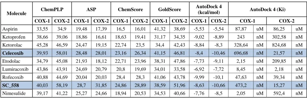
To find better and highly selective Cox-2 inhibitor than inhibitors found currently in the market, Aspirin, ketoprofen, ketorolac, celecoxib, etodolac, lumiracoxib, rofecoxib, sc_558 and nimesulide were used as a control in a simultaneously run docking to discuss program‘s validity.. Score values for each drug is given in Table 4.2.
25
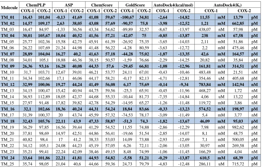
Table 4. 1: Docking scores of each candidate retrieved from GOLD; Ki and Free energy (kcal/mol) values retrieved from AutoDock 4
Molecule ChemPLP ASP ChemScore GoldScore AutoDock4(kcal/mol) AutoDock4(Ki)
COX-1 COX-2 COX-1 COX-2 COX-1 COX-2 COX-1 COX-2 COX-1 COX-2 COX-1 COX-2
TM_01 16,43 101,04 -0,13 41,69 41,08 59,67 -100,67 34,81 -2,64 -14,82 11,55 mM 13,79 pM TM_02 14,57 109,17 2,63 38,05 43,08 57,69 -90,57 75,8 -3,98 -12,52 1,21 mM 662,85 pM TM_03 16,47 84,97 -1,33 36,56 43,34 54,62 -89,89 32,57 -8,67 -13,97 438,07 nM 57,98 pM TM_04 30,01 105,67 10,04 40,52 41,56 57,21 -42,07 75 -0,85 -13,87 238 mM 67,58 pM TM_05 25,77 102,36 15,58 42,75 45,91 61,39 -26,92 75,35 -3,65 -14,03 2,11 mM 51,86 pM TM_06 26,22 107,69 21,24 44,98 41,48 56,22 -4,28 80,59 -3,63 -12,72 2,2 mM 475,46 pM TM_07 28,89 104,04 16,27 40,2 41,63 57,18 -44,28 75,02 -1,87 -13,35 42,6 mM 164,57 pM TM_08 34,01 105,1 18,88 46,36 38,15 50,57 -1,59 76,66 -2,29 -14,25 20,82 mM 35,84 pM TM_09 26,36 93,16 16,28 40,08 44,33 57,6 -29,45 66,81 -1,08 -12,96 161,81 mM 314,51 pM TM_10 31,7 103,71 12,67 39,01 46,21 53,77 24,11 67,01 -0,43 -10,46 483,48 mM 21,51 nM TM_11 34,34 102,66 17,1 46,06 44,17 58,21 -0,17 82,13 -4,71 -12,81 354,46 uM 405,68 pM TM_12 32,99 100,06 19,27 44,24 41,49 56,08 6,17 75,69 -0,14 -9,34 783,84 mM 142,94 nM TM_13 34,15 100,47 15,42 40,94 44,75 59,56 -25,3 65,91 -0,45 -11,96 468,27 mM 1,72 nM TM_14 36,93 112,89 18,05 42,37 44,68 55,33 -10,44 81,73 -7,35 -14,84 4,06 uM 13,22 pM TM_15 27,97 91,48 17,82 39,82 42,78 54,29 -14,95 65,27 -1,26 -11,48 119,72 mM 3,86 nM TM_16 32,1 102,66 18,36 40,24 44,31 54,24 18,84 83,66 -0,33 -13,23 574,52 mM 198,97 pM TM_17 31,39 100,37 20 43,74 45,59 57,32 -74,53 78,17 -3,09 -11,49 5,4 mM 3,77 nM TM_18 32,43 105,76 22,11 43,9 47,33 58,87 -51,3 74,3 -1,82 -13,67 46,09 mM 95,03 pM TM_19 36,29 97,85 16,56 39,44 41,29 54,52 11,55 74,88 -2,86 -12,29 7,98 mM 982,62 pM TM_20 37,81 98,69 14,97 42,51 44,86 56,41 -19,66 51,54 -2,85 -14,07 8,1 mM 48,75 pM TM_21 40,82 98,4 17 41,2 38,33 53,03 14,09 73,88 -2,93 -12,69 7,1 mM 500,05 pM TM_22 34,12 105,1 24,08 44,23 45,19 57,05 6,26 72,11 -2,06 -13,05 30,97 mM 269,58 pM TM_23 35,21 99,41 22,24 42,09 38,46 49,15 8,48 74,99 -1,06 -11,45 166,29 mM 4,04 nM TM_24 33,64 101,86 22,21 41,81 44,93 54,82 -5,58 51,21 -0,29 -13,87 610,5 mM 68,39 pM TM_25 35,74 98,05 21,04 40,6 44,66 59,36 24,73 79,79 -4,83 -12,48 286,11 uM 715,72 pM
26
Molecule ChemPLP ASP ChemScore GoldScore AutoDock4(kcal/mol) AutoDock4(Ki)
COX-1 COX-2 COX-1 COX-2 COX-1 COX-2 COX-1 COX-2 COX-1 COX-2 COX-1 COX-2
TM_26 34,59 89,41 19,68 44,84 46,13 60,7 -18,4 37,56 -2,94 -13,75 7,05 mM 84,01 pM TM_27 42,92 107,54 19,21 44,66 47,24 59,49 21,98 73,39 -0,82 -13,31 248,87 mM 175,25 pM TM_28 43,18 106,3 21,01 45,14 44,9 57,74 -12,8 65,7 -2,13 -13,88 27,44 mM 66,58 pM TM_29 37,77 102,16 20,69 44,99 48,69 57,82 -10,09 50,85 -2,58 -13,61 12,78 mM 105,59 pM TM_30 41,38 99,57 21,45 41,65 45,02 57,12 -16,82 34,86 -0,89 -12,03 223,76 mM 1,52 nM TM_31 42 96,54 17,08 38,39 46,26 53,43 -36,44 72,69 -0,51 -12,53 420,36 mM 653,39 pM TM_32 39,21 89,25 15,44 25,95 41,64 51,8 -46,22 56,2 -5,35 -12,19 119,38 uM 1,15 nM TM_33 44,57 79,57 20,44 34,46 33,33 48,76 9,27 44,45 -5,82 -9,9 53,88 uM 55,16 nM TM_34 43,84 78,67 30,73 43,85 38,28 52,58 0 52,2 -1,42 -8,69 91,02 mM 429,15 nM TM_35 37,96 75,69 26,69 32,3 36,79 46,37 18,34 42,95 -6,19 -9,76 29,04 uM 69,61 nM TM_36 43,13 84,69 37,88 42,99 33,79 44,39 0 0 -2,84 -10,85 8,33 mM 11,15 nM TM_37 53,13 80,55 39,92 54,67 43,43 51,51 0 14,65 -2,28 -6,54 21,16 mM 15,96 uM TM_38 47,25 72,93 27,58 32,47 36,86 44,72 28,2 37,99 -7,84 -9,08 1,8 uM 222,37 nM TM_39 77,66 97,73 15,76 38,01 46,33 51,41 25,65 51,84 -3,26 -9,59 4,07 mM 93,36 nM TM_40 30,3 53,64 5,95 7,86 43,95 42,61 0 0 -0,79 -8,04 262,11 mM 1,28 uM TM_41 65,5 84,48 49,52 52,35 42,51 54,88 88,49 81,13 -12,81 -14,64 404,47 pM 18,42 pM TM_42 64,17 79,92 25,55 33,73 42,12 45,05 48,85 60,36 -8,39 -9,96 704,53 nM 50,31 nM TM_43 69,24 85,76 27,74 40,02 50,55 49,61 40,83 73,11 -7,39 -8,97 3,84 uM 266,68 nM TM_44 64,98 80,2 27,05 33,51 42,42 45,06 47,89 59,98 -8,59 -9,96 503,62 nM 49,85 nM TM_45 60,29 86,91 41,39 35,27 44,81 50,37 68,91 77,66 -11,43 -13,3 4,15 nM 176,83 pM TM_46 78,48 102,55 29,85 33,35 50,57 50,99 70,88 59,12 -8,18 -12,95 1,01 uM 321,73 pM TM_47 88,42 98,31 38,2 43,3 34,07 47,38 0 59,3 -5,41 -10,58 107,55 uM 17,45 nM TM_48 57,52 75,76 33,82 37,26 40,22 41,15 51,25 64,9 -6,7 -8,66 12,22 uM 450,88 nM TM_49 65,24 81,14 30,08 35,21 40,92 42,97 2,6 61,41 -1,16 -11,57 140,92 mM 3,31 nM TM_50 74,92 96,42 42,1 38,04 45,62 55,1 71,64 82,37 -10,31 -12,25 27,77 nM 1,05 nM TM_51 81,33 97,15 43,48 39,06 46,22 60,82 84,73 79,38 -11,47 -13,97 3,89 nM 57,83 pM
27
Table 4. 2: Scores of known COX-1 or COX-2 inhibitors. Selective drugs are marked with bold.
Molecule ChemPLP ASP ChemScore GoldScore
AutoDock 4
(kcal/mol) AutoDock 4 (Ki)
COX-1 COX-2 COX-1 COX-2 COX-1 COX-2 COX-1 COX-2 COX-1 COX-2 COX-1 COX-2
Aspirin 33,55 34,9 19,48 17,39 16,5 16,01 41,32 38,69 -5,53 -5,54 87,87 uM 86,25 uM Ketoprofen 38,66 39,06 18,86 16,61 18,63 19,41 31,17 34,35 -9,02 -8,89 243 nM 302,58 nM Ketorolac 45,28 46,59 24,47 19,15 22,74 23,5 34,4 42,43 -8,84 -8,3 328,64 nM 824,68 nM Celecoxib 39,93 58,01 28,48 28,01 23,16 26,34 41,15 46,81 -8,4 -10,46 696,68 nM 21,57 nM Etodolac 34,79 45,08 21,93 18,12 22,71 23,96 38,31 47,86 -7,73 -9,11 2,15 uM 209,85 nM Lumiracoxib 43,86 43,91 24,69 20,79 20,8 19,69 34,01 33,58 -6,92 -7,72 8,45 uM 2,18 uM Rofecoxib 40,88 44,69 20,04 20,03 28,4 28,3 41,06 43,78 -9,99 -10,1 47,63 nM 39,34 nM SC_558 40,03 58,19 28,7 31,85 24,86 28,89 38,59 51,96 -8,63 -10,66 473,2 nM 15,27 nM Nimesulide 39,17 41,22 25,27 24,66 18,94 20,53 34,53 40,66 -7,76 -8,5 2,05 uM 592,4 nM
Table 4. 3: Folds of ratios calculated from Table 4.2 and resulting COX-2/1 inhibitor case GOLD
COX-2/COX-1
AUTODOCK COX-2/COX-1
(kcal/mol)
AutoDock 4 (Ki) AUTODOCK COX-2/COX-1 (Ki) GOLD COX-2/COX-1 AUTODOCK COX-2/COX-1 COX-1 COX-2
Aspirin 0,98 1,00 87,87 uM 86,25 uM 0,98 COX-1 COX-2
Ketoprofen 0,99 0,99 243 nM 302,58 nM 1,25 COX-1 COX-1
Ketorolac 0,96 0,94 328,64 nM 824,68 nM 2,51 COX-1 COX-1
Celecoxib 1,23 1,25 696,68 nM 21,57 nM 0,03 COX-2 COX-2
Etodolac 1,10 1,18 2,15 uM 209,85 nM 0,10 COX-2 COX-2
Lumiracoxib 0,94 1,12 8,45 uM 2,18 uM 0,26 COX-1 COX-2
Rofecoxib 1,04 1,01 47,63 nM 39,34 nM 0,83 COX-2 COX-2
SC_558 1,27 1,24 473,2 nM 15,27 nM 0,03 COX-2 COX-2
28
Celecoxib is the drug, which is used for inhibiting COX-2 and can be found currently in the market; however, it has many long term use side effect.
Elimination of ligands with COX-2/COX-1 inhibition ratio, in order to study in more detail, celecoxib scores were taken as a baseline. Ligands with better scores than celecoxib are highlighted in Table 4.1. In the following Table 4.4, COX-2 GOLD score folds against COX-1, COX-2 AutoDock 4 free energy folds against COX-1 and COX-2 ChemPLP scores against COX-1 retrieved from GOLD were collected. 13 COX-2 targeted de novo designed ligands were found, namely; TM_01, TM_02, TM_04, TM_07, TM_09, TM_12, TM_16, TM_18, TM_24, TM_27, TM_28, TM_31, TM_34. Their COX-2/COX-1 inhibition ratios are higher than any other known COX-2 selective inhibitors, according to calculation by ChemPLP, AutoDock and Gold.
29
Table 4. 4: COX-2 based designed drug’s folds
Molecule GOLD COX-2/COX-1 AutoDock4 (kcal/mol) COX-2/COX-1 AutoDock4 (Ki) COX-2/COX-1 ChemPLP COX-2/COX-1 TM_01 3,53 5,61 1,19E-09 6,15 TM_02 3,40 3,15 5,48E-07 7,49 TM_04 2,49 16,32 2,84E-10 3,52 TM_07 2,32 7,14 3,86E-09 3,60 TM_09 2,19 12,00 1,94E-09 3,53 TM_12 2,14 66,71 1,82E-07 3,03 TM_16 2,08 40,09 3,46E-10 3,20 TM_18 2,05 7,51 2,06E-09 3,26 TM_24 1,97 47,83 1,12E-10 3,03 TM_27 1,94 16,23 7,04E-10 2,51 TM_28 1,92 6,52 2,43E-09 2,46 TM_31 1,79 24,57 1,55E-09 2,30 TM_34 1,55 6,12 4,71E-06 1,79 TM_39 1,34 2,94 2,29E-05 1,26 TM_49 1,17 9,97 2,35E-08 1,24
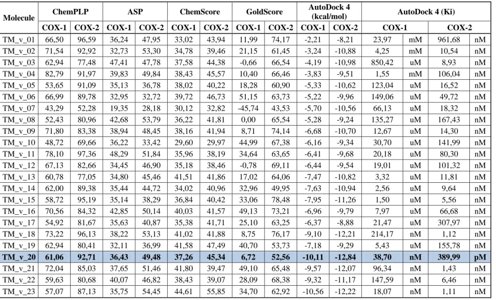
From another perspective, COX-1 de novo ligands were designed to inhibit COX-1. However, here failed COX-1 based de novo ligands were docked into COX-2. 23 ligands in Table 4.5 have 1.15 fold and above ratio of COX-2/COX-1. After further elimination, scores were compared to celecoxib and SC_558, eventually 6 ligands remained. However, surprisingly only TM_v_20 has the best fold among all. Therefore, only TM_v_20 was selected from this group since its ChemPLP, AutoDock and Gold folds are higher than any other known COX-2 selective inhibitors. Ratios are compared to values listed in Table 4.7 since it contains known inhibitor score values retrieved from GOLD and AutoDock 4.
30
Table 4. 5: COX-1 based de novo designed drug scores
Molecule ChemPLP ASP ChemScore GoldScore
AutoDock 4
(kcal/mol) AutoDock 4 (Ki)
COX-1 COX-2 COX-1 COX-2 COX-1 COX-2 COX-1 COX-2 COX-1 COX-2 COX-1 COX-2
TM_v_01 66,50 96,59 36,24 47,95 33,02 43,94 11,99 74,17 -2,21 -8,21 23,97 mM 961,68 nM TM_v_02 71,54 92,92 32,73 53,30 34,78 39,46 21,15 61,45 -3,24 -10,88 4,25 mM 10,54 nM TM_v_03 62,94 77,48 47,41 47,78 37,58 44,38 -0,66 66,54 -4,19 -10,98 850,42 uM 8,93 nM TM_v_04 82,79 91,97 39,83 49,84 38,43 45,57 10,40 66,46 -3,83 -9,51 1,55 mM 106,04 nM TM_v_05 53,65 91,09 35,13 36,78 38,02 40,22 18,28 60,90 -5,33 -10,62 123,04 uM 16,52 nM TM_v_06 66,99 89,78 32,95 32,72 39,72 46,73 51,15 63,73 -5,22 -9,96 149,06 uM 49,72 nM TM_v_07 43,29 52,28 19,35 28,18 30,12 32,82 -45,74 43,53 -5,70 -10,56 66,13 uM 18,32 nM TM_v_08 52,43 80,96 42,68 53,79 36,22 41,81 0,00 65,54 -5,28 -9,24 135,27 uM 167,43 nM TM_v_09 71,80 83,38 38,94 48,45 38,16 41,94 8,71 74,14 -6,68 -10,70 12,67 uM 14,30 nM TM_v_10 48,72 69,66 36,22 33,42 29,60 29,97 44,99 67,38 -6,16 -9,34 30,70 uM 141,99 nM TM_v_11 78,10 97,36 48,29 51,84 35,96 38,19 34,64 63,65 -6,41 -9,68 20,18 uM 80,30 nM TM_v_12 67,13 82,66 34,45 46,90 35,18 38,46 -0,78 69,11 -6,44 -9,54 19,01 uM 101,32 nM TM_v_13 60,78 77,05 34,80 45,46 41,51 41,86 17,02 64,06 -7,47 -10,82 3,32 uM 11,81 nM TM_v_14 62,00 89,38 35,44 44,72 34,02 40,96 32,96 49,95 -7,63 -10,94 2,56 uM 9,64 nM TM_v_15 58,72 95,19 35,14 38,29 36,84 40,42 33,06 78,48 -7,95 -11,26 1,50 uM 5,56 nM TM_v_16 70,56 84,32 42,85 50,14 40,03 41,57 49,13 73,21 -6,96 -9,79 7,97 uM 66,68 nM TM_v_17 54,92 81,67 35,63 40,87 35,38 41,71 25,10 63,25 -6,37 -8,88 21,47 uM 307,97 nM TM_v_18 73,22 96,13 38,22 53,13 41,02 41,88 8,75 76,17 -9,10 -12,21 214,17 nM 1,12 nM TM_v_19 62,94 80,41 32,11 36,99 41,58 47,49 40,70 53,73 -7,18 -9,29 5,43 uM 155,78 nM TM_v_20 61,06 92,71 36,43 49,48 37,26 45,34 6,72 52,56 -10,11 -12,84 38,70 nM 389,99 pM TM_v_21 72,04 85,03 37,65 51,46 41,80 39,47 49,10 65,48 -9,57 -12,07 96,34 nM 1,43 nM TM_v_22 59,63 80,68 40,07 46,82 38,43 39,07 28,09 68,38 -9,32 -11,17 147,59 nM 6,46 nM TM_v_23 57,07 87,13 35,75 54,45 44,61 55,85 34,70 62,92 -10,56 -12,22 18,07 nM 1,11 nM
31
Table 4. 6:COX-1 based designed drug’s folds
Molecule Name Total Gold Score of COX-2/COX-1 Autodock4 Energy (kcal/mol) COX-2/COX-1 Autodock4 (Ki) COX-2/COX-1 ChemPLP COX-2/COX-1 TM_v_01 1,39 3,71 4,01E-05 1,45 TM_v_05 1,33 1,99 1,34E-04 1,70 TM_v_08 1,34 1,75 1,24E-03 1,54 TM_v_15 1,33 1,42 3,71E-03 1,62 TM_v_17 1,30 1,39 1,43E-02 1,49 TM_v_20 1,39 1,27 1,01E-02 1,52
Table 4. 7: Fold of Known Drugs
Molecule Name Total Gold Score of COX2/COX1 Autodock4 Energy (kcal/mol) COX2/COX2 Autodock4 (Ki) COX-2/COX-1 ChemPLP COX2/COX1 SC_558 1,39 1,23 3,37E-02 1,57 Celecoxib 1,28 1,25 2,77E-02 1,49
32
14 ligands were derived from 3 main scaffold and namely they are; TM_2013_04_07_151011_894 (COX-2 based), TM_2013_05_17_230538_994 (COX-2 based), TM_2013_05_17_231259_735 (COX-1 based). Detailed explanation of elimination and also from which library de novo designed drugs originate from is summarized below:
TM_2013_04_07_151011_894 de novo receptor 1384 fragment was created (instock_lead_21_p1_1) de novo evolution 50 ligand (instog_frag_link_1_0) obabel autodock and GOLD selection
TM_2013_05_17_230538_994 de novo receptor 601 fragment (instock_lead_21_p1_1) de novo evolution 50 ligand (instog_frag_link_1_0) and 7 ligand (peptide_fragsx) obabel autodock and GOLDselection
TM_2013_05_17_231259_735 de novo receptor 3 fragment (instock_lead_21_p1_1) de novo evolution 50ligand (instog_frag_link_1_0)and 1 ligand (peptide_fragsx) obabel autodock and GOLD selection
Library names were expressed in parenthesis above.
2-D, 3-D structures, 2-D interaction map and COX-2 enzyme surface around ligand and for the ligands that passed ADMET, interacting residues of COX-2 with ligand can be found in Figure 4.1-4.43.
33
Molecule Open Formula Molecule Name Molecular
Weight (Da)
TM_01 352,9
TM_02 360,75
34
TM_07
328,17
TM_09 342,52
35
TM_16 323,35
TM_18 347,11
36
TM_27 341,06
TM_28 346,86
TM_31
37
TM_34 374,22
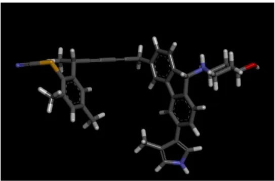
TM_v_20
341,42
38
For Figure 4.1 - 4.51 each color stands for a specific atom: Red: Oxygen, White: Hydrogen, Yellow: Sulphur, Blue: Nitrogen, Green: Chloride, Black: Carbon,
Mahogani: Bromide.
Figure 4. 2: 3-D molecular view of TM_01
39
Figure 4. 4: 3-D molecular view of TM_04
40
Figure 4. 6: 3-D molecular view of TM_09
41
Figure 4. 8: 3-D molecular view of TM_16
42
Figure 4. 10: 3-D molecular view of TM_24
43
Figure 4. 12: 3-D molecular view of TM_28
44
Figure 4. 14: 3-D molecular view of TM_34
45
For 2-D interaction map figures, legend is as follows;
Figure 4. 16: 2-D interaction diagram of ligand TM_01 with COX-2
46
Figure 4. 18: Interacting residues of COX-2 with TM_01
47
Figure 4. 20: COX-2 enzyme surface around ligand TM_02
48
Figure 4. 22: COX-2 enzyme surface around ligand TM_04
49
Figure 4. 24: 2-D interaction diagram of ligand TM_07 with COX-2
50
Figure 4. 26: Interacting residues of COX-2 with TM_07
51
Figure 4. 28: COX-2 enzyme surface around ligand TM_09
52
Figure 4. 30: 2-D interaction diagram of ligand TM_12 with COX-2
53
Figure 4. 32: Interacting residues of COX-2 with TM_12
54
Figure 4. 34: COX-2 enzyme surface around ligand TM_16
55
Figure 4. 36: 2-D interaction diagram of ligand TM_18 with COX-2
56
Figure 4. 38: 2-D interaction diagram of ligand TM_24 with COX-2
57
Figure 4. 40: 2-D interaction diagram of ligand TM_27 with COX-2
58
Figure 4. 42: 2-D interaction diagram of ligand TM_28 with COX-2
59
Figure 4. 44: Interacting residues of COX-2 with TM_28
60
Figure 4. 46: COX-2 enzyme surface around ligand TM_31
61
Figure 4. 48: 2-D interaction diagram of ligand TM_34 with COX-2
62
Figure 4. 50: 2-D interaction diagram of ligand TM_v_20 with COX-2
63 4.3 ADMET results
Figure 4. 52: ADMET results
ADMET test filters poor candidates with undesirable chemical groups according to published SMARTS, Lipinski and Veber rules. Currently available ADMET models expect human intestinal absorption (HIA) after oral administration. ADMET aqueous solubility: predicts the solubility of each compound in water at 25°C. ADMET blood brain barrier: predicts the ratio of concentrations of compound on both sides of the blood brain membrane after oral administration. ADMET plasma protein binding: predicts whether or not a compound is prone to be highly bound to carrier proteins in the blood. ADMET CYP2D6 binding: predicts cytochrome P450 2D6 enzyme inhibition. ADMET hepatotoxicity: predicts dose-dependent human, hepatoxicity of compounds. Atom based Log P98 (A LogP 98), ADMET 2D polar surface area (ADMET 2D PSA), Blood Brain Barrier penetration (BBB-95 and BBB-99).
ADMET experiment was calculated for ADMET_AlogP98, Absoption-99, Absorbtion-95, BBB-95 and BBB-99. As shown in Figure 4.52, 3 ligands are
64
rounded by a green circle (ABP 99), 2 are encircled by BBB-95 and BBB-99. Remaining 8 are all in the circles, which is the safe area. Namely these compounds are; TM_01, TM_04, TM_07, TM_09, TM_12, TM_16, TM_27, TM_28, and TM_31.
This shows the usability of ligands against COX-2 enzyme as drugs. This means that 8 of the selected results are perfect drug candidates according to ADMET simulation. Detailed explanation for the names that either passed or failed ADMET test can be found in Table 4.8.
Original names of ligands were expressed below. Red highlight means ligands that could not pass ADMET and green highlight means ligands that passed ADMET.
Table 4. 8: Result of ADMET
COX-1 based ligands
TM_v_20 TM_2013_04_07_151011_894-7-35 COX-2 based ligands
TM_01 TM_2013_05_17_230538_994-10--1.pdb TM_02 TM_2013_05_17_230538_994-10--33.pdb TM_04 TM_2013_05_17_230538_994-10--22.pdb TM_07 TM_2013_05_17_230538_994-10--23.pdb TM_09 TM_2013_05_17_230538_994-10--18.pdb TM_12 TM_2013_05_17_230538_994-10--41.pdb TM_16 TM_2013_05_17_230538_994-10--43.pdb TM_18 TM_2013_05_17_230538_994-10--19.pdb TM_24 TM_2013_05_17_230538_994-10--44.pdb TM_27 TM_2013_05_17_230538_994-10--36.pdb TM_28 TM_2013_05_17_230538_994-10--25.pdb TM_31 TM_2013_05_17_230538_994-10--5.pdb TM_34 TM_2013_05_17_231259_735-1---4.pdb