Ipek Yonal-Hindilerden,
1Fehmi Hindilerden,
2Sanem Bulut-Dereli,
3Eren Y
JldJz,
4Ibrahim Oner Dogan,
5and Meliha Nalcaci
11Istanbul Medical Faculty, Department of Internal Medicine, Division of Hematology, Istanbul University, 34104 Istanbul, Turkey 2Hematology Clinic, Bakırkoy Sadi Konuk Training and Research Hospital, 34147 Istanbul, Turkey
3Department of Radiology, Istanbul Bilim University, 34394 Istanbul, Turkey
4Istanbul Medical Faculty, Department of Orthopaedics and Traumatology, Istanbul University, 34104 Istanbul, Turkey 5Istanbul Medical Faculty, Department of Pathology, Istanbul University, 34104 Istanbul, Turkey
Correspondence should be addressed to Ipek Yonal-Hindilerden; [email protected] Received 19 October 2015; Accepted 1 December 2015
Academic Editor: Ramon Tiu
Copyright © 2015 Ipek Yonal-Hindilerden et al. This is an open access article distributed under the Creative Commons Attribution License, which permits unrestricted use, distribution, and reproduction in any medium, provided the original work is properly cited.
We describe an unusual case of hairy cell leukemia (HCL) in a 55-year-old male presenting with isolated skeletal disease as the initial manifestation without abnormal peripheral blood counts, bone marrow involvement, or splenomegaly. To the best of our knowledge, there have been only two previous reports of a similar case. The patient presented with pain in the right femur. Anteroposterior radiographs of both femurs revealed mixed lytic-sclerotic lesions. PET scan showed multiple metastatic lesions on axial skeleton, pelvis, and both femurs. Histopathological examination of the bone biopsy revealed an infiltrate of HCL. Localized radiation therapy to both proximal femurs and subsequently 4 weeks later, a 7-day course of 0.1 mg/kg/day cladribine provided complete remission with relief of symptoms and resolution of bone lesions. We addressed the manifestations and management of HCL patients with skeletal involvement.
1. Introduction
Hairy cell leukemia (HCL) is an uncommon indolent B-cell lymphoproliferative disease that typically affects middle-aged men with a median age of diagnosis of 52 years. HCL is char-acterized by sensitivity to treatment with either𝛼-interferon or purine analog-based therapy such as cladribine. In 1958, HCL was first defined as a distinct clinicopathologic entity by Bouroncle et al. [1]. HCL represents about two percent of all leukemias and is characterized by infiltration of peripheral blood, bone marrow, and spleen by malignant cells with prominent irregular cytoplasmic projections, namely, “hairy cells” [2]. Usually, patients with HCL respond well to purine analog-based therapy. HCL patients may be asymptomatic or develop symptoms of cytopenia, particularly infections
[3]. Patients usually present with splenomegaly (80– 90%), pancytopenia (60–80%) and hepatomegaly (40–50%). Unusual sites of involvement include abdominal lymph nodes (10%), skin, serosa, meninges, kidney, eye, and pancreas [1, 4–6]. Skeletal involvement is a rare complication of HCL. The incidence of skeletal involvement in HCL is reported as 3% in the literature [7, 8]. Skeletal lesions are commonly lytic and the most common sites of involvement are the femoral head and neck [9]. Lembersky et al. reported that skeletal involvement is typically associated with high tumor burden [8]. While bone marrow infiltration is the rule in HCL, we reported a rare case of HCL in a patient with isolated skeletal disease and no splenomegaly or bone marrow involvement. To the best of our knowledge, there are only several case reports in the literature of a similar case [10, 11].
(a) (b)
Figure 1: Anteroposterior radiographs of both femurs. (a) Mixed lytic-sclerotic lesions on the right femur. (b) Mixed lytic-sclerotic lesions on the left femur and a lytic destructive process with eccentric localization and a narrow zone of transition at diaphysis of the left femur extending to the left subtrochanteric region.
(a) (b)
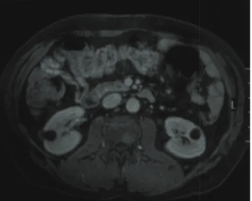
Figure 2: MRI scan of pelvis and both femurs showed multiple metastatic bone lesions dominantly located in both femoral heads, necks, and trochanters with a largest of diameter 2 cm. (a) Multiple hypointense metastatic bone lesions on T1-weighted coronal imaging. (b) Multiple hyperintense metastatic bone lesions on T2-weighted coronal imaging.
2. Case Presentation
A 55-year-old male was admitted to the orthopaedics and traumatology clinic with a 3-month history of severe pain in the right femur extending to the right leg. His medical history was insignificant. On physical examination, the patient’s right leg was tender to palpation, particularly in the area of the proximal femur, and there was limited range of motion of the right hip. Blood count was as follows: haemoglobin: 13 g/dL, total leukocyte count: 6030/mm3 (neutrophil 58%, lymphocyte 34%), and platelet: 267000/mm3. Biochemical tests showed increased erythrocyte sedimentation rate (ESR) of 50 mm/h and hypergammaglobulinemia (gamma glob-ulin 1.56 g/dL); lactate dehydrogenase (LDH) was normal. Anteroposterior radiographs of both femurs revealed mixed lytic-sclerotic lesions (Figures 1(a) and 1(b)) and a lytic destructive process with eccentric localization and a narrow zone of transition at diaphysis of left femur extending to left
subtrochanteric region (Figure 1(b)). On MRI scan, multiple hypointense metastatic bone lesions on T1-weighted coronal imaging and hyperintense metastatic lesions on T2-weighted coronal imaging dominantly located in both femoral heads, necks, and trochanters with largest of diameter 2 cm were present (Figures 2(a) and 2(b)). Abdominal MRI on T1-weighted axial imaging revealed a 1 cm wide heterogeneous metastatic lesion at left side of posterior L2 and L3 vertebrae corpus (Figure 3). Histopathological examination of the biopsy from right femoral lesion showed diffuse neoplastic infiltration consisting of cells with round, oval, regular nuclei, and medium-sized, clear cytoplasm (Figure 4). Neoplastic cells expressed CD20, LCA, CD79 alpha, tartrate-resistant acid phosphatase (TRAP), CD11c, CD68, and annexin (Fig-ure 5). BRAF V600E mutation was positive. Diagnosed with HCL, the patient was referred to our hematology depart-ment. Blood smear showed 54% neutrophils, 40% lympho-cytes, and 6% monocytes. Lymphocytes appeared normal.
L2 and L3 vertebrae corpus.
Figure 4: The biopsy specimen of the right femoral lesion showed diffuse neoplastic infiltration consisting of cells with round, oval, regular nuclei, and medium-sized, clear cytoplasm (H&E,×400).
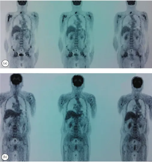
Bilateral bone marrow aspirates revealed normal bone mar-row elements with no hairy cells. Flow cytometry performed on bilateral bone marrow aspirates revealed no abnormal clone of lymphoid cells or aberrant antigen expression. Bilat-eral bone marrow trephine biopsies showed a normocellular bone marrow with no abnormal infiltrates. On PET scan, there was increased FDG uptake on lateral sides of right 2nd and 3rd ribs, posterior sides of right 9th and 10th ribs, lateral sides of left 2nd, 5th, 6th, and 7th ribs, corpus sterni, corpus of D1, D5, D6, L1, and L2 vertebrae, spinous processes of D12, L3, and L4 vertebrae, right pedicle of C6 and D11 vertebrae, both femoral heads, necks and diaphyseal regions, sacrum, and pelvic bones (Figure 6(a)). Patient underwent radiotherapy to bilateral proximal femur at a dose of 20 Gy and obtained a complete clinical response with resolution of pain. Four weeks after completion of radiotherapy, a 7-day course of 0.1 mg/kg/day cladribine was administered. Follow-up PET scan performed 8 weeks after chemotherapy showed marked metabolic response with decrease in uptake of multiple metastatic lesions on both proximal humeri, right lamina of C6 vertebrae, and both first ribs (Figure 6(b)). Repeat PET scan 12 months after chemotherapy showed complete recovery of skeletal disease with no evidence of residual
numbers of mononuclear cells with cytoplasmic projections in the blood and/or bone marrow [13]. Bone marrow is hypercellular in most cases and hairy cell infiltration may be diffuse, focal, or interstitial. Hairy cells strongly express pan-B-cell antigens including CD19, CD20, CD22, and CD25 and characteristically express CD11c, CD103, CD123, cyclin D1, and annexin A1 and usually lack expression of CD5, CD10, CD21, and CD23 [14, 15]. BRAF is a commonly mutated gene in a variety of cancers. BRAF V600E is the most common mutation reported in exon 15 of BRAF [16, 17]. Although BRAF V600E mutation is not detected in other B-cell malignancies, until now almost all cases of HCL display BRAF V600E mutation. Therefore, it is thought that BRAF V600E mutation may be considered as a hallmark of HCL [18]. Recently, CDNK1B was reported as the second most common mutated gene in HCL. Somatic CDKN1B (p27) mutations were identified in 16% of HCL patients and coexist with BRAF V600E mutations [19].
Approximately one-quarter of HCL patients are asymp-tomatic. Some patients can be observed for months or years after the initial diagnosis, which can be made after an incidental finding of splenomegaly or cytopenias during evaluation for an unrelated cause. One-quarter of patients present with abdominal fullness due to splenomegaly [14]. In 14% of the patients, the spleen is moderately enlarged and in 42%, it is massive [20]. Another one-quarter of patients present with systemic complaints such as weight loss, weakness, and fatigue. Fever or night sweats are unusual symptoms. Leukopenia is seen in 58% of patients and marked thrombocytopenia (<100,000/mm3) in 46% of patients [20].
One-quarter of patients present either with recurrent infec-tions secondary to leukopenia or with bruising and bleeding secondary to severe thrombocytopenia. Hairy cells typically infiltrate the bone marrow, peripheral blood, spleen, and liver. Most common findings are splenomegaly (60–70%) and hepatomegaly (40–50%). Lymphadenopathy present in about 10 percent of patients is not a major feature of HCL [3]. Other reported uncommon features of disease include skin [21] and neurologic involvements [22], rheumatologic dis-eases/vasculitis [23], gastric infiltration, ascites [5], nephrotic syndrome [24], scleroderma [25, 26], sarcoidosis [27], and retinal vasculitis/uveitis [28].
Skeletal involvement is an uncommon disease compli-cation. Clinical features of reported cases of HCL with
(c) (d)
(a) (b)
Figure 5: Immunohistochemical features of the infiltrated cells in the right femur expressed CD20 ((a)×200), TRAP ((b) ×200), CD11c ((c)×400), and annexin ((d) ×400).
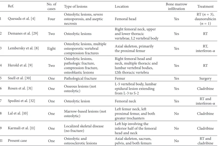
skeletal involvement, including our case, are summarized in Table 1. Quesada et al. reported four patients with osseous involvement among 46 patients with HCL [4]. Manifestations included osteolytic lesions, severe osteoporosis, and aseptic necrosis of the femoral head. Demanes et al. reported two patients, one of whom initially presented with lytic lesions in the right femoral neck and the other is characterized by development of lytic lesions in the right femoral neck, upper and lower thoracic vertebrae, and L2 vertebral body 3 years after initial diagnosis [29]. Lembersky et al. identified eight patients with osseous complications associated with HCL [8]. The lesions were primarily lytic and located in the proximal femur. All patients had bone marrow infiltration with hairy cells. This study indicated that bone involvement is associated with high tumor burden in the bone marrow and suggested that, in such cases in addition to local radiation therapy, systemic treatment with interferon should be considered. In our case, however, there was skeletal involvement without bone marrow infiltration. Herold et al. defined two patients: one with lytic lesions of the right femoral head and neck complicated by a pathologic fracture and the other with osteolytic and osteoblastic lesions developed in multiple thoracic and lumbar vertebral bodies and a compression
fracture of the 12th thoracic vertebra [9]. In our case, lesions were mixed lytic-sclerotic and sites of involvement included axial skeleton, sacrum, pelvis, and both femurs. Snell et al. reported a patient who presented with a pathological femoral fracture 22 years after HCL diagnosis, which is the longest documented period between HCL diagnosis and subsequent bony involvement [30]. Rosen et al. reported a 54-year-old man who presented with a L5 vertebral body lesion and a lumbar epidural lesion extending from L3 to S2 [31]. Biopsy sampling of the epidural mass demonstrated CD20-positive B cells that expressed TRAP and CD25, consistent with a diagnosis of HCL. Bone marrow biopsy of the same patient demonstrated focal and interstitial involvement by HCL. After treatment with a 5-day course of cladribine, the patient’s symptoms resolved. After a follow-up of 8 months, the patient was clinically stable with normal hematological parameters and no neurological symptoms. Spedini et al. described a 71-year-old man presenting with a lytic lesion in the femoral neck. His bone marrow biopsy showed nodular pattern of involvement with intracellular TRAP positivity suggestive of HCL. The patient was successfully treated with radiother-apy and interferon-𝛼 [32]. Contrary to the aforementioned reports in line with our observations, Lal et al. reported
(b)
Figure 6: PET scan at diagnosis and 8 weeks after chemotherapy. (a) There was increased FDG uptake on axial skeleton, pelvis, and both femurs at diagnosis. (b) Follow-up PET scan demonstrated marked metabolic response with decrease in uptake of multiple metastatic lesions on both proximal humeri, the right lamina of C6 vertebra, and both first ribs.
a rare HCL patient presenting with isolated skeletal disease occupying the left femur neck, left proximal femur, and both greater trochanters yet without bone marrow involvement or splenomegaly [10]. After a seven-day infusion of 2-chlorodeoxyadenosine, the femoral lesions showed marked improvement [10]. Karmali et al. described an unusual case presenting with isolated left hip involvement and no bone marrow infiltration, who was treated successfully with a 5-day course of cladribine [11]. Filippi et al. reviewed the role of radiation therapy for skeletal localizations of HCL. Skeletal lesions in HCL, with an estimated incidence of 3%, are mainly osteolytic, can occur at various sites, and are almost always symptomatic. Localized radiation therapy has been extensively used as effective palliative treatment in such cases, with different total doses and fractionation schedules [33]. Demanes et al. reported that radiation therapy in moderate doses (approximately 5000 rad in five weeks) is useful in patients with symptoms or with bone lesions at critical locations [29]. Quesada et al. reported that radiotherapy or chemotherapy provides palliation and may prevent further morbidity from bone lesions [4].
Treatment indications in HCL are cytopenias and infec-tious complications, bleeding, or painful splenomegaly. The mainstay of HCL treatment is two nucleoside analogues: pentostatin and cladribine [34–39]. These agents induce complete remission (CR) in more than 80% of patients with a median duration of progression-free survival (PFS) of over 10 years [40–43]. If the remission lasts more than two years, it is recommended to retreat with the same agent. If the remission duration is short, the use of an alternative agent is recommended. Combination of either of the nucleoside analogues with rituximab is suggested for those relapsing before two years [43, 44]. Also, rit-uximab 375 mg/m2 given weekly for 8 weeks is the best choice for patients refractory to treatment with nucleoside analogues [45]. Splenectomy can be considered if patients have symptomatic splenomegaly (>10 cm below costal mar-gin) in the presence of minimal bone marrow involve-ment [3]. Choice of agent at relapse depends on duration of first remission. Asymptomatic patients or those with minimal cytopenias may not require immediate therapy at relapse.
Table 1: Clinical features of reported cases of HCL with skeletal involvement.
Ref. No. of
cases Type of lesions Location
Bone marrow
infiltration Treatment 1 Quesada et al. [4] Four
Osteolytic lesions, severe osteoporosis, and aseptic necrosis
Femoral head Yes
RT (𝑛 = 3), daunorubicin
(𝑛 = 1) 2 Demanes et al. [29] Two Osteolytic lesions
Right femoral neck, upper and lower thoracic vertebrae, L2 vertebral body
Yes RT
3 Lembersky et al. [8] Eight
Osteolytic lesions, multiple osteoporotic vertebral compression fractures
Axial skeleton, primarily
the proximal femur Yes
RT, interferon-𝛼 4 Herold et al. [9] Two
Osteolytic lesions, pathologic fracture, compression fracture, osteoblastic lesions
Right femoral head and neck, multiple thoracic and lumbar vertebral bodies, 12th thoracic vertebra
Yes RT
5 Snell et al. [30] One Pathological fracture Femur Yes Surgery 6 Rosen et al. [31] One Osseous lesions (notosteolytic)
L-5 vertebral body, lumbar epidural lesion extending from L-3 to S-2
Yes Cladribine 7 Spedini et al. [32] One Osteolytic lesion Femoral neck Yes interferon-𝛼RT and
8 Lal et al. [10] One Marrow-based lesions (not osteolytic)
Left femur neck, left proximal femur, and both greater trochanters
No Cladribine
9 Karmali et al. [11] One Localized skeletal disease (no fracture)
Left hip involving the inferior half of the femoral head and neck
No Cladribine 11 Present case One Osteolytic andosteosclerotic lesions Axial skeleton, sacrum,pelvis, and both femurs No cladribineRT and
Ref.: references, No.: number, and RT: radiation therapy.
The current case represents an unusual presentation of severe bone pain and multiple metastatic lesions. The B cells in the bone biopsy specimen were positive for CD20, LCA, CD79 alpha, TRAP, CD11c, CD68, and annexin. Also, a molecular marker, BRAF V600E mutation, was detected in our case and distinguished the disease from other B-cell malignancies. Bilateral posterior iliac crest bone marrow biopsies showed no disease infiltration. Also, no organomegaly was present on physical examination and MRI imaging. The final diagnosis was HCL with isolated skeletal involvement. To the best of our knowledge, there are limited HCL reports in the literature with isolated skeletal involvement [10, 11]. Therefore, there is no stan-dard established treatment for this group. Our patient first underwent radiotherapy to both proximal femurs as palliative treatment as suggested before [29, 33]. Four weeks after completion of radiotherapy, we also administered a 7-day course of 0.1 mg/kg/day cladribine because of the presence of multiple metastatic lesions as in the case of isolated skeletal involvement by Lal et al. [10]. Our patient is still in continued clinical or radiographic remission 14 months after completion of chemotherapy. To conclude, skeletal involvement by HCL without coexisting bone marrow involvement should be included in the differential diagnosis of lytic-sclerotic bone lesions.
Conflict of Interests
The authors declare that there is no conflict of interests regarding the publication of this paper.
References
[1] B. A. Bouroncle, B. K. Wiseman, and C. A. Doan, “Leukemic reticuloendotheliosis,” Blood, vol. 13, no. 7, pp. 609–630, 1958. [2] L. Bernstein, P. Newton, and R. K. Ross, “Epidemiology of hairy
cell leukemia in Los Angeles County,” Cancer Research, vol. 50, no. 12, pp. 3605–3609, 1990.
[3] G. Jones, N. Parry-Jones, B. Wilkins, M. Else, and D. Catovsky, “Revised guidelines for the diagnosis and management of hairy cell leukaemia and hairy cell leukaemia variant,” British Journal
of Haematology, vol. 156, no. 2, pp. 186–195, 2012.
[4] J. R. Quesada, M. J. Keating, H. I. Libshitz, and L. Llamas, “Bone involvement in hairy cell leukemia,” The American Journal of
Medicine, vol. 74, no. 2, pp. 228–231, 1983.
[5] B. A. Bouroncle, “Unusual presentations and complications of hairy cell leukemia,” Leukemia, vol. 1, no. 4, pp. 288–293, 1987. [6] B. Bertram, K. Schulte, S. Wolf, and W. M. Gl¨ockner, “A visual
disorder as the initial symptom of hairy cell leukemia,” Deutsche
Medizinische Wochenschrift, vol. 116, no. 4, p. 157, 1991.
[7] U. W. Jehn, R. Bartl, H. Dietzfelbinger, T. Haferlach, and V. Heinemann, “An update: 12-year follow-up of patients
Clinical Advances in Hematology and Oncology, vol. 6, no. 4, pp.
290–294, 2008.
[12] R. Schrek and W. J. Donnelly, “‘Hairy’ cells in blood in lym-phoreticular neoplastic disease and ‘flagellated’ cells of normal lymph nodes,” Blood, vol. 27, no. 2, pp. 199–211, 1966.
[13] H. M. Golomb, R. Braylan, and A. Polliack, “’Hairy’ cell leukaemia (leukaemic reticuloendotheliosis): a scanning elec-tron microscopic study of eight cases,” British Journal of
Haema-tology, vol. 29, no. 3, pp. 455–460, 1975.
[14] M. R. Grever, “How I treat hairy cell leukemia,” Blood, vol. 115, no. 1, pp. 21–28, 2010.
[15] B. A. Robbins, D. J. Ellison, J. C. Spinosa et al., “Diagnostic application of two-color flow cytometry in 161 cases of hairy cell leukemia,” Blood, vol. 82, no. 4, pp. 1277–1287, 1993.
[16] H. Davies, G. R. Bignell, C. Cox et al., “Mutations of the BRAF gene in human cancer,” Nature, vol. 417, no. 6892, pp. 949–954, 2002.
[17] T. Tadmor, E. Tiacci, B. Falini, and A. Polliack, “The BRAF-V600E mutation in hematological malignancies: a new player in hairy cell leukemia and Langerhans cell histiocytosis,” Leukemia
and Lymphoma, vol. 53, no. 12, pp. 2339–2340, 2012.
[18] E. Tiacci, V. Trifonov, G. Schiavoni et al., “BRAF mutations in hairy-cell leukemia,” The New England Journal of Medicine, vol. 364, no. 24, pp. 2305–2315, 2011.
[19] S. Dietrich, J. H¨ullein, S. C. Lee et al., “Recurrent CDKN1B (p27) mutations in hairy cell leukemia,” Blood, vol. 126, no. 8, pp. 1005–1008, 2015.
[20] M. A. Hoffman, “Clinical presentations and complications of hairy cell leukemia,” Hematology/Oncology Clinics of North
America, vol. 20, no. 5, pp. 1065–1073, 2006.
[21] M. C. Finan, W. P. D. Su, and C.-Y. Li, “Cutaneous findings in hairy cell leukemia,” Journal of the American Academy of
Dermatology, vol. 11, part 1, no. 5, pp. 788–797, 1984.
[22] D. W. Wolfe, J. A. Scopelliti, and B. D. Boselli, “Leukemic meningitis in a patient with hairy cell leukemia. A case report,”
Cancer, vol. 54, no. 6, pp. 1085–1087, 1984.
[23] P. Hasler, H. Kistler, and H. Gerber, “Vasculitides in hairy cell leukemia,” Seminars in Arthritis & Rheumatism, vol. 25, no. 2, pp. 134–142, 1995.
[24] K. P. Schofield, N. Vites, C. G. Geary, R. Gokal, and M. P. Mallick, “Nephrotic syndrome and hairy cell leukaemia,” British
Journal of Haematology, vol. 60, no. 2, pp. 389–390, 1985.
[25] G. B. Cavallero, M. Bonferroni, A. Gallamini, M. Grasso, and A. Carbone, “Scleroderma and hairy-cell leukemia,” European
Journal of Haematology, vol. 52, no. 3, pp. 189–190, 1994.
cell leukemia: case report and literature review,” Western Journal
of Medicine, vol. 170, no. 3, pp. 172–174, 1999.
[31] D. S. Rosen, S. Smith, S. Gurbuxani, and B. Yamini, “Extranodal hairy cell leukemia presenting in the lumbar spine,” Journal of
Neurosurgery: Spine, vol. 9, no. 4, pp. 374–376, 2008.
[32] P. Spedini, M. Tajana, and C. Bergonzi, “Unusual presentation of hairy cell leukemia,” Haematologica, vol. 85, no. 5, p. 548, 2000. [33] A. R. Filippi, P. Franco, C. Marinone, C. Tarella, and U. Ricardi, “Treatment options in skeletal localizations of hairy cell leukemia: a systematic review on the role of radiation therapy,”
American Journal of Hematology, vol. 82, no. 11, pp. 1017–1021,
2007.
[34] D. Catovsky, E. Matutes, J. G. Talavera et al., “Long term results with 2’deoxycoformycin in hairy cell leukemia,” Leukemia &
Lymphoma, vol. 14, supplement 1, pp. 109–113, 1994.
[35] H. M. Golomb, R. Dodge, R. Mick et al., “Pentostatin treatment for hairy cell leukemia patients who failed initial therapy with recombinant alpha-interferon: a report of CALGB study 8515,”
Leukemia, vol. 8, no. 12, pp. 2037–2040, 1994.
[36] M. Grever, K. Kopecky, M. K. Foucar et al., “Randomized comparison of pentostatin versus interferon alfa-2a in previ-ously untreated patients with hairy cell leukemia: an intergroup study,” Journal of Clinical Oncology, vol. 13, no. 4, pp. 974–982, 1995.
[37] M. A. Hoffman, D. Janson, E. Rose, and K. R. Rai, “Treatment of hairy-cell leukemia with cladribine: response, toxicity, and long-term follow-up,” Journal of Clinical Oncology, vol. 15, no. 3, pp. 1138–1142, 1997.
[38] F. Lauria, D. Rondelli, P. L. Zinzani et al., “Long-lasting complete remission in patients with hairy cell leukemia treated with 2-CdA: a 5-year survey,” Leukemia, vol. 11, no. 5, pp. 629–632, 1997. [39] A. Saven, C. Burian, J. A. Koziol, and L. D. Piro, “Long-term follow-up of patients with hairy cell leukemia after cladribine treatment,” Blood, vol. 92, no. 6, pp. 1918–1926, 1998.
[40] F. Maloisel, L. Benboubker, M. Gardembas et al., “Long-term outcome with pentostatin treatment in hairy cell leukemia patients. A French retrospective study of 238 patients,”
Leukemia, vol. 17, no. 1, pp. 45–51, 2003.
[41] P. L. Zinzani, M. Tani, E. Marchi et al., “Long-term follow-up of front-line treatment of hairy cell leukemia with 2-chlorodeoxyadenosine,” Haematologica, vol. 89, no. 3, pp. 309– 313, 2004.
[42] P. Chadha, A. W. Rademaker, P. Mendiratta et al., “Treatment of hairy cell leukemia with 2-chlorodeoxyadenosine (2-CdA): long-term follow-up of the Northwestern University experi-ence,” Blood, vol. 106, no. 1, pp. 241–246, 2005.
[43] M. Else, C. E. Dearden, E. Matutes et al., “Long-term follow-up of 233 patients with hairy cell leukaemia, treated initially with pentostatin or cladribine, at a median of 16 years from diagnosis,” British Journal of Haematology, vol. 145, no. 6, pp. 733–740, 2009.
[44] M. Else, C. E. Dearden, E. Matutes et al., “Rituximab with pentostatin or cladribine: an effective combination treatment for hairy cell leukemia after disease recurrence,” Leukemia &
Lymphoma, vol. 52, supplement 2, pp. 75–78, 2011.
[45] J. Nieva, K. Bethel, and A. Saven, “Phase 2 study of rituximab in the treatment of cladribine-failed patients with hairy cell leukemia,” Blood, vol. 102, no. 3, pp. 810–813, 2003.
Submit your manuscripts at
http://www.hindawi.com
Stem Cells
International
Hindawi Publishing Corporationhttp://www.hindawi.com Volume 2014
Hindawi Publishing Corporation
http://www.hindawi.com Volume 2014
Behavioural
Neurology
Endocrinology
International Journal ofHindawi Publishing Corporation
http://www.hindawi.com Volume 2014
Hindawi Publishing Corporation
http://www.hindawi.com Volume 2014
BioMed
Research International
Oncology
Journal ofHindawi Publishing Corporation
http://www.hindawi.com Volume 2014
Hindawi Publishing Corporation
http://www.hindawi.com Volume 2014
Oxidative Medicine and Cellular Longevity
Hindawi Publishing Corporation
http://www.hindawi.com Volume 2014
PPAR Research
Immunology ResearchHindawi Publishing Corporation
http://www.hindawi.com Volume 2014
Journal of
Obesity
Journal ofHindawi Publishing Corporation
http://www.hindawi.com Volume 2014
Hindawi Publishing Corporation
http://www.hindawi.com Volume 2014
Computational and Mathematical Methods in Medicine
Ophthalmology
Journal ofHindawi Publishing Corporation
http://www.hindawi.com Volume 2014
Hindawi Publishing Corporation
http://www.hindawi.com Volume 2014
Research and Treatment
AIDS
Hindawi Publishing Corporation
http://www.hindawi.com Volume 2014