i
DEVELOPMENT OF PEPTIDE NANOMATERIALS FOR NEURAL REGENERATION
A DISSERTATION SUBMITTED TO
THE GRADUATE SCHOOL OF ENGINEERING AND SCIENCE OF BILKENT UNIVERSITY
IN PARTIAL FULFILLMENT OF THE REQUIREMENTS FOR THE DEGREE OF
DOCTOR OF PHILOSOPHY IN
MATERIALS SCIENCE AND NANOTECHNOLOGY
By
BÜŞRA MAMMADOV May, 2015
ii
DEVELOPMENT OF PEPTIDE NANOMATERIALS FOR NEURAL REGENERATION
By Büşra Mammadov May, 2015
We certify that we have read this dissertation and that in our opinion it is fully adequate, in scope and in quality, as a dissertation for the degree of Doctor of Philosophy.
………. Asst. Prof. Dr. Ayşe Begüm Tekinay (Advisor) ………. Assoc. Prof. Dr. Mustafa Özgür Güler (Co-advisor)
………. Asst. Prof. Dr. Aykutlu Dana ………. Assoc. Prof. Dr. Michelle Adams ………. Prof. Dr. Ümit Hıdır Ulaş ………. Assoc. Prof. Dr. Bahri Aydın
Approved for the Graduate School of Engineering and Science:
………. Prof. Dr. Levent Onural Director of the Graduate School
iii ABSTRACT
DEVELOPMENT OF PEPTIDE NANOMATERIALS FOR NEURAL REGENERATION
Büşra Mammadov
PhD in Materials Science and Nanotechnology Advisor: Asst. Prof. Dr. Ayşe Begüm Tekinay Co-advisor: Assoc. Prof. Dr. Mustafa Özgür Güler
May, 2015
Nervous system consists of a dense network of cells and their connections and exhibits a high level of complexity. This complexity arises from the high variety of cell types with very specific functions, the high number of cells along with the abundance of connections between these cells. When combined with the nonproliferative nature of neural cells and inhibitory nature of the pathological extracellular matrix (ECM), this complexity leads to a very limited regenerative potential. Thus neurodegenerative disorders and traumatic injuries of neural tissues lead to lifelong disabilities due to the poor success of current therapies. Novel therapeutic approaches which can overcome barriers that impede neural regeneration are therefore required to be developed. Smartly designed nanomaterials that can direct cells towards desired functions can improve the regeneration of neural tissues.
Herein, I have described my work on development of peptide nanofibers for neuroregeneration and biological applications of these nanomaterials. To achieve the regeneration of the nervous system, the composition of the neural ECM under healthy conditions and during early development was mimicked through structural resemblance and bioactive epitope presentation using nanofibers. Laminin derived IKVAV peptide sequence and glycosaminoglycan mimicking, growth factor-binding sulfonated peptide sequence were presented on peptide nanofiber scaffolds. Differentiation of PC-12 cells, a model cell system for neuroregenerative studies, was found to be improved on these nanofiber scaffolds when compared to the cells on epitope free control scaffolds. Cells could even extend neurites on these scaffolds in the presence of inhibitory chondroitin sulfate proteoglycans. These nanofibers also proved to be efficient in sciatic nerve regeneration after injury. When injected into the lumen of polymeric nerve guidance channels, this bioactive nanofiber system provided guidance to the elongating axons and resulted in better axonal regeneration that was evident both from histological analysis and electromyography results. Results of in vitro and in vivo experiments were correlated and indicated the neuroregenerative potential of these peptide nanofibers. In addition, semiconductive oligothiophene was encapsulated in peptide nanofibers without compromising the biocompatibility. These hybrid nanofiber scaffolds can potentially be used for electrical stimulation of neurons that can further boost regeneration.
iv ÖZET
SİNİRSEL REJENERASYON AMAÇLI PEPTİT NANOMALZEMELERİN GELİŞTİRİLMESİ
Büşra Mammadov
Malzeme Bilimi ve Nanoteknoloji, Doktora Tez danışmanı: Yrd.Doç.Dr. Ayşe Begüm Tekinay
Eş Danışman: Doç.Dr. Mustafa Özgür Güler Mayıs, 2015
Sinir sistemi hücrelerin birbiriyle olan bağlantılarından oluşan yoğun ve kompleks bir ağa sahiptir. Sinir sisteminde farklı fonksiyonlara sahip çeşitli hücrelerin varlığı, hücre sayısının ve hücrelerin yaptığı bağlantıların fazla oluşu bu sistemi oldukça karmaşık kılmaktadır. Bu kompleksiteye ek olarak sinir hücrelerinin çoğalmaması ve patolojik durumlarda oluşan hücreler arası matriksin inhibe edici doğası hasar sonrası rejenerasyon imkanını sınırlamaktadır. Güncel tedavi seçeneklerinin düşük başarı oranı nörodejeneratif hastalıklar ve travmatik hasarlar nedeni ile birçok hastayı ömür boyu bakıma muhtaç bırakmaktadır. Bu nedenle yeni tedavi yaklaşımlarına ihtiyaç duyulmaktadır. Hücrelerin kaybedilen fonksiyonlarını yerine getirmeye yönlendirmek üzere tasarlanmış nanomalzemerin geliştirilmesi sinir rejenerasyonunda umut verici olabilir.
Bu çalışmada sinirsel rejenerasyon amaçlı geliştirilmiş peptit nanofiberler ve bu malzemelerin biyolojik uygulamaları anlatılmıştır. Sinir sisteminde rejenerasyon sağlayabilmek amacıyla sağlıklı doku ve embriyonik dokulardaki hücreler arası matriksi yapı ve biyolojik sinyaller yönünden taklit eden nanofiberler üretilmiştir. Bu nanofiberler laminin proteinine has bir sekans olan „IKVAV‟ ve büyüme faktörlerine bağlanma özelliği olan glikozaminoglikan taklidi sülfonatlı bir diziden oluşmaktadır. Biyoaktif peptit nanofiber iskeleler üzerinde sinirsel yönde başkalaştırılan PC-12 hücrelerinin kontrol nanofiberler üzerindeki hücrelere göre daha iyi başkalaştığı görülmüştür. Aksonal büyümeyi inhibe edici kondroitin sülfat proteoglikanların varlığında dahi nörit uzatımı etkin bir şekilde devam etmiştir. Hücreler arası matriks taklidi bu nanofiberlerin siyatik sinir rejenerasyonunda da etkili olduğu görülmüştür. Polimerik sinir tüpünün içine enjekte edilen nanofiberlerle tedavi edilen hayvanlarda aksonal rejenerasyonun daha iyi olduğu histolojik analizler ve elektromiyografi sonuçları ile gösterilmiştir. Birbirini destekleyen in vitro ve in vivo sonuçlar, bu çalışmada kullanılan peptit nanofiberlerin sinir rejenerasyonu için etkili bir malzeme olduğunu göstermektedir. Ayrıca bu nanofiberlerin içine yarı iletken oligotiyofen enkapsüle edilmiş ve bu modifikasyonun hücre canlılığını etkilemediği gözlemlenmiştir. Yeni geliştirilen bu multifonksiyonel peptit nanofiberler daha iyi bir rejenerasyon için sinir hücrelerinin elektriksel uyarımına imkan verecektir.
v
ACKNOWLEDGEMENTS
I have learned quite valuable things during this journey of PhD that envisioned my view of perspective both in science and everyday life. I owe many thanks to a lot of people who helped me find my way in the jungle of scientific questions, supported me during times of demotivation and treated me with patience and surrounded me with their tenderness in my hard times. This thesis could not arise without their support and I acknowledge their invaluable support.
I would like to thank my advisors Prof. Tekinay and Prof. Güler for all the scientific support they provided me. I would like to thank Prof. Dr. Ümit Hıdır Ulaş, Assoc. Prof. Dr. Fatih Zor and Assist. Prof. Dr. Hakan Akgün for their collaboration in the
in vivo experiments. I also would like to express my gratitudes to Zeynep Erdoğan
for her technical support in chemical characterizations which boosted my knowledge on these techniques, smoothened my work and saved my time with her perfect assistance. I am thankful to Prof. Dr. Salim Çıracı for his guidance and support at the hardest part of my PhD. I acknowledge all BML and NBT lab members for the fruitful scientific discussions and for their friendship. I appreciate the contributions of Ruslan Garifullin and Aref Khalily; they always helped me whenever I ended up with a question in chemistry during the research I conducted. I would like to thank Melike Sever and Mevhibe Yakut with my deepest gratitudes for always being nice to me while working together and for the times they backed me up especially after my daughter‟s birth.
I would like to thank TUBITAK for the financial support. I received my stipend from TÜBİTAK BİDEB 2211-C programme during the PhD period. Also, our
vi
experimental work was supported by TUBITAK projects 111M410, 113Z495 and 113S038.
Some friends made even the hard times enjoyable with their emotional support and cheerfulness. Yelda Ertaş, Aslı Çelebioğlu, Fatma Kayacı and Zeynep Aytaç; thank you girls for the every limited time we could share. I enjoyed every second and those times provided the fuel to work after long times of demotivation.
My family deserves a lot more than acknowledgement for their support and guidance in my academic success starting from very early ages. It would not be possible to complete the work published in here without their endless support after the birth of my daughter.
I would love to thank for every support he provided along with his great patience during most stressfull days to Reşad. I also want to thank my little cutie Gülnare for the warmth of her smiles which provided me whole the energy I required.
vii
TABLE OF CONTENTS
TABLE OF CONTENTS ... vii
LIST OF FIGURES ... x
LIST OF TABLES ... xiii
LIST OF ABBREVIATIONS ... xiv
CHAPTER 1 ... 1
INTRODUCTION ... 1
1.1. Neurodegenerative disorders and neural injuries ... 2
1.2. Role of ECM in poor regenerative capacity of central nervous system ... 4
1.3. Role of ECM in regenerative capacity of the peripheral nervous system ... 10
1.4. Mimicking healthy ECM with synthetic scaffolds ... 13
1.4.1. Different approaches in the design of synthetic materials for neural differentiation ... 15
1.4.2. Peptide nanofibers as scaffolds for neural regeneration ... 18
CHAPTER 2 ... 25
ECM MIMETIC PEPTIDE NANOFIBERS FOR NEURAL DIFFERENTIATION 25 2.1. INTRODUCTION ... 26
2.2. MATERIALS and METHODS ... 29
2.2.1. Materials ... 29
2.2.2. Synthesis and purification of peptide amphiphiles ... 30
2.2.3. SEM imaging of PA nanofiber matrices ... 31
2.2.4. AFM imaging of PA nanofiber matrices ... 31
2.2.5. Circular dichroism ... 32
2.2.6. Oscillatory rheology ... 32
2.2.7. NGF release assay by ELISA ... 32
2.2.8. PA charge optimization for PC-12 differentiation... 33
2.2.9. PA concentration optimization for PC-12 differentiation ... 34
2.2.10. Optimization of neural induction medium ... 35
2.2.11. Optimization of cell density ... 36
viii
2.2.13. Optimization of PA coating ... 37
2.2.14. Analysis of cell viability on PA nanofiber coated surfaces ... 37
2.2.15. SEM imaging of PC-12 cells on PA substrates ... 38
2.2.16. BrdU assay for analysis of cell proliferation ... 38
2.2.17. Immunofluorescent staining against SYN1 ... 39
2.2.18. Neurite outgrowth inhibition assay ... 39
2.3. RESULTS ... 40
2.3.1. Peptide amphiphile design and characterization... 40
2.3.2. Neural differentiation of PC-12 cells on PA scaffolds ... 52
2.3.3. Investigation of NGF/PA-GAG interaction ... 65
2.3.4. Neurite inhibition assay by CSPGs... 67
2.4. CONCLUSIONS ... 72
CHAPTER 3 ... 74
BIOFUNCTIONALIZATION OF POLYMERIC NERVE GUIDANCE CONDUITS WITH PEPTIDE NANOFIBERS FOR SCIATIC NERVE REGENERATION ... 74
3.1. INTRODUCTION ... 75
3.2. MATERIALS AND METHODS ... 79
3.2.1. Materials ... 79
3.2.2. Synthesis and purification of peptide amphiphiles ... 79
3.2.3. Scanning electron microscope and transmission electron imaging of peptide nanofibers ... 79
3.2.4. Surgical procedure ... 80
3.2.5. Walking track analysis ... 82
3.2.6. Electrophysiological measurements ... 82
3.2.7. Histological analysis of sciatic nerves ... 83
3.3. RESULTS ... 84
3.3.1. Characterization of peptide nanofiber gels ... 84
3.3.2. Functional evaluation of transected sciatic nerves after repair ... 86
3.3.3. Histological analysis of regenerating nerve morphology ... 92
3.4. CONCLUSION ... 103
ix
ENCAPSULATION OF OLIGOTHIOPHENE DERIVATIVE IN PEPTIDE NANOFIBER SCAFFOLDS FOR ELECTRICAL STIMULATION OF NEURAL
CELLS ... 105
4.1. INTRODUCTION ... 106
4.2. MATERIALS AND METHODS ... 112
4.2.1. Optimization of DH4T amount for encapsulation into PA molecules .. 112
4.2.2. Calculation of encapsulated DH4T amounts from UV measurements .. 113
4.2.3. Fluorescence measurement of DH4T encapsulated PAs ... 114
4.2.4. Viability assay of PC-12 cells on PA substrates ... 114
4.3. RESULTS ... 115
4.3.1. Optimization of DH4T amount for encapsulation ... 116
4.3.2. Encapsulation of DH4T in different PA molecules ... 122
4.3.3. Viability assay for PC-12 cells ... 124
4.4. CONCLUSION ... 126
CHAPTER 5 ... 127
CONCLUSIONS AND FUTURE PERSPECTIVES ... 127
x
LIST OF FIGURES
Figure 1.1 . Neural stem cell fate determination is mainly guided by extracellular
matrix molecules, soluble factors and cell-to-cell interactions. ... 15
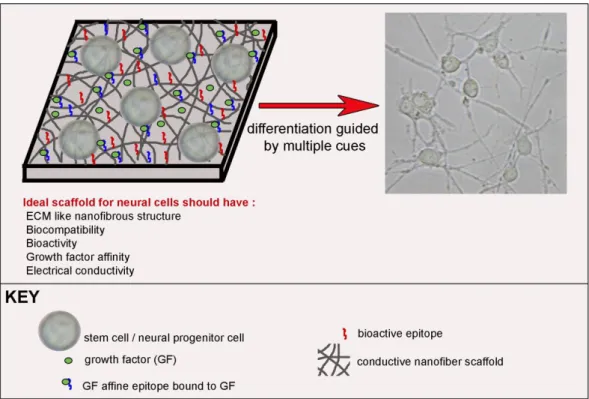
Figure 1.2. Bioactive scaffold design for neural differentiation of stem cells. ... 18
Figure 1. 4. Immunohistochemical analysis of neural progenitor cell differentiation on IKVAV-PA gels ... 22
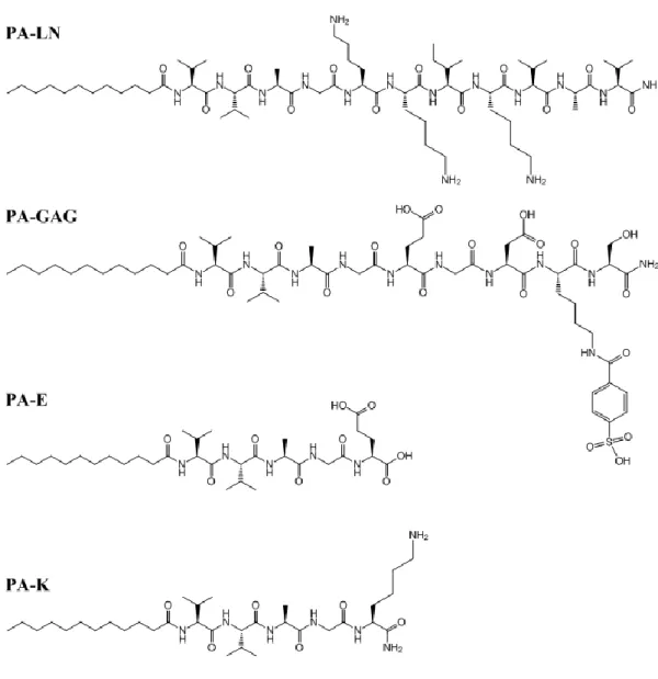
Figure 2. 1. Chemical structures of the PA molecules used in this study. ... 42
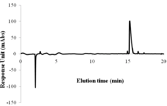
Figure 2. 2. Liquid chromatography analysis of PA-LN. ... 43
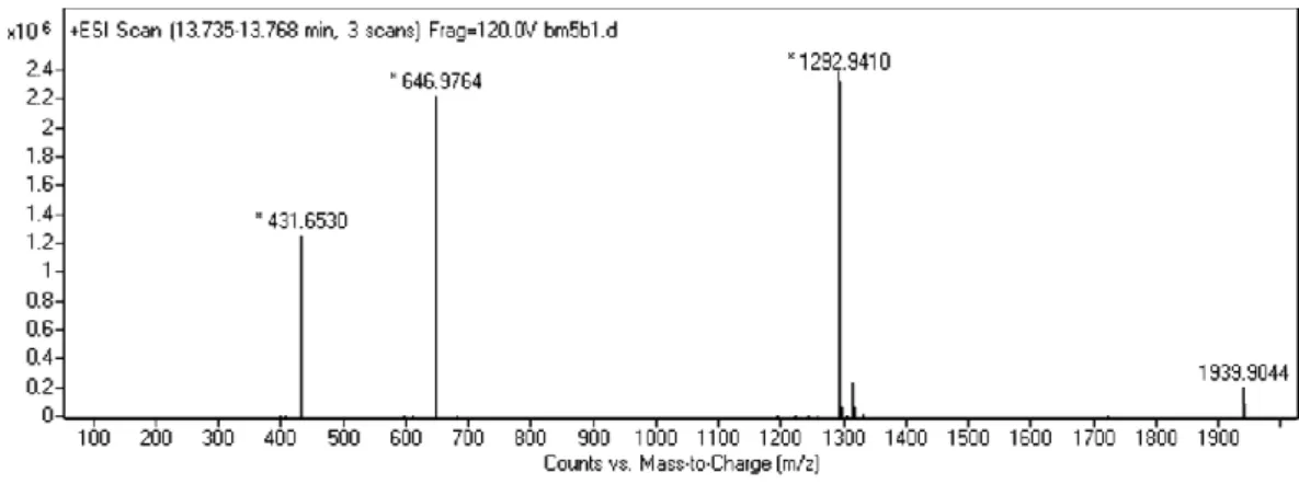
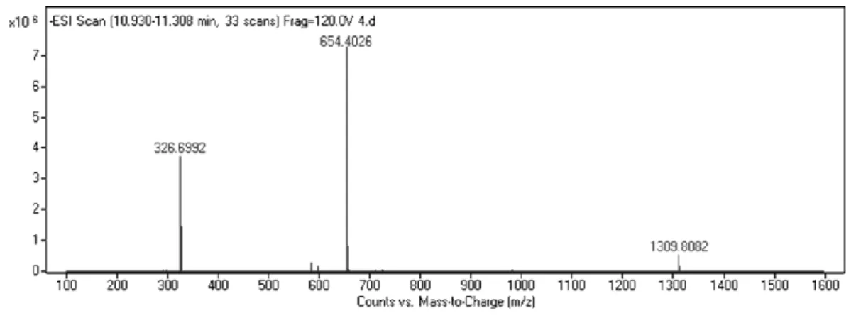
Figure 2. 3. Mass spectrometry analysis of PA-LN. ... 43
Figure 2. 4. Liquid chromatography analysis of PA-GAG. ... 44
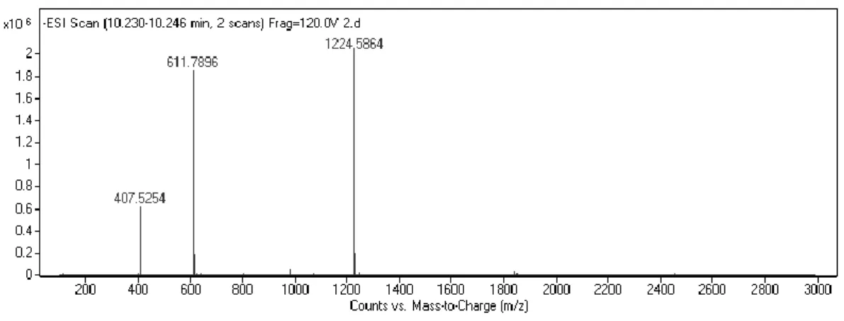
Figure 2. 5. Mass spectrometry analysis of PA-GAG... 44
Figure 2. 6. Liquid chromatography analysis of PA-K. ... 45
Figure 2. 7. Mass spectrometry analysis of PA-K. ... 45
Figure 2. 8. Liquid chromatography analysis of PA-E. ... 46
Figure 2. 9. Mass spectrometry analysis of PA-E. ... 46
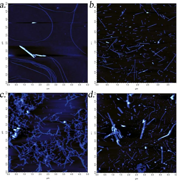
Figure 2. 10. AFM images of peptide amphiphile nanofibers ... 48
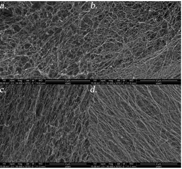
Figure 2. 11. SEM images of peptide amphiphile gels ... 49
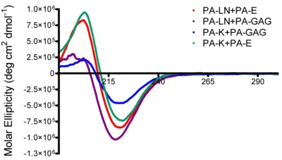
Figure 2. 12. Characterization of secondary structure of peptide amphiphiles by CD. ... 50
Figure 2. 13. Measurement of stiffness of PA gels by oscillatory rheology ... 52
Figure 2. 14. Effect of final charge of PA substrate on neurite outgrowth by PC-12 cells ... 54
Figure 2. 15. Effect of PA concentration on neurite outgrowth by PC-12 cells. ... 54
Figure 2. 16. Effect of induction medium on neurite outgrowth by PC-12 cells ... 55
Figure 2. 17. Effect of NGF amount on neurite outgrowth by PC-12 cells ... 56
Figure 2. 18. Results of live-dead assay on PA substrates and PDL. ... 57
Figure 2. 19. SEM images of PC-12 cells cultured on PA gels ... 58
Figure 2. 20. Proliferation assay results of PC-12 cells on day 7 after neural induction. ... 60
Figure 2. 21. Neurite outgrowth of PC-12 cells on PA scaffolds. ... 61
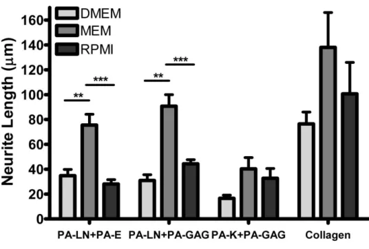
Figure 2. 22. Quantified results of neurite length on PA substrates at day 7. ... 62
Figure 2. 23. Quantified results of percentage of cells extending neurites on PA substrates at day 7. ... 62
Figure 2. 24. Immunostaining of PC-12 cells cultured on PA nanofiber scaffolds against synaptophysin 1 ... 64
Figure 2. 25. Time-dependent NGF release from PA gels... 67
Figure 2. 26. Optical images of cells on substrates with CSPG... 70
Figure 2. 28. Effect of CSPG inhibition on percentage of cells extending neurites at day 2 ... 71
xi
Figure 2. 29. Percentage of neurite length retained after CSPG treatment for 2 days.
... 72
Figure 3. 1. Images of surgical operation shows conduit sutured to the excised sciatic nerve (a-c) and application of the PA gel in the conduit (d). ... 81
Figure 3. 2. SEM (a, b) and TEM (c, d) images of LN+GAG (a, c) and PA-K+PA-E (b, d). ... 86
Figure 3. 3. Footprints of rats obtained during walking track analysis at week 12 after operation. ... 88
Figure 3. 4. Sciatic functional index measurement results showing better, yet nonsignificant scores in rats treated with PA-LN+PA-GAG biofunctionalized nerve guidance conduits. ... 88
Figure 3. 5. Sciatic functional index measurement results showing performance of individual rats within groups. ... 89
Figure 3. 6. Recovery of lost amplitude at week 12 as measured by EMG. ... 92
Figure 3. 7. Percentage increase in latency in operated nerves when compared to the healthy unoperated nerves at week 12, as measured by EMG. ... 92
Figure 3. 8. Excised nerve tissue parts used for different histological analysis ... 93
Figure 3. 9. H&E stained nerve tissues harvested at 12 weeks after operation ... 95
Figure 3. 10. Magnified images of H&E stained nerve tissues harvested at 12 weeks after operation. ... 97
Figure 3. 11. Nerve tissues harvested at 12 weeks after operation are immunostained against β-III-tubulin. ... 99
Figure 3.12. Nerve tissues harvested at 12 weeks after operation were immunostained against S100.. ... 102
Figure 4.2. Chemical structure of commonly used conducting polymers ... 110
Figure 4. 3. Chemical structure of DH4T molecule. ... 116
Figure 4. 4. Chemical structure of PA-KK and PA-EE molecules ... 116
Figure 4. 5. Absorbance spectrum of standard DH4T samples of known concentrations. ... 117
Figure 4. 6. DH4T standard curve plotted by using absorbance values at 410 nm.. 118
Figure 4. 7. Absorbance spectrum of samples with different DH4T amounts used for encapsulation. ... 119
Figure 4. 8. Absorbance spectrum of 0.5 mg DH4T encapsulated PA-GAG diluted in DMF or ddH2O showing the shift absorption spectra upon encapsulation. ... 121
Figure 4. 9. Fluorescence measurement of 0.5 mg DH4T encapsulated PA-GAG samples excited at 400 nm. ... 121
Figure 4. 10. Absorbance spectrum of DH4T encapsulated PA samples diluted with DMF. ... 123
Figure 4. 11. Absorbance spectrum of DH4T encapsulated PA samples diluted with ddH2O. ... 123
xii
Figure 4. 12. Viability assay results of PC-12 cells cultured on PA substrates for 24 h ... 125
xiii
LIST OF TABLES
Table 2.1. Charge, sequence and molecular weight of PA molecules used in the
study. ... 34
Table 4. 1. Properties of scaffolds used for viability assay... 115
Table 4. 2. Optimization of DH4T amount for encapsulation. ... 119
Table 4. 3. Results of DH4T encapsulation in different PA molecules. ... 124
Table 4. 1. Properties of scaffolds used for viability assay... 115
Table 4. 2. Optimization of DH4T amount for encapsulation. ... 119
xiv
LIST OF ABBREVIATIONS
ALS : amyotrophic lateral sclerosis SCI : spinal cord injury CNS : central nervous system PNS : peripheral nervous system
ECM : extracellular matrix PG : proteoglycan
CSPG : chondroitin sulfate proteoglycan HSPG : heparan sulfate proteoglycan DSPG : dermatan sulfate proteoglycan
KSPG : keratan sulfate proteoglycan GAG : glycosaminoglycan
HA : hyaluronic acid
MAG : myelin-associated glycoprotein OMgP : oligodendrocyte myelin glycoprotein
cAMP : Cyclic adenosine monophosphate CREB : cAMP response element binding protein
NGF : nerve growth factor
BDNF : brain derived neurotrophic factor GDNF : glial derived neurotrophic factor
IGF : insulin like growth factor CNTF : ciliary neurotrophic factor FGF-2 : fibroblast growth factor 2 TNF-a : tumor necrosis factor alpha
xv
LIF : leukocyte inhibitory factor IL : interleukin
tPA : tissue plasminogen activator NT-3 : neurotrophin-3
RA : retinoic acid PA : peptide amphiphile SEM : scanning electron microscope TEM : transmission electron microscope
AFM : atomic force microscopy SHH : sonic hedgehog
CN : cavernous nerve PDL : poly-D-Lysine CD : circular dichroism
DMEM : Dulbecco‟s Modified Eagles Medium RPMI : Roswell Park Memorial Institute Medium
MEM : Minimal Essential Medium EthD-1 : ethidium homodimer I
SYN1 : synaptophysin 1 BSA : bovine serum albumin PBS : phosphate buffered saline
HPLC : high performance liquid chromatography LC-MS : liquid chromatography-mass spectroscopy
ANOVA : analysis of variance PNI : peripheral nerve injury NGC : nerve guidance conduit
xvi
HRP : horseradish peroxidase SFI : sciatic functional index
EMG : electromyography HE : hematoxylin-eosin
CaMK : calcium/calmodulin dependent protein kinase MEK : mitogen activated protein kinase
ERK : extracellular signal regulated protein kinase DRG : dorsal root ganglion
DH4T : 3,3′′′-Dihexyl-2,2′:5′,2′′:5′′,2′′′-quaterthiophene THF : tetrahydrofuran
1
CHAPTER 1
INTRODUCTION
2
1.1. Neurodegenerative disorders and neural injuries
Neurodegeneration is the common term for defining progressive loss of structure and function of nerve cells. It generally occurs due to the accumulation of misfolded aggregated proteins in some parts of the aging brain, which results in cell death and inflammatory damage in those regions1. Clinically, there are different types of neurodegeneration in distinct neurodegenerative diseases. Therefore, there are some variances in the proximal triggers and pathological markers of neurological diseases such as Lewy bodies in Parkinson‟s disease, plaques and tangles in Alzheimer‟s disease, demyelination in multiple sclerosis and motor-neuron death in ALS. Although initiated by different initial triggering factors, these diseases share some overlapping downstream and secondary pathways such as neuroinflammation2. Prevalence of neurodegenerative disorders is on the rise due to the increase in the number of aged population. In a report published in 2012, it was stated that there exists 5.2 million people living with Alzheimer‟s in US. Prevalence above age 65 is quite high, with one in eight people suffering from the disease3. The number of patients suffering worldwide was reported to be 35.6 million in 2010 and this number is expected to double every 20 years due to the increase in life expectancy4. Approximately 50000 patients are diagnosed with Parkinson‟s disease each year5. Neurodegenerative disorders are not the only reason of neural cell loss; neuronal loss from traumatic injuries of brain and spinal cord also result in destructive effects on motor, sensory and autonomic functions. Spinal cord injury (SCI), for example, generally causes permanent loss of sensation and motor function below the site of injury. Mainly caused by motor vehicle and sports accidents, spinal cord injuries lead
3
to lifelong disabilities if not fatal. According to the 2014 report of National Spinal Cord Injury Statistical Center, approximately 12500 new injuries occur each year and there exists approximately 276000 patients suffering from SCI living in US. Less than 1% of patients could recover completely after injury6.
Peripheral nervous system (PNS) is even more open to injuries due to the absence of the cranium and spine which protect central nervous system (CNS) from external damages. PNS injuries result in a high number of patients suffering from neuropathic pain which brings an economic burden resulting from health care costs. 20 million patients suffer from peripheral neuropathy and approximately $150 billion is spent annually for health care due to nerve injuries in United States7. Approximately a hundred thousand patients undergo peripheral nerve surgeries per year in United States and Europe8.
The number of patients suffering from neurological disorders and traumatic injuries demand the development of successful therapies for the restoration of lost neurological functions. Although the demand is high mostly due to age-related neurodegenerative disorders and injuries resulting from motor vehicle accidents, such efficient therapies are not present, yet. In order to develop effective therapies for the induction of neural regeneration, one must first understand the reasons behind the poor regenerative capacity of neural tissues, especially through the intrinsic properties of extracellular matrix (ECM) in health and disease state. Different properties of the ECM in CNS and PNS should also be considered when designing therapies, since such differences lead to differing regenerative capacity in these tissues.
4
1.2. Role of ECM in poor regenerative capacity of
central nervous system
Fully restoring functional capacities of neurons after damage due to traumatic injuries or neurodegenerative disorders is still not possible. Although conventional therapies provide neural regeneration up to a certain level, they are not as efficient as desired. The term “neural regeneration” refers to several different mechanisms for the restoration of the functions of degenerated neural tissue. It either occurs by the generation of new neurons from the progenitor cells residing near the damaged area, generation of new synapses by the neurons that survive after the damage or by repair of the damaged axons/myelin sheaths to prevent secondary cell loss after injury. These strategies for regeneration of the nervous system are ideally combined to achieve a functional outcome. However, due to some intrinsic properties such as the unproliferative nature of neurons, presence of progenitors in localized areas and in low numbers together with the upregulation of inhibitory elements upon injury leading to glial scar formation, regeneration capacity of the central nervous system is quite limited.
Central nervous system consists of a dense network of cells leaving a smaller volume for the extracellular matrix components, 10-20% for the brain unlike most other tissues9. Before 1971, brain was thought to be totally free of ECM and consisting of only a very dense network of cells10. Yet, this small amount of ECM is sufficient to modulate the survival, neurite outgrowth and synaptic plasticity of neurons. Neural ECM undergoes prominent changes during the maturation of the nervous system.
5
Embryonic and postnatal ECM is rather a loose network, while some condensation occurs through adulthood starting from the postnatal period, according to specific needs of neural cells11-12. In fact, the balance among ECM components is so important that any imbalance leads to massive degeneration in the CNS.
Being quite different from that of other tissues, ECM content also differs dramatically in between specific locations in the CNS. Three different ECM types are deposited in basement membrane, perineuronal nets (PNN) and the gap filling interstitial matrix. Basement membrane (basal lamina) is more similar to other tissues in terms of the ECM components it contains. Collagen, entactin (laminin– nidogen complexes), fibronectin, dystroglycan and perlecan are the predominant proteins and proteoglycans of basement membrane13. This structure is associated with blood vessels and is important in the stabilization of the blood-brain barrier14.
Perineuronal nets are the dense network of matrix around cell bodies and mainly responsible for the maintenance of synaptic plasticity. Main components of PNN are hyaluronan, chondroitin sulfate proteoglycans (CSPGs) and tenascins13, 15. Being absent in embryonic tissue, these structures form a barrier around the cells in mature tissue and prevent the formation of new synaptic connections while preserving the ones that are already formed. Mammalian nervous system has a considerable capacity for plasticity that continues through postnatal development until maturation of the CNS. During this period, named as critical period, nervous system continues to develop and produces new neural connections that are influenced by experience. Plasticity declines upon the closure of the critical period, the time of which coincides
6
with the formation of perineuronal nets indicating these structures as barriers to the establishment of new connections15.
Being absent from perineuronal nets, small amounts of fiber forming collagens and elastins along with fibronectin and laminin are present in the interstitial matrix. Sulfated proteoglycans and hyaluronan along with tenascins are the main components of this ECM13.
Proteoglycans (PGs) are the predominant components of the central nervous system ECM. Proteoglycans are classified into four main groups depending on the glycosaminoglycan (GAG) chain; chondroitin sulfate proteoglycans (CSPGs), heparan sulfate proteoglycans (HSPGs), dermatan sulfate proteoglycans (DSPGs) and keratan sulfate proteoglycans (KSPGs). CSPGs and HSPGs are the main PGs of the CNS, CSPGs being the most abundant15. Each group is also composed of a variety of PGs due to the different protein cores carrying GAG chains. Lecticans are the most abundant CSPG type in the brain and are classified into four subgroups. Neurocan and brevican are neural tissue specific lecticans while aggrecan and versican are common to the ECM of other tissues16. Besides sulfated GAGs that are found attached to protein cores, hyaluronic acid (HA) is also abundant in the CNS tissues. HA is a large polymeric carbohydrate chain that is nonsulfated and does not contain a protein core. It binds to other proteoglycans and forms a dense mesh-like ECM structure in perineuronal nets17.
Depending on the protein core and GAG chain, proteoglycans in the CNS are permissive or non-permissive to neurite extension. GAGs can exert their effects on
7
cells either by inducing direct signaling through cell surface receptors (like HA achieves through CD44 receptor) or by their affinity to growth activating molecules. Affinity of HSPGs to growth factors is quite important in the modulation of neuronal function, especially through FGF signaling (improved upon syndecan binding to FGF18). CSPGs can also bind a range of molecules including semaphorin 5A, binding to which is found to change permissive nature to inhibitory19. Binding of CSPGs to other ECM proteins, such as tenascin and laminin blocks the axon growth promoting ability of these proteins and thus inhibits axon growth20. While binding of CSPG to laminin is inhibitory, binding by HSPG improves axonal elongation21. Binding of HSPGs to the same receptor in a competitive manner (as in the case of semaphorin 5A) abolishes the inhibitory effect of CSPG on neurotrophin responsiveness19. CSPGs can also inhibit neural regeneration by limiting calcium availability either by directly binding to extracellular calcium or binding to calcium channels, thereby preventing its uptake by neurons22. Binding of CSPGs to PTPsigma receptor on neurons leads to the phosphorylation of neurotrophin receptors TrkB and TrkC, possibly leading to a downregulation of responsiveness to neurotrophins23. Inhibitory effects of CSPGs are either due to the protein core or sulfated GAG chain, depending on the CSPG type. For versican, neurocan and phosphacan, the protein core is indispensable for axonal inhibition, while the GAG chain is responsible for inhibition in aggrecan and brevican. Chondroitinase treatment, which degrades the GAG chains, eliminates the inhibitory effect of aggrecan and brevican while being ineffective in interfering with the function of the other CSPGs24-29.
8
Although they limit the regenerative capacity of CNS with their inherent inhibitory properties, CSPGs have indispensible roles in neural development, both for cell migration, and formation and stabilization of neural connections. Neural crest cells avoid migrating through local environments that are abundant in CSPG. Presence of CSPGs along their path therefore limits their movements and creates a trajectory for them to migrate to their defined positions30. Repulsive nature of CSPGs for growth cones is also quite important in axonal path finding and the correct formation of neural networks during embryonic development as well as postnatal development in critical period. Treating developing nervous system with chondroitinase ABC leads to various axon path finding errors, verifying the importance of CSPGs in the process31. CSPGs are also important in adult CNS for proper stabilization of synaptic connections and prevention of improper axonal sprouting and formation of disorganized neural networks32.
Having a regulatory function on neural network formation in development and keeping connections stable, CSPGs are quite important in healthy CNS. However they should be kept in balance in order not to over-inhibit neural functions. This balance becomes disorganized upon a trauma leading to production of reactive astrocytes. Astrocytes start to proliferate and deposit dense ECM in the injury site and fill the gap caused by the injury. This preserves the overall structure of the tissue, stabilize blood brain barrier and limits the damage to injury site, thereby preventing secondary damage33. Although important for the overall health of CNS, this hastily-deposited ECM is responsible for the absence of regeneration in CNS injuries. This
9
sprouting due to the abundance of CSPGs. Hence, nearby healthy cells that have the potential to make new synaptic connections to tolerate the neural cell loss in the injury site cannot enter into the area. Besides, synaptic connections cannot be established by undamaged cells in the injury site due to the inhibitory effect of CSPGs on plasticity.
Besides CSPGs, myelin components are also inhibitory for neural regeneration after CNS injury. The major inhibitory myelin components can be listed as myelin-associated glycoprotein (MAG), Nogo-A, oligodendrocyte myelin glycoprotein (OMgP), and myelin lipid sulfatide34-35. Myelin inhibitors MAG, Nogo-A and OMgP are found to exert their inhibitory effect through binding to Nogo 66 receptor 1 (NgR1) on neurons36-39. Presence of NgR1 is essential for growth cone collapse upon encountering myelin inhibitors40-41. Paired immunoglobulin-like receptor B (PirB), and its human homolog (LILRB2) are recently identified as a receptor for these three myelin inhibitors and blocking this receptor along with NgR1 largely eliminates myelin inhibition of neurite outgrowth42.
A third type of CNS inhibitory molecules includes the axon guidance molecules semaphorin and ephrin. Most semaphorins are inhibitory for neurons and play a role in network stabilization by limiting neurite outgrowth. In the glial scar, class 3 semaphorins (Sema3s) contribute to the inhibitory environment in a CSPG dependent manner. Interfering with the CSPG-Sema3s interaction eliminates the inhibitory nature of this molecule in vitro43. Besides Sema3s, membrane associated semaphorins like Sema4D are also upregulated in the glial scar and inhibit neurite outgrowth44. Ephrins exert their inhibitory effect by binding to EphA and EphB
10
receptor tyrosine kinase on neural cell surface. EphrinB3 is expressed in CNS myelin, ephrin B2 is upregulated upon injury by reactive astrocytes and ephrinA5 expression is found to be increased in cortical lesion and they all inhibit neurite outgrowth45-47.
1.3. Role of ECM in regenerative capacity of the
peripheral nervous system
Reaction of the CNS to any injury leading to scar tissue formation acts as a barrier for tissue regeneration, while it is the opposite in PNS. Different types of glial cells in CNS and PNS, astrocytes and Schwann cells, lead to formation of completely different environments for axons struggling to heal after an injury. While astrocytes produce a growth inhibitory environment consisting of chemorepulsive guidance molecules, Schwann cells produce a growth promoting environment by the secretion of supportive ECM molecules, neurotrophins, as well as dealing with the clearance of inhibitory myelin products after injury. A chain of events occurring after injury in the peripheral nerve leads to formation of a permissive environment for axonal regeneration. Wallerian degeneration in the distal stump of the injured nerve leads to fragmentation of the axon and associated myelin products. Fragmented products are then cleared by macrophages that are recruited to the injury site48. Myelin associated proteins are known to inhibit axonal elongation and their clearance in PNS after injury forms a permissive environment for regeneration as opposed to that in CNS. While the distal stump undergoes Wallerian degeneration, axonal regeneration starts from the proximal end. Schwann cells dedifferentiate and start to proliferate. They
11
myelinate newly elongating axon thus providing an electrical insulation to the regenerating axon49.
Permissive environment, axon guidance cues and intrinsic growth capacity of peripheral nerves are crucial for the successful regeneration of the PNS. Unlike their CNS counterparts, peripheral nerves have an intrinsic growth capacity that is activated upon injury. Cyclic adenosine monophosphate (cAMP) level is upregulated upon peripheral nerve injury which activates protein kinase A50-51. Protein kinase A is thought to take role in cytoskeletal reorganization by inhibiting RhoA. RhoA is normally activated by the presence of myelin debris and its activation halts the cytoskeletal reorganization required for axonal elongation. Hence, its inhibition by protein kinase A makes axon‟s cytoskeletal dynamics favorable for regeneration52
. In addition, cAMP has a transcriptional role by activating cAMP response element binding protein (CREB) which in turn enables cell to overcome myelin associated inhibition of neurite outgrowth53. Upregulation of intracellular cAMP levels also leads to an increase in cell-surface localization of TrkB receptors, which in turn increases the axon‟s responsiveness to neurotrophins54-55.
Unlike the CNS, PNS is abundant in basal lamina. Secreted by Schwann cells, the basal lamina of peripheral nerve tissue contains laminin, type IV collagen, and heparan sulfate proteoglycans56. Being present in healthy peripheral nerve, basal lamina production is upregulated in the injured nerve48. In addition to their importance in the myelination of nerves by Schwann cells, basal lamina components are also important for axonal elongation. Laminin is a positive regulator of neurite outgrowth and has been found to be over expressed after peripheral nerve injury.
12
Abolishing the expression of laminin by Schwann cells severely impairs axon regeneration. Besides their role in neurite outgrowth, laminins are also important for the proper differentiation of Schwann cells and myelination of growing axons57. In addition to laminin, NGF, BDNF, GDNF, IGF, CNTF and FGF-2 are also upregulated in the injured peripheral nerve. Increased amount of these growth factors in the injury site provide a better axonal elongation along with proper myelination by Schwann cells58.
Myelin inhibition, which severely contraints the regeneration of the CNS, is not a major issue in the PNS. There are several routes by which peripheral nerves deal with this problem. Responsiveness of the axon to myelin products is thought to be different in the PNS when compared to the CNS, which is actually related to the intrinsic growth capacity of peripheral nerves. Intracellular cAMP levels, for example, determine whether MAG will act as growth inhibitory or not. Besides, lack of some myelin proteins in PNS myelin is thought to make PNS myelin less inhibitory though major components such as MAG still exist in its structure48. Clearance of myelin from the injury site, which does not take place in CNS, is another important concern. Myelin is rapidly cleared away from the injured peripheral nerve by the coordinated action of Schwann cells and macrophages during Wallerian degeneration. In the early stages after injury, Schwann cells are the main phagocytic cells in the injured nerve and they start myelin clearance as early as 5 hours after injury. They secrete phospholipase A2 which degrades phosphotidylcholines that are abundant in myelin. Lysophosphatidylcholine is produced by the hydrolysis of phosphotidylcholine by phospholipase A2 and it has a
13
myelinolytic activity59. Schwann cells also recruit macrophages to the injured peripheral nerve by secreting several chemokines including TNF-a, LIF, IL-1a and IL-1b, IL-660. Infiltration of macrophages to the nerve is essential in clearing myelin debris from the distal stump during Wallerian degeneration. Inhibitory myelin proteins are not cleared in CNS due to the lack of macrophages in the lesion site, infiltration of which is restricted by the blood-brain barrier.
Following peripheral nerve injury, fibrinogen from blood starts to infiltrate into the nerve where it is converted to fibrin. Accumulated fibrin prevents Schwann cell migration to the injury site and prevents remyelination61. Schwann cells upregulate production of tissue plasminogen activator (tPA) and thereby activate fibrinolytic pathway. Lysis of fibrin enables Schwann cell to enter the injured nerve and start myelination of the growing axon62.
1.4. Mimicking healthy ECM with synthetic scaffolds
Limited capacity of regeneration in neural tissues is aimed to be improved by materials designed for stem cell culture, differentiation and transplantation, which are called „scaffolds‟. Besides being used as a vehicle for transplantation of cells, scaffolds can also be designed to present biological signals specific to the tissue of interest or to provide slow release of encapsulated therapeutical small molecules.
Stem cells are promising candidates for treatment of neurological disorders since they can differentiate into neural cells when induced appropriately. Their differentiation can be enhanced by using bioactive materials, which can also be used
14
as vehicles for cell transplantation to damage site. Various synthetic materials have been studied as scaffolds for their in vitro and in vivo potential in neural differentiation and nerve regeneration63-66.
Natural cues for directing cell fate consist of different chemical, physical and biological signals in the extracellular matrix, neighboring cell interactions, soluble factors such as chemokines, growth factors and hormones, and the inherited potency of the cell67. Combination of these factors and their interactions affects ultimate cellular differentiation mechanisms (Figure 1.1). Understanding this complex environment and mimicking it as efficiently as possible will enable directing cell differentiation in a desired manner. One way of mimicking the natural environment of cells in vitro is to supply soluble factors in the culture media. This approach is relatively simple but costly since it requires high concentrations of soluble factors. Also it is not very efficient since it does not mimic the mechanical and structural properties of the cells‟ natural environment. Cells are embedded in tissues in a three-dimensional (3D) manner and they can migrate or grow extensions such as axons and dendrites of neural cells. Physical properties of the 3D network including stiffness, roughness and pore size are also important and should be considered while designing a synthetic system that mimics the native tissue. As conventional in vitro cell culture surfaces cannot provide an appropriate environment to cells, 3D scaffolds combined with soluble signals or bioactive signals tethered to the surface are used to mimic the natural neural niche.
Natural extracellular matrix proteins or synthetic scaffolds have been used for developing 3D scaffolds to mimic biological, chemical and mechanical properties of
15
natural matrices of the cells. Although natural proteins are biocompatible, it is not easy to functionalize these scaffolds with desired bioactive groups, or to manipulate their mechanical properties such as stiffness. Thus, these scaffolds might not be sufficient to direct the differentiation of cells especially into complex cells like neurons68-72. On the other hand, synthetic materials can be synthesized with desired functionalities such as specific bioactive groups, stiffness and roughness in order to mimic the natural environment of cells.
Figure 1.1 . Neural stem cell fate determination is mainly guided by extracellular matrix molecules, soluble factors and cell-to-cell interactions. Reproduced by permission of The Royal Society of Chemistry73.
1.4.1. Different approaches in the design of synthetic materials for
neural differentiation
A variety of different methods can be used to develop scaffold materials with desired properties including support of cell survival and induction of differentiation
16
into desired cell type. Besides changing material type, nerve regeneration studies have also focused on designing scaffolds with incorporated bioactivity for induction of neurogenesis. One approach to induce differentiation is adding soluble inducers while culturing cells on non-bioactive scaffolds. In this approach, scaffold is a better environment for cell survival compared to the tissue culture plate. However, as the scaffold itself does not include any signals for differentiation, differentiation is induced solely by the soluble inducers added to the growth media. Soluble inducers commonly used for neural differentiation include neurotrophic factors including nerve growth factor (NGF), brain-derived neurotrophic factor (BDNF), neurotrophin-3 (NT-3) and retinoic acid (RA).
Another approach is embedding soluble inducers mentioned above into the scaffold in order to provide gradual release or tethering these inducers to the surface of the scaffold through functional groups in order to provide spatial organization to cells. In the case of gradual release, scaffold is not bioactive but has a role in differentiation by releasing inducers at desired concentrations in a longer time period. When tethering the inducers to the surface, the scaffold is bioactive; however, its bioactivity is not directly related to differentiation process. Bioactive groups are presented on the materials‟ surfaces so that they bind to the soluble inducers and present them similarly to the extracellular matrix components (such as heparan sulfate proteoglycans) presenting growth factors to the cells. Soluble factors can also be immobilized on materials' surfaces directly by covalent attachment without the use of growth factor affine proteoglycans74. Loss of biological function
17
due to the hindering of bioactive part might pose a problem in this approach and it should be carefully analyzed to be sure to detect any loss of function.
Functionalization of the surface with bioactive signals is a method used for differentiation induction directed by scaffold. Bioactive groups can be either chemical groups inspired from the natural environment of cells in a specific tissue, like brain, or functional groups of natural inducers such as peptides found in neural differentiation inducing proteins. Besides addition of bioactive signals, scaffolds can also be functionalized by tuning their mechanical properties and physical morphology in order to support neural cell survival and differentiation. Since they present bioactive groups, these scaffolds can be used alone for induction of differentiation, depending on the complexity of the final cell type and potency of the starting cell for differentiation into the desired cell phenotype (Figure 1.2). Such a scaffold can induce differentiation into different neuronal subtypes. However, it might not be sufficient for maturation of the induced cells into functional cells. Additional inducers can be added to the culture medium to promote maturation or the scaffold can be functionalized with multiple bioactive groups to overcome maturation problem.
18
Figure 1.2. Bioactive scaffold design for neural differentiation of stem cells. Reproduced by permission of The Royal Society of Chemistry73.
1.4.2. Peptide nanofibers as scaffolds for neural regeneration
Self-assembled peptide nanofiber scaffolds are powerful tools for producing bioactive scaffolds that are tailored according to the demands of specific cells. Peptide amphiphiles (PA), which are molecules consisting of a hydrophobic alkyl chain conjugated to a peptide sequence, can be used as building blocks of these scaffolds (Figure 1.3). Peptide part of the PA molecules usually contains an amino acid sequence containing β-sheet forming amino acids such as valine or alanine along with some charged amino acids and an epitope sequence for bioactivity75. Charged amino acids are important both for solubility of the peptide and for
19
nanofiber formation. PA molecules of opposite charges come together by electrostatic interactions in aqueous environment along with the hydrophobic interactions of alkyl groups. Through these interactions, peptide nanofibers, that are 8-10 nm in diameter, are produced. These nanofibers are actually cylindrical micelles where hydrophobic tails are embedded in the nanofibers and hydrophilic peptide sequences containing the epitope remains on the periphery of the nanofibers. During formation of the nanofibers, water is trapped in the peptide nanofiber networks leading to formation of a hydrogel system. Physical structures of these nanofiber networks are quite similar to the structure of fibrous extracellular matrix75-76. Besides having a similar physical structure to ECM, these scaffolds can be produced with similar bioactivity by incorporating short peptide sequences in the epitope region, which are derived from the cell surface receptor interacting domains of ECM proteins. RGDS77, KRSR78, YIGSR79, DGEA78, REDV80 and IKVAV81-82 are some of these sequences that have been previously derived from ECM proteins and used to obtain bioactive peptide nanofiber scaffolds.
20
Figure 1. 3. Chemical structure, SEM and TEM images of a peptide amphiphile molecule. Functional units of a representative peptide amphiphile and an illustration of self assembly are given A and B. SEM image of peptide nanofiber networks and TEM image of individual nanofibers are given in C and D. From Silva G.A et al, Science 2004, 303(5662): 1352-1355. Reprinted with permission from AAAS82.
Peptide amphiphile nanofibers have been widely used for tissue engineering applications in different tissues. Using the same core structure while changing the epitope sequence enables direction of different cell types into desired functions. Use of Tenascin-C derived epitopes for example directed osteogenic differentiation of mesenchymal stem cells while use of glycosaminoglycan mimicking sequences lead angiogenesis by HUVECs in vitro and when applied in vivo by boosting the effect of VEGF83-84. Pre-chondrogenic cells cultured on these GAG mimetic PA nanofibers were found to better differentiate into chondrocytes when compared to those on epitope free scaffols85. Peptide amphiphile nanofibers can also be used as a template
21
for formation of mineralized tissue. Dopa containing PA nanofibers were found to initiate hydroxyapatite formation which derived differentiation of mesenchymal stem cells into mature osteoblasts86.
Extracellular matrix derived PA nanofibers are also used for engineering neural tissues. PA nanofibers with laminin derived epitope (IKVAV) were found to induce selective differentiation of neural progenitor cells into neurons while suppressing astrocyte differentiation (Figure 1.4)87. IKVAV presenting PA nanofibers were also used in spinal cord injury (SCI) model in mice. IKVAV-PA injection led to a reduction in astrogliosis and cell death while increasing the number of oligodendroglia at the site of injury. IKVAV peptide alone was not able to promote recovery, which showed that both nanofiber structure of the PA and the IKVAV sequence were required88. Anatomical basis of the behavioral recovery coming from the injection of IKVAV-PA was analyzed in a separate study and the major factor for this improvement was found to be the increased density of serotonergic fibers close to the lesion89.
22
Figure 1. 4. Immunohistochemical analysis of neural progenitor cell differentiation on IKVAV-PA gels. Cells are stained against neural marker β-III-tubulin (green) and
23
astrocyte marker GFAP (red). Cells in PA gels apparently undergo differentiation towards neural lineage at day 7 (C) when compared to those on laminin (E). Images of cells at day 1 in PA gels (A, B) and on laminin (D) are also given. Quantifications given in F, G and H clearly demonstrate preference of neural lineage over glial lineage in IKVAV-PA gel when compared to cells on laminin and poly-D-lysine (PDL). EQS-PA is epitope free control peptide amphiphile. From Silva G.A et al, Science 2004, 303(5662): 1352-1355. Reprinted with permission from AAAS82.
Mechanical properties of PA nanofiber scaffolds can also be modified by using different proportions of signal incorporated PAs and non-bioactive but mechanically more stable molecules. Such an approach leads to formation of more stable and stiffer gels90. In addition, their gelation kinetics can be modified by changing amino acid sequences that are important for structural properties of the fibers. By including more hydrophilic and bulky amino acids, gelation can be slowed down without disrupting bioactivity, which can be important for in vivo applications such as injections91.
The PA gels can also be used for efficient delivery of biological molecules to the treatment site. One such example is the delivery of Sonic hedgehog (SHH) via monodomain gels containing aligned PA nanofibers. SHH protein has important roles in peripheral nerve regeneration. In this animal model, SHH signaling was inhibited in rats and their cavernous nerve (CN) was crushed to form peripheral nerve damage. SHH was then delivered to crushed CN from PA gel and promoted
24
CN regeneration, suppressed penile apoptosis and improved erectile function. Such a treatment might be crucial in regeneration of the CN in prostatectomy and diabetic patients where SHH levels are decreased92.
Peptide amphiphiles can be mixed with biological materials also. A hybrid matrix approach with the combination of type I collagen and IKVAV containing peptide amphiphile nanofibers was shown to support neuronal survival, morphogenesis, and fine control over dendrite and axon growth of Purkinje cells. While collagen provided favorable mechanical support, laminin epitope concentration was adjusted to control matrix bioactivity by using PAs. Therefore, this system enabled the adjustment of laminin epitope density to control bioactivity without affecting its structural integrity79.
25
CHAPTER 2
ECM MIMETIC PEPTIDE NANOFIBERS FOR
NEURAL DIFFERENTIATION
This work is partially described in the following publication:
Mammadov B, Mammadov R, Guler MO, & Tekinay AB (2012) Cooperative effect of heparan sulfate and laminin mimetic peptide nanofibers on the promotion of neurite outgrowth. Acta Biomater 8(6):2077-2086.
26
2.1. INTRODUCTION
Traumatic nerve injuries including the peripheral nerve injuries and spinal cord injuries lead to disability of more than 17,000 people in the United States every year93-94. Although some of the peripheral injuries can partially recover spontaneously, some injuries do not recover even after painful surgeries. Poor regenerative capacity of neural tissue results in loss of motor abilities in patients leading to serious economical loss due to patient care expenses. For a successful therapeutic approach, the natural environment of cells that contains inducers of neural differentiation and neurite outgrowth should be considered. Neural extracellular matrix is composed of a variety of proteins and proteoglycans that are either permissive, some being instructive in fact, or restrictive for neurite outgrowth. Balance between the abundance of permissive and restrictive components is essential for the central nervous system to function accurately in terms of neurite outgrowth and synaptic activity. This balance becomes chaotic after CNS injury by upregulation of chondroitin sulfate proteoglycans, the main inhibitory ECM component, in glial scar which leads to poor regeneration95-97. Hence, any attempt aiming neural regeneration after injury should consider this unbalanced environment in which neurons that still survive reside in. In order for these remaining neurons to regain functionality, a properly designed extracellular niche with neurite outgrowth promoting clues should be provided. This designed niche should also aid neurons to overcome the pathologically abundant inhibitory components and make new synaptic connections to tolerate the loss of neurons caused by the injury98.
27
Laminin is the major ECM element in CNS and PNS basement membranes and it is essential for axonal growth as well as for myelination of axons by Schwann cells99. Laminin works as an effective neurite outgrowth promoting molecule in vitro as well100-104. Besides interacting with neurons through integrin receptors, laminin also interacts with other ECM components like heparan sulfate proteoglycans105-109. Laminin-HSPG complex formation upon their noncovalent interaction was observed to enhance neurite promoting activity in vitro21, 110. Heparan sulfate and heparin are highly sulfated glycosaminoglycans that are known by their high affinity to growth factors. Growth factor-GAG interactions stabilize growth factors, increase their local concentration in the ECM and provide proper conformation for receptor interaction21. Hence, laminin-HSPG and laminin-heparin complexes have potential of being a dual inducer of neurite outgrowth by inducing cells through integrin receptors and HSPG stabilized growth factors simultaneously. Although affinity of HSPG and heparin to neurotrophins such as nerve growth factor (NGF) is moderate, their affinity to heparin enables their slow release in the abundance of heparin111. Binding of NGF to heparin also enhances bioactivity of NGF111. In addition, heparin stabilizes laminin networks and enhances their functions leading to improved neuritogenesis by heparin-laminin complexes112-113.
Bioactive epitopes from neurite inducer proteins have previously been used for decoration of scaffold surfaces instead of their full-length versions in order to obtain biofunctional materials. These synthetic materials can mimic the natural environment of neural cells and enhance neurite outgrowth as well as neural differentiation of stem cells. Peptide amphiphile (PA) nanofibers are among the most versatile
28
scaffolds because of their ease of modification by using simple chemistry. These nanofiber scaffolds can be designed according to the needs of a variety of tissues just by changing the biofunctional peptide sequence. PA molecules form nanofibers by internalization of hydrophobic alkyl tails in aqueous environment where epitope forming amino acids are presented on the surface of the nanofibers. Laminin epitope (IKVAV) carrying peptide amphiphiles were shown to efficiently induce neural differentiation of stem cells82. These laminin-derived PAs were also tested in vivo and found to promote axonal growth, serotonergic fiber plasticity and inhibit glial scar formation after spinal cord injury88-89.
In this study, heparan sulfate mimicking PA (PA-GAG) molecules were used for the first time for promoting neurite outgrowth. Heparan sulfate interacts with a variety of ECM molecules as well as growth factors in microenvironment of cells. Herein, a self-assembling peptide scaffold was formed by heparan sulfate mimicking peptide nanofibers along with laminin derived peptide nanofibers. Heparan sulfate mimicking functional groups on this scaffold provide a close similarity to permissive elements of neural ECM where laminin interacts with HSPG. Full-length laminin-HSPG complexes were shown to be more effective in neurite outgrowth when compared to laminin alone. In accordance with this finding, heparan sulfate mimicking moieties and laminin derived epitopes presented on PA nanofibers were analyzed to find any cooperative work in neurite outgrowth process resulting in a more effective neural induction when compared to these epitopes alone. In addition, cells cultured on these PA scaffolds were analyzed under inhibitory conditions provided by chondroitin sulfate proteoglycans in order to find any effect of PA
29
nanofibers in overcoming inhibition of axonal growth. Inhibition of axonal regeneration by extracellular environmental elements in CNS is the main problem leading poor regeneration after any damage to CNS and overcoming this barrier is a promising approach for therapy if it can be achieved.
2.2. MATERIALS and METHODS
2.2.1. Materials
9-fluorenylmethoxycarbonyl (Fmoc) and tert-butoxycarbonyl (Boc) protected amino acids, [4-[a-(20,40-dimethoxyphenyl)Fmocaminomethyl]enoxy]acetamidonorleucyl-MBHA resin(Rink amide [4-[a-(20,40-dimethoxyphenyl)Fmocaminomethyl]enoxy]acetamidonorleucyl-MBHA resin), Fmoc-Asp(OtBu)-Wang resin and 2-(1H-Benzotriazol-1-yl)-1,1,3,3-tetramethyluronium hexafluorophosphate (HBTU) were purchased from NovaBiochem and ABCR. The other chemicals were purchased from Fisher, Merck, Alfa Aesar or Aldrich. NGF was purchased from Thermo Scientific (RP-86969). Poly-D-Lysine (PDL) and collagen type I (C7661) were purchased from Sigma. Chondroitin sulfate proteoglycan mixture (CSPG) was purchased from Millipore (CC117). Live/Dead Assay (L3224) and other cell culture materials were purchased from Invitrogen. Proliferation assay was purchased from Roche (11647229001). Antibodies and reagents for ELISA assay were purchased from R&D.
30
2.2.2. Synthesis and purification of peptide amphiphiles
PAs were constructed on either Rink Amide MBHA resin or Fmoc- Glu (OtBu)-Wang resin. Amino acid couplings were performed with two equivalents of Fmoc protected amino acid, 1.95 equivalents HBTU and three equivalents of N,N-diisopropylethylamine (DIEA) for 2 h. Fmoc removal was performed with 20% piperidine/dimethylformamide solution (DMF) for 20 min. 10% acetic anhydride solution in DMF was used to block remaining free amine groups after amino acid coupling. After each step, resin was washed with 3 times DMF, 3 times DCM and 3 times DMF again, respectively. Sulfobenzoic acid was added to the side chain of lysine to synthesize sulfonated PA (PA-GAG). A lysine residue with 4-methytrityl (Mtt) side chain protection was used for selective deprotection of amine groups. Mtt removal was performed by shaking resins for 5 min with TFA:TIS:H2O:DCM in the ratio of 5:2.5:2.5:90. Cleavage of the PAs from the resin was carried out with a mixture of TFA:TIS:H2O in the ratio of 95:2.5:2.5 for 2 h. Excess TFA was removed by rotary evaporation. The remaining viscous PA solution was triturated with ice-cold ether and the resulting white precipitate was dried under vacuum. PAs were characterized by liquid chromatography–mass spectrometry (LC–MS). Mass spectrum was obtained with Agilent LC-MS equipped with Agilent 6530 Q-TOF with an ESI source and Zorbax Extend-C18 2.1 x 50 mm column for basic conditions and Zorbax SB-C8 4.6 mm x 100 mm column for acidic conditions. A gradient of (a) water (0.1% formic acid or 0.1% NH4OH) and (b) acetonitrile (0.1% formic acid or 0.1% NH4OH) was used. In order to purify the peptides, Agilent preparative reverse-phase HPLC system equipped with Zorbax Extend-C18 21.2 x 150 mm column was
31
used for basic conditions and Zorbax SB-C8 21.2 x 150 mm column was used for acidic conditions. A gradient of (a) water (0.1% TFA or 0.1% NH4OH) and (b) acetonitrile (0.1% TFA or 0.1% NH4OH) was used.
2.2.3. SEM imaging of PA nanofiber matrices
PA nanofiber networks were observed by imaging with a scanning electron microscope. Oppositely charged PA solutions (1 wt %) were mixed in appropriate volume ratio (final volume is 50 µL) to produce gels with neutral charge. Gels were dehydrated by incubating sample in 20%, 40%, 60%, 80% and 100% v/v ethanol/water sequentially. They were critical point-dried by using Autosamdri®-815B equipment from Tousimis. Dried PA gels were coated with 9 nm Au/Pd and SEM (FEI Quanta 200 FEG) images were taken by using an Everhart–Thornley Detector (ETD) at high vacuum mode with 15 keV beam energy.
2.2.4. AFM imaging of PA nanofiber matrices
PA nanofibers were also observed by imaging with atomic force microscopy (AFM). Oppositely charged PA solutions (0.04 wt%) were mixed in appropriate volume ratios (final volume being 100 µL) to produce nanofibers with neutral charge. PA solutions were drop-cast onto a 1 cm2 silicon wafer. After 1 min, solvent on the wafer was removed by using dust-free tissue paper, and the rest was air-dried. Non-contact mode AFM was performed by using model MFP-30 from Asylum Research. Resonance frequency and spring constant of AFM tips used in this study were 150 kHz and 5 N m-1, respectively. All images were taken with a 0.5–1 Hz scan rate.
32
2.2.5. Circular dichroism
A JASCO J815 CD spectrometer was used at room temperature. Oppositely charged 2.5x10⁻⁴ M PA solutions were mixed in appropriate volume ratios (final volume being 500 µL) to produce nanofibers with net neutral charge. Measurements were carried out from 300 nm to 190 nm; data interval and data pitch being 0.1 nm, and scanning speed being 100 nm min-1, all measurements with three accumulations. Digital Integration Time (DIT) was selected as 1 s, band width as 1 nm, and the sensitivity was standard.
2.2.6. Oscillatory rheology
Oscillatory rheology measurements were performed with Anton Paar Physica RM301 Rheometer operating with a 25 mm parallel plate configuration at 25 °C. Each sample of 250 µL total volumes with a final PA concentration of 1 wt% and equimolar concentrations of each PA component was carefully loaded onto the center of the lower plate and incubated for 10 min before measurement. After equilibration, the upper plate was lowered to a gap distance of 0.5 mm. Storage moduli (G') and loss moduli (G'') values were scanned from 100 rad s-1 to 0.1 rad s-1 of angular frequency, with a 0.5% shear strain. Three samples were measured for each PA gel.
2.2.7. NGF release assay by ELISA
500 ng NGF was encapsulated into PA-LN+PA-E (n = 5) or PA-LN+PA-GAG (n=4) gels. Molar ratios of PAs were adjusted to neutralize the final charge, similar to cell culture experiments. 25 µL of positively charged PA (PA-LN) was mixed with 1 µL