CHARACTERIZATION OF INNATE AND ADAPTIVE
IMMUNE RESPONSES OF TWO RARE PRIMARY
IMMUNE DEFICIENCIES: CTPS1 AND CD55
A THESIS SUBMITTED TO
THE GRADUATE SCHOOL OF ENGINEERING AND SCIENCE
OF BILKENT UNIVERSITY
IN PARTIAL FULLFILMENT OF THE REQUIREMENTS FOR
THE DEGREE OF
MASTER OF SCIENCE
IN
MOLECULAR BIOLOGY AND GENETICS
By
Göksu Gökberk Kaya
SEPTEMBER 2019
i
CHARACTERIZATION OF INNATE AND ADAPTIVE IMMUNE
RESPONSES OF TWO RARE PRIMARY IMMUNE
DEFICIENCIES: CTPS1 AND CD55
By Göksu Gökberk Kaya
September 2019
We certify that we have read this thesis and that in our opinion it is fully adequate, in scope and in quality, as a thesis for the degree of Master of Science.
________________________ İhsan Gürsel (Advisor)
_______________________ Ahmet Oğuzhan Özen
________________________ Ali Osmay Güre
Approved for the Graduate School of Engineering and Science
________________________ Ezhan Karaşan
ii
I dedicate this work to the patients and their
families….
iii
ABSTRACT
CHARACTERIZATION OF INNATE AND ADAPTIVE IMMUNE
RESPONSES OF TWO RARE PRIMARY IMMUNE
DEFICIENCIES: CTPS1 AND CD55
Göksu Gökberk Kaya
M.Sc. in Molecular Biology and Genetics
Advisor: İhsan Gürsel
September 2019
Primary Immune deficiencies (PIDs) are disorders of immune system caused by mutated genes. There are approximately 350 different disorders and each day novel ones are being defined. They can be categorized based on part of immune system harboring mutation; that is, they can be divided into disorders of innate and adaptive immune system. Each of them represents itself distinctly. In that perspective, studies based on characterization of PIDs enable us to comprehend how immune system works. Herein, we characterized innate and adaptive immune responses of two rare immune deficiencies: CTPS1 and CD55 which are novel examples of disorders of adaptive and innate immune system, respectively. CTPS1 is an enzyme functioning in
de novo synthesis of nucleotide, CTP. Defective CTPS1 enzyme impairs lymphocytes
to proliferate, however, other aspects of this deficiency still remain elusive. Since patients are prone to viral infections, we first explored functionality of cytotoxic T-cells through assessing STAT1 phosphorylation levels and expression of activation markers. Even though flow cytometry analyses revealed that CTPS1 deficient CD8+ T-cells had
normal phospho-STAT1 levels, degranulation marker confined to surface of CD8+
T-cells were found to be elevated. Next, we investigated CD4+ T-cells with cytokines that
are crucial for differentiation and fate. We detected that patient CD4+ T-cells had low
phospho-STAT3 and phospho-STAT5 levels. Then, we checked the cytokine production profiles of CD4+ T-cells. Data indicated that percentages of 17a and
IL-iv
10 secreting cells are reduced in patient whereas Th1 and Th2 signatures were similar to healthy controls. Moreover, IFN-a levels of PBMCs upon TLR3, TLR7 and TLR9 ligand stimulations were found to be similar to healthy responses. Notably, patient had slightly reduced TLR7 and IFI16-STING mediated type II IFN secretion. We further showed that CTPS1 PBMCs had normal IL-12 levels, implying that the reduction in IFN-g was not due to either dysfunction of innate immune cells or by aberrant APC function. Surprisingly, patient PBMCs had higher number of granulocytes and flow cytometry analyses revealed that these granulocytes were CD14- CD15+ low-density
granulocytes. This prompted us to assess the NETotic tendencies of CTPS1 neutrophils and we observed via microscopic and spectrofluorometric investigations that they underwent spontaneous NETosis. In the second part of this study, we worked with CD55 deficient patient PBMCs. CD55 is a complement regulatory protein and it inhibits formation of C3-convertase in classical and alternative complement pathways. Thus, patients suffer from aberrant complement activation in its absence as well as severe bowel inflammation and recurrent infections along with nutrient loss leading to malnutrition and growth deprivation. Eculizumab therapy was initiated to these patients in order to neutralize their pathologic C5 levels. We attempted to investigate effect of CD55 deficiency on PRR-complement cross-talk, recurrent infections and checked the contribution of Eculizumab therapy to their immune status. PBMCs of 4 patients, i) before (BT) and ii) after (AT) a single dose of Eculizumab administration were isolated. BT PBMCs had significantly reduced IFN-a and IP-10 secretions upon endosomal (TLR3, TLR7 and TLR9) TLRs and nucleic acid sensors (STING, DAI, RIG-I & MAVS) stimulations. Moreover, single Eculizumab therapy did not alter this innate immune dysfunction. Furthermore, we assessed levels of TNF-a, IL-6 productions from PBMCs stimulated with same ligands and observed that IL-6 but not TNF-a was reduced after PRR stimulations. Next, immunomodulatory effects of CD55 EVs before and after Eculizumab therapy was sought. ELISA results demonstrated that AT EV incubation on CD55-/- PBMCs lead to reduced TNF-a, IL-1b and IFN-g production. Meanwhile, AT
EVs increased IL-10 production from patient PBMCs. Lastly, we assessed cytokine levels after healthy PBMCs were incubated with BT and AT EVs and found that PBMCs that incubated with BT EVs had elevated levels of IP-10 cytokine. When taken together, progression of PIDs might have been contributed by extracellular vesicles.
Keywords: Primary Immune Deficiencies, CTPS1, CD55, complement, innate immune
v
ÖZET
İKİ ADET NADİR GÖRÜLEN PRİMER İMMÜN YETMEZLİKLERİN
DOĞAL VE EDİNSEL BAĞIŞIKLIK CEVAPLARININ
KARAKTERİZE EDİLMESİ: CTPS1 VE CD55
Göksu Gökberk Kaya
Moleküler Biyoloji ve Genetik, Yüksek Lisans
Tez Danışmanı: İhsan Gürsel
Eylül 2019
Immün sistemde kalıtsal olarak bulunan mutasyonların sonucunda ortaya çıkan hastalıklar, Primer İmmün Yetmezlik (PİY) olarak bilinmektedir. Yaklaşık 350’den fazla PİY tespit edilmiş olmasına rağmen, her geçen gün yeni PİY vakaları tanımlanarak bu liste büyümektedir. PİY’ler, immün sistemi etkiledikleri alanlara göre iki farklı kategoriye ayrılır: Doğal ve edinsel bağışıklık immün yetmezlikleri. Bundan dolayı, her bir immün yetmezlik çeşidinin semptomları birbirinden farklıdır. Bunu göz önünde bulundurarak, PİY üzerine yapılan çalışmalar, immün sistemin nasıl çalıştığını göstermede önemli rol oynamaktadır. Bu çalışmada, iki adet nadir görülen monogenik eksikliğin immün yanıtlarını araştırdık. CTPS1 eksikliği edinsel bağışıklık yetmezliklerinin, CD55 eksikliği ise doğuştan gelen immün yetmezliklerinin en yeni örneklerindendir. CTPS1, sitidin trifosfat (CTP) de novo üretimin yolağında yer alan enzimlerden biridir ve eksikliği lenfositlerin çoğalmasını etkilediği bilinen bir PIY’dir. Fakat, bu hastalığın diğer immün parametreleri nasıl etkilediği hâlâ tam olarak bilinmemektedir. Hastaların viral enfeksiyona yatkınlığı sebebiyle, ilk olarak hastadan izole edilen sitotoksik T-hücrelerin STAT1 fosforlanma seviyelerini ve etkinleşme yüzey belirteç miktarını akan hücre ölçer ile tespit ettik. STAT1 fosforlanma düzeyi sağlıklı bireylerle aynı düzeyde seyretse de sitotoksik hücrelerin yüzeyinde ifade edilen etkinleşme belirteçlerinin hastada arttığını bulduk. Bunun üzerine, CD4+ T-hücrelerinin farklılaşmasında önemli rol oynayan
vi
sitokinlerin etkisini araştırdık. CD4+ T-hücrelerinin sağlıklı bireylere oranla daha düşük
düzeyde STAT3 ve STAT5 fosforlanması gerçekleştirebildiğini tespit ettik. Sonrasında, yardımcı T-hücrelerinden üretilen sitokin seviyelerini inceledik. Sonuçlar, 17a ve IL-10 sitokinlerinin seviyelerinin de sağlıklılara kıyasla azalmış, Th1 ve Th2 yanıtlarının ise benzer seviyelerde olduğunu göstermiştir. Ayrıca, TLR3, TLR7 ve TLR9 almaçları uyarıldıklarında CTPS1 periferik mononükleer hücreleri (PMBCs) tarafından üretilen IFN-a yanıtlarının sağlıklılara göre benzer seviyede olduğunu bulduk. Bunun yanı sıra, hasta PBMC’lerinin TLR7 ve IFI16-STING yolakları uyarıldığında, tip iki IFN yanıtlarının az miktarda azaldığını elde ettik. Bu yolakların uyarımı sonucunda üretilen IL-12 yanıtlarının normal seviyelerde olduğunu bulduk. Bu sonuçlar bize, azalmış IFN-g yanıtlarının doğal bağışıklık sisteminin düzgün fonksiyon gösterememesinden ya da anormal APC yanıtlarından kaynaklanmadığını göstermiş oldu. Beklenmedik şekilde, hastanın PBMC’lerinde yüksek miktarda granüllü hücrelerin bulunduğunu gözlemleyince, akan hücre ölçer ile yaptığımız incelemeler bu hücrelerin CD14- CD15+
ifade eden düşük yoğunluklu granülositler olduğunu belirledik. Bu bulgu bizi, hastanın nötrofillerinin NETotik eğilimlerini araştırmaya itti. Mikroskopik ve spektroflorometrik incelemeler bize CTPS1 eksikliği bulunan hasta nötrofilerinin spontan NETosiz gerçekleştirdiğini gösterdi. Bu tezin ikinci kısmında, CD55 eksikliği bulunan hastaların PBMC’leri üzerine çalışmalar yaptık. CD55, kompleman yolağında bulunan bir düzenleyici proteindir. Bu protein, klasik ve alternatif kompleman yolaklarında bulunan C3 Konvertaz’ın etkinliğini düzenlemektedir. Bunun doğal sonucu olarak, CD55 eksikliği bulunan hastalarda anormal düzeyde kompleman etkinleşmesi gerçekleşmektedir. Bununla beraber, hastalarda şiddetli karın iltihabı, sık enfeksiyonlar ve besin kaybı sonucu gerçekleşen yetersiz beslenme ve büyüme bozukluğu görülmektedir. Hastalardaki yüksek miktarda seyreden C5a aktif protein seviyelerinin azaltılması amacıyla, Eculizumab adlı ilaç hastalara verilmeye başlanmıştır. Bu nedenle bu çalışmamızda, CD55 eksikliğinin PRR-kompleman çapraz konuşması ve tekrarlayan enfeksiyonlar üzerindeki etkisini araştırmayı amaçladık. Bununla beraber, Eculizumab terapisinin immün sistemin üzerine olan etkisini de araştırdık. Dört hastanın PBCM’leri tek doz Eculizumab i) terapisi öncesi (BT) ve ii) terapisi sonrası (AT) izole edildi. BT PBMC’leri, endozomal TLR (TLR3, TLR7 ve TLR9) ve hücre içi nükleik asit sensör (STING, DAI, RIG-I & MAVS) yolakları uyarıldığında, sağlıklılara oranla anlamlı derecede düşük seviyelerde IFN-a ve IP-10 üretirlerken, Eculizumab terapisinin, doğal bağışıklık yanıtlarındaki bozukluğu geri döndürmeye yeterli olmadığını bulduk. Aynı almaçlar uyarılıp BT ve AT PBMC’lerinden üretilen TNF-a, ve
vii
IL-6 sitokin miktarları da tayin edildi. Sonuçlar bize sadece IL-6 sitokininin sağlıklılarla karşılaştırıldığında anlamlı derecede düşük seviyelerde üretildiğini gösterdi. CD55 eksikliği bulunan hastalarının hücre dışı keseciklerinin (EVs), immün sistem üzerine etkisini Eculizumab terapisi öncesinde ve sonrasında inceledik. Sonuçlar bize, AT EV’lerinin, CD55 hastalarının PBMC’leri ile beraber bekletilmesi sonucunda, BT EV’lere oranla daha düşük seviyede TNF-a, IL-1b and IFN-g ürettirdiklerini gösterdi. Aynı zamanda AT EV’lerinin PBMC’lerden üretilen IL-10 miktarını artırdığını de tespit ettik. Son olarak, BT ve AT EV’leri, sağlıklı bireylerden izole edilen PBMC’lerle inkübe edildiğinde, BT EV’lerin, PBMC’lere daha fazla IP-10 salgılattığını tespit ettik. Bütün bu sonuçlar birlikte ele alındığında, EV’lerin, PIY’lerin hastalık sürecine katkıda bulunabileceğini önermektedir.
Anahtar Kelimeler: Primer immün yetmezlik, CTPS1, CD55, kompleman, doğal bağışıklık sistemi, edinsel bağışıklık sistemi, Eculizumab, hücre dışı kesecikler.
viii
ACKNOWLEDGEMENTS
First and foremost, I would like to express my sincere gratitude to my advisor Prof. Dr. İhsan Gürsel and the person, that introduced immunology field to me, Prof. Dr. Mayda Gürsel, for giving me chance to work with them. While they have been supporting and encouraging me with their patience and invaluable ideas, they have provided their life experiences.
I would like to express my deepest appreciation to members of my thesis committee: Prof. Dr. Ahmet Özen, not only for his continuous support, suggestions, contribution but also by providing CHAPLE blood samples to this study. Moreover, I would like to thank Assoc. Prof. Dr. Ali Osmay Güre for contributing this study by valuable suggestions and accepting to become part of this thesis jury.
I would like to thank all past and current Thorlab members: Gizem, Naz, İrem, Özlem, Pınar, Fehime, Banu, Tamer and Troll Lab members: Emre Mert, Başak, Büşra, Emre, Esin, Naz, İsmail Cem for their support and help. Furthermore, I am in-depth to my beloved friends Bilgehan, Tuğçe and Muzaffer, Ceren for their endless friendships and supports. I thank all seniors and interns, as colleagues, for helping me, especially Önay, Ceren, Arlind and Toros. Meanwhile, I would like to express my sincere gratitude to İhsan Cihan Ayanoğlu. He conveyed his scientific knowledge, academic experiences as well as supported me in life issues that I have come across. He became like my big brother and I will forever be in his depth.
Without the closest friends of mine, I cannot survive throughout years of undergraduate and masters. Therefore, I would like to thank my dearest friends Umut Can Serçe and Görkem İzel Gülcan for their support and friendship.
Last but not least, I would like to express my heartful contentment to Ayşenur Çenesiz. During writing process of this thesis, she lightened the darkest hours and days with beauties that we have been sharing.
I would like to thank TÜBİTAK for providing financial support throughout this study with project 315S131.
ix
Contents
ABSTRACT ... III ÖZET ... v ACKNOWLEDGEMENTS ... viii CONTENTS ... IX LIST OF FIGURES ... XII LIST OF TABLES ... XV ABBREVIATIONS ... xvi1. INTRODUCTION ... 1
1.1 IMMUNESYSTEM ... 1
1.1.1 Innate Immune System ... 2
1.1.1.1 Complement System ... 4
1.1.1.2 Pathogen Recognition Receptors (PRRs) ... 8
1.1.1.2.1 Toll-Like Receptors (TLRs) ... 8
1.1.1.2.2 TLR Signaling Pathway ... 8
1.1.1.2.2.1 Cell Surface Toll-Like Receptors ... 10
1.1.1.2.2.2 Endosomal Toll-Like Receptors ... 11
1.1.1.2.3 Cytosolic Nucleic Acid Sensors ... 12
1.1.1.2.4 RIG-I Like Receptors ... 14
1.1.1.2.5 DNA-dependent activator of interferon regulatory genes ... 14
1.1.1.3 Neutrophils, Low-Density Granulocytes (LDGs) and Neutrophil Extracellular Traps (NETs) 15 1.1.2 Immune Deficiencies ... 18
1.1.2.1 Primary Immune Deficiency Disorders ... 20
1.1.2.2 Cytidine Triphosphate Synthase 1 (CTPS1) Deficiency ... 24
1.1.2.3 CD55 Deficiency ... 26
1.2EXTRACELLULARVESICLES ... 29
1.2.1 Types and biogenesis of EVs ... 29
1.2.2 EVs and their impact on immune system ... 30
1.3 SUBJECTANDOUTLINEOFTHETHESIS ... 32
2. MATERIALS AND METHODS ... 35
2.1MATERIALS ... 35
2.1.1 Cell Culture Media and Buffers ... 35
2.1.2 PRR and Cytokine Receptor ligands used in in vitro stimulation experiments ... 35
x
2.1.4 Antibodies used in Flow Cytometry ... 40
2.1.5 Antibodies used in Western Blotting ... 42
2.2METHODS ... 43
2.2.1 Patients and Controls ... 43
2.2.1.1 CHAPLE Patients and Controls ... 43
2.2.1.2 CTPS1 Deficient Patient and Controls ... 43
2.2.2 Cell Culture ... 44
2.2.2.1 Isolation of Peripheric Blood Mononuclear Cells (PBMCs) from Whole Blood ... 44
2.2.2.2 Purification of Polymorphonuclear Neutrophils from Whole Blood ... 44
2.2.3 In vitro Stimulation of PBMCs ... 45
2.2.4 In vitro Assessments of Neutrophil Activities ... 45
2.2.4.1 Visualization of Neutrophil Extracellular Traps (NETs) ... 45
2.2.4.2 Quantification Neutrophil Extracellular Traps (NETs) ... 45
2.2.5 Cytokine Enzyme Linked Immunosorbent Assay (Cytokine ELISA) ... 46
2.2.6 Flow Cytometry Methods ... 47
2.2.6.1 Cell Counting ... 47
2.2.6.2 Cell Surface Staining ... 47
2.2.6.3 Assessment of STAT Phosphorylation Levels of T-Cells ... 48
2.2.6.4 Intracellular Cytokine Staining (ICS) of T-Cells ... 48
2.2.6.5 Nuclear Transcription Factor Staining of PBMCs ... 49
2.2.7 Extracellular Vesicle Isolation and Characterization ... 49
2.2.7.1 Extracellular Vesicle Purification From Human Plasmas ... 49
2.2.7.2 Determination of Size and Concentrations of EVs by qNano Gold ... 50
2.2.7.3 Verification of EV-specific Surface Markers by Bead-based Characterization Method ... 50
2.2.8. Quantification of Protein Concentration by BCA Assay ... 51
2.2.9. SDS-Page and Western Blot ... 51
2.2.9.1 Protein Isolation from Cell Lysates and EVs/Exosomes ... 51
2.2.9.2 Denaturation of Proteins and SDS-Page Running ... 52
2.2.9.3 SDS-Page to and PDVF Membrane Transfer ... 52
2.2.9.4 Blotting and Imaging ... 52
2.2.10. Statistical Analyses ... 53
3. RESULTS ... 54
3.1.CHARACTERIZATIONOFIMMUNERESPONSESOFCTPS1DEFICIENCY ... 54
3.1.1. Functional studies of adaptive immune cells of CTPS1 deficient patient ... 54
3.1.1.1 CTPS1 deficient patient CD8+ T-cells had normal STAT1 phosphorylation level but increased CD107a expression on their surface ... 54
xi
3.1.1.2 Assessment of STAT phosphorylation levels within patient CD4+ T-cells revealed reduced
STAT3 and STAT5 phosphorylation ... 57
3.1.1.3 Intracellular cytokine staining of patient T-helper cells demonstrated impaired Th17 and Treg signatures…….………..……….. 61
3.1.2. Functional studies of innate immune cells of CTPS1 deficient patient ... 63
3.1.2.1 Responses of CTPS1 PBMCs in the context of PRR ligand stimulations ... 63
3.1.2.1.1 IFN-g but not IFN-a secretion from CTPS1 deficient PBMCs is reduced upon TLR and Cytosolic ligand stimulation… ... 63
3.1.2.1.2 Healthy and CTPS1 PBMCs have similar IL-10 cytokine secretion levels upon PRRs engagements ... 66
3.1.2.2 CTPS1 deficient patient had Low-Density Granulocytes (LGDs) in peripheral blood ... 67
3.1.2.3 Investigation of Neutrophils in CTPS1 deficiency ... 70
3.1.2.3.1 Neutrophils of CTPS1 deficient patient had higher but non-significant spontaneous NETotic tendencies ... 70
3.1.3. Effects of CTPS1 deficient EVs on immune system ... 73
3.1.3.1 CTPS1 and healthy EVs have similar effect on healthy PBMCs ... 73
3.2. CHARACTERIZATIONOFCD55DEFICIENCYBEFOREANDAFTERECULIZUMABTHERAPY ... 81
3.2.1. EV-dependent immune modification of CD55 deficient PBMCs before and after Eculizumab therapy………...86 4. DISCUSSION ... 91 5. REREFENCES ... 99 6. APPENDICES ... 133 6.1.APPENDIXA ... 133 6.2. APPENDIX B ... 137
xii
List of Figures
Figure 1.1 Origins of PAMPs, DAMPs and their recognition by innate immunity. ... 3
Figure 1.2 Cross-talk between complement pathway and physiological systems. ... 4
Figure 1.3 Activation of systemic complement cascade. ... 6
Figure 1.4 Mammalian TLRs (1-13) recognize different ligands and propagates through different pathways. ... 10
Figure 1.5 Cytosolic nucleic acids sensors of innate immunity. ... 13
Figure 1.6 Molecular signaling of NETosis. ... 16
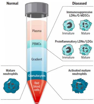
Figure 1.7 Subsets of neutrophils and their distribution through whole blood layering during health and disease. ... 18
Figure 1.8 Major factors causing Secondary Immune Deficiencies. ... 20
Figure 1.9 Prevalence (in 100,000) of PIDs in different countries across the world. .. 21
Figure 1.10 Classifications and examples of PIDs. ... 22
Figure 1.11 Illustration of mammalian de novo pyrimidine synthesis pathway. ... 24
Figure 1.12 CTPS1 deficiency results in defective T-cell and B-cell proliferation. ... 25
Figure 1.13 Eculizumab mechanism of action. ... 28
Figure 1.14 Mechanisms of microvesicle (upper) and exosome (lower) biogenesis. . 30
Figure 3.1 STAT1 phosphorylation levels of control and patient CD8+ T-Cells in response to IFN-b stimulation. ... 55
Figure 3.2 CD107a surface expression of unstimulated CD8+ T-cells. ... 56
Figure 3.3 STAT phosphorylation levels of CD4+ T-Cells of healthy donors and CTPS1 patient. ... 58
Figure 3.4 Percentages of pSTAT positive CD4+ T-cells from healthy and CTPS1 -/-individuals following cytokine incubations. ... 60
Figure 3.5 Cytokine production profiles of CD4+ T-cell in response to PMA/Ionomycin stimulation. ... 62
xiii
Figure 3.6 IFN-a, IFN-g and IL-12 cytokine production profiles from healthy and patient PBMCs upon multiple PRR ligand stimulations. ... 66 Figure 3.7 Effect of PRR engagement on healthy and CTPS1-/- PBMCs in the context
of IL-10 secretion. ... 67 Figure 3.8 Identification of Low-Density Granulocytes within patient’s PBMCs fraction. ... 69 Figure 3.9 Representative immunofluorescent images of healthy and CTPS1
-/-neutrophils undergoing NETosis. ... 71 Figure 3.10 Quantification of released dsDNA from neutrophils. ... 72 Figure 3.11 Verification of extracellular vesicle isolation from control and CTPS1
-/-plasmas. ... 74 Figure 3.12 Differential activation capacities of healthy and CTPS1-deficient patient plasma-derived EVs on healthy PBMCs. ... 76 Figure 3.13 Differential activation capacities of healthy and CTPS1-deficient patient plasma-derived EVs on healthy PBMCs. ... 78 Figure 3.14 Effect of CTPS1-/- EV pre-treatment on surface expression of HLA-DR,
CD86 and PD-L1 molecules from healthy donor PBMCs. ... 80 Figure 3.15 Confirmation of the absence of CD55 protein in patient cells by Flow cytometry and immunoblotting assays. ... 82 Figure 3.16 Production of IFN-𝛼 and IP-10 cytokines from healthy, BT and AT of CHAPLE (CD55-/-) patient PBMCs upon stimulation with various PRR ligands. ... 83
Figure 3.17 Effect of PRR stimulations on pro-inflammatory and anti-inflammatory cytokine secretion profiles of healthy, BT or AT of CHAPLE (CD55-/-) patient PBMCs
upon stimulation with various PRR ligands. ... 85 Figure 3.18 Verification of extracellular vesicle isolation from control and BT or AT CHAPLE (CD55-/-) patients. ... 87
Figure 3.19 Cytokine secretion profiles of before or after therapy EVs on BT or AT CD55 deficient patients PBMCs. ... 89 Figure 3.20 Induction profiles of EVs of CHAPLE patients (BT or AT) from healthy PBMCs. ... 90
xiv
Figure B1.1 Immunophenotyping of circulating lymphocytes revealed CTPS1-/- patient
had inverted CD4/CD8 ratio. ... 137 Figure B1.2 Healthy and CTPS1-/- patient CD4+ T-cells’ cytokine production in
response to PMA/Ionomycin stimulation. ... 138 Figure B1.3 Transcription Factor staining revealed reduced Foxp3 CD4+ T-cells in CTPS1 patient’s circulation. ... 139 Figure B1.4 Pro-inflammatory cytokine production from healthy and patient PBMCs upon PRR ligand stimulations. ... 140 Figure B1.5 Confirmation of Low-Density Granulocytes within patient’s PBMCs fraction. ... 141
xv
List of Tables
Table 1.1 Soluble and membrane bound regulators of complement cascade. ... 7 Table 2.1. Mitogens, ligands and cytokines used in this study. ... 36 Table 2.2. Antibodies and recombinant proteins used in cytokine ELISA throughout this study. ... 39 Table 2.3. Fluorochrome labeled recombinant antibodies used in flow cytometry analyses throughout this study methods. ... 41 Table 2.4. Features of Western Blot antibodies used throughout the thesis. ... 42
xvi
ABBREVIATIONS
ALRs AIM2-like receptors AIM2 Absent in Melanoma 2 APC Antigen Presenting Cell
aHUS atypical Hemolytic Uremic Syndrome
ATP Adenosine Triphosphate
AT After Eculizumab Therapy
BCRs B-cell Receptors
BT Before Eculizumab Therapy
C1INH C1 inhibitor
CHAPLE Hyperactivation of Complement Angiopathic Thrombosis and Protein Losing Enteropathy
CLRs C-type Lectin Receptors
CMV Cytomegalovirus
CTP Cytidine Triphosphate
CTPS1 Cytidine Triphosphate Synthase 1 CID Combined Immunodeficiency DAF Decay-Accelerating Factor
DAMPs Damage-Associated Molecular Patterns
DNA Deoxyribonucleic Acid
dsDNA Double-Stranded DNA
dsRNA Double-Stranded RNA
EBV Epstein-Barr virus
ESCRT Endosomal sorting complex required for transport EVs Extracellular vesicles
FBS Fetal Bovine Serum
xvii GPI Glycosylphosphatidylinositol
HAE Hereditary Angioedema
HIV Human Immunodeficiency Virus
HMBG1 High mobility binding group protein 1 HSCT Hematopoietic-stem Cell Transplantation HSV Herpes Simplex virus
IKK IkB kinase
iNKT invariant Natural Killer Cells IP-10 IFN-g Inducible Protein-10 IRAK IL-1-receptor associated kinase IRFs Interferon Regulatory Factors IVIG Intravenous Immunoglobulin LDNs Low Density Neutrophils LGDs Low Density Granulocytes LPS Lipopolysaccharide LRRs Leucine Rich Repeats
MAC Membrane Attack Complex
MAIT Mucosal Associated invariant T-cells MAPK Mitogen-activated protein kinase MASP MBL-associated serine proteases
MBL Mannose-Binding Lectin
MD-2 Myeloid Differentiation protein-2 MDA5 Melanoma differentiation associated 5
MFI Mean Fluorescent Intensity
MPO Myeloperoxidase
MVB Multi vesicular bodies
xviii
NADPH Nicotinamide adenine dinucleotide phosphate
NE Neutrophil Elastase
NETosis NET formation
NETs Neutrophil Extracellular Traps NK Cells Natural Killer Cells
NLRs NOD Like Receptors
NOD Nucleotide Oligomerization Domain ODNs Oligodeoxynucleotides
PAMPs Pathogen-Associated Molecular Patterns PBMCs Peripheral Blood Mononuclear Cells pDCs Plasmacytoid Dendritic Cells
PKC Protein Kinase C
PNH Paroxysmal Nocturnal Hemoglobinuria poly(dA:dT) Poly(deoxyadenylic-deoxythymidylic) poly(I:C) Polyinosinic-Polycytidylic acid
PIDs Primary Immune Deficiencies PRRs Pathogen Recognition Receptors RIG-I Retinoic Acid Inducible Gene I
RLRs RIG Like Receptors
RNA Ribonucleic Acid
ROS Reactive Oxygen Species
SCID Severe Combined Immunodeficiency SLE Systemic Lupus Erythematosus
ssRNA Single-Stranded RNA
STING Stimulator of IFN genes
TANK TRAF-family member associated NFkB activator TBK1 Tank Binding Kinase Protein 1
xix
TCC Terminal Complement Complex
TCRs T-cell Receptors
Th T-helper
TLRs Toll Like Receptors TNF Tumor Necrosis Factor TIR Toll/IL-1 receptor
TRAF TNF-receptor associated factor Tregs Regulatory T-cells
TRIF (TIR)-domain-containing adapter-inducing IFN-b VZV Varicella Zoster virus
1
1. INTRODUCTION
1.1. IMMUNE SYSTEM
Throughout evolution, all organisms were subjected to threats of pathogenic microorganisms as invaders. Hence, evolutionary parameters selected organisms possessing survival against them. As a consequence of this, complex and unique network of defense responses were evolved which is called as immune system [1]. Although invertebrates have primitive immune responses, as complexity of immune system gets higher, species possessing it become more convoluted in the context of evolutionary order [2]. In that essence, most appreciated immune responses are the ones within class of mammalians. Mammalian immune system can be fundamentally divided into i) innate and ii) adaptive arms. These distinct yet interconnected array of responses are composed of special cells, receptors and molecules residing in different layers and levels of physiological barriers [3].
Innate immunity is regarded as first line of defense of the whole immune system. This arm adopted certain levels of strategies, which are anatomic barriers, complement and antimicrobial proteins, innate immune cells, to protect host from invaders [4]. In order to successfully invade, pathogens must pass through anatomic barriers such as skin, intestinal epithelium, oral mucosa, then, they dodge to overcome against complement proteins. If they still prevail, tissue resident innate immune cells can recognize and generate quick responses. Meanwhile, the responses of innate immune cells are achieved through germline encoded receptors that are able to recognize evolutionarily conserved non-self-motifs on pathogens. Even though innate immune cells were used to be considered as having no memory, growing bodies of evidences challenge it; that is, innate immune cells indeed confer a memory through epigenetic reprogramming and ‘Trained Immunity’ is coined to define this novel aspect of innate immunology field [5–6]. Responses elicited from activated innate immune cells, specifically phagocytes and antigen-presenting cells (APCs), are destined to license adaptive immune cells. While innate cells provide fast, non-specific immunity, adaptive immunity is established through slow, vast but specific responses against antigenic peptides of invaders. In order to achieve that, two broad categories of adaptive immune system are needed to
2
be involved. These are cell-mediated immunity and humoral immunity, comprise from subsets of T-cells and B-cell produced antibodies, respectively [7–8]. Even though all T-cells are primed in secondary lymphoid organs by activated and migrated APCs, their classification is generally divided into CD8+ and CD4+ T-cells regarding to their
composition of T-cell Receptors (TCRs). CD8+ T-cells (or cytotoxic T-cells) generate crucial and powerful immune responses in the context of infection of intracellular pathogens and cancer cells [9]. CD4+ T-cells or helper T-cells (Th), however, are further categorized into subtypes according to their different orchestration of the immune system. As their names imply, they are differentiated into several subtypes; so as to provide assistance to clearance of pathogens. Similarly, B-cells and their subsets, bearing B-cell Receptors (BCRs), are able to be primed through T-helper cells or directly by pathogens per se. Regardless of activation route, they proliferate and differentiate to become plasma cells and secrete specific subtypes of immunoglobulins such as IgA, IgG, IgE [10–11]. Unlike memory mechanism of innate cells, cells of adaptive immunity become quiescent until reinvasion of pathogens and this process is building block of vaccination [12].
1.1.1. Innate Immune System
Tissues of hosts are constantly being received threats from pathogenic and even commensal organisms. In most of the conditions, they reside on external surfaces, barriers of the mammalian tissues. These barriers in frontiers are epithelial tissues that are composed of skin, covering internal linings such as respiratory, urogenital and gastrointestinal tracks of host. Not only they cover surroundings but also, they harbor special cells that are evolved to secrete antimicrobial proteins and peptides. To illustrate, Paneth cells residing within the base of epithelial crypts of small intestines secrete a-defensins and RegIIIg; furthermore, differentiated keratinocytes in skin produce b-defensins and cathelicidins [13]. These peptides are evolutionarily conserved to kill efficiently and directly the invading pathogens through integrating themselves into membranes of microorganism.
Yet, microorganisms are evolved to penetrate through these barriers. As a result of this, they face another ancient component of innate immunity which is called as complement pathway. The complement system is array of soluble and membrane proteins circulating throughout extracellular fluids and blood within host. Upon an
3
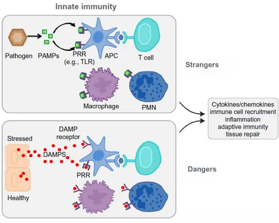
encounter with a pathogen, they get activated and interact with each other to kill and/or facilitate phagocytosis of invaders [14–15]. Nonetheless, pathogens might be equipped to escape complement pathway as well [16]. Then, innate immune cells; for example, monocytes, macrophages, dendritic cells, neutrophils and their receptors, Pathogen Recognition Receptors (PRRs), step in. Innate cells are able to discriminate non-self from self-motifs through 5 distinct PRR families which are: i) Toll-like Receptors (TLRs), ii) Nucleotide Oligomerization Domain (NOD)-like Receptors (NLRs), iii) Retinoic Acid Inducible Gene I (RIG-I) like Receptors (RLRs), iv) C-type Lectin Receptors (CLRs) and v) Absent In Melanoma 2 (AIM2)-like receptors (ALRs) [17–18]. They initiate signals when they sense Pathogen-Associated Molecular Patterns (PAMPs); similarly, they can even recognize sterile inflammatory signals which are Danger-Associated Molecular Patterns (DAMPs) [19]. While PAMPs are component of pathogens such as bacterial LPS, flagellin, unmethylated CpG-rich DNA, host cells, that underwent into necrosis or necroptosis due to stress, release DAMPs such as high mobility binding group protein 1 (HMBG1), DNA, histones, and ATP (Figure 1.1).
Figure 1.1 Origins of PAMPs, DAMPs and their recognition by innate immunity.
4
1.1.1.1 Complement System
Harboring over then 30 distinct proteins and being discovered 110 years ago, complement system, is one of the most ancient arm of innate immune system [21]. These, membrane bound and soluble proteins get activated upon presence of PAMPs and/or DAMPs and immune complexes (IgM) [22]. Although complement cascade can be activated systemically and locally they all are synthesized as pro-enzymes [22–23]. In order to become active in the cascade they need to be cleaved by other complement proteins. In contrast to common appreciated roles of complements; for example, opsonization, facilitating phagocytosis and direct lysing of pathogens, recent discoveries demonstrated that activated complement proteins are able to cross-talk and influence PRR signaling, especially TLRs, RIG-MAVS and inflammasomes, regulate tissue damage repair in cancer and kidney diseases, influence metabolic activities of cells and modulate responses of adaptive immune cells [21–24, 30] (Figure 1.2).
Figure 1.2 Cross-talk between complement pathway and physiological systems.
(Adopted from [21])
Systemic complement activation is composed of three distinct cascades: i) Classical, ii) Lectin iii) Alternative pathways. Even though they all lead to same outcome,
5
generation of anaphylatoxins alongside Membrane Attack Complex (MAC), each pathway utilizes different combinations of cascade proteins including their respective regulators (Figure 1.3).
Lectin cascade of complement pathway is initiated via oligomeric receptors called mannose-binding lectin (MBL) and Ficolins. While MBLs are evolved to recognize mannose or fucose carbohydrate residues that are only presented on microorganisms, Ficolins recognize acetylated oligosaccharide motifs. Moreover, MBLs and Ficolins form complexes with soluble, inactive MBL-associated serine proteases (MASP) as MASP-1, MASP-2 and MASP-3 circulate throughout blood. Upon binding surface of invaders, MBLs and Ficolins undergo conformational change which in turn activates MASPs. Classical pathway utilizes C1q, C1r and C1s comprising C1 complex. While C1q, functionally and structurally, resembles to MBLs and Ficolins, C1r and C1s can be regarded as MASPs of classical pathway. In contrast to MBLs and Ficolins, C1q recognize pathogens through 3 distinct ways: i) Direct, ii) through C-reactive Protein and iii) constant part of antibodies bound to pathogens. Nevertheless, either activated MASPs or C1r-C1s cleaves C4 complement protein in plasma, eventually leading to generation of C4b and C4a [14–15]. While C4b confined to surface of pathogen, C4a floats in plasma. Then, C4b recruits another cardinal complement protein, C2 and enables its cleavage by MASPs into C2a and C2b. C4bC2a complex is formed and called as C3 convertase or classical C3 convertase. As its name implies, C3 convertase, converts C3 into C3a and C3b. Consequently, C3b remains on (opsonize) pathogen surface by covalent bound and together with C4bC2a, C3b becomes C5 convertase, cleaving soluble C5 protein into C5a and C5b. Meanwhile C3a and C5a becomes one of the anaphylatoxins and contributes to phagocytosis and inflammation. Membrane-bound C5b, however, recruits C6, C7 and C8. C7 undergoes conformational change and inserts its hydrophobic sites into membranes of pathogens while recruiting C8. These series of recruitments eventually lead to incorporation of C9 proteins into membranes. Polymerization of C9 proteins form MAC complex (also called as Terminal Complement Complex (TCC) [31]) which destroy pathogens via loss of intracellular elements and allowing penetration of antimicrobial peptides and lysosomal enzymes into pathogens.
Alternative pathway, rather than directly recognizing the pathogens, is activated by already deposited C3b proteins from alternative and lectin pathway or spontaneous hydrolysis. In the case of former, after factor B binds to cleaved C3b, factor D cleaves
6
it into membrane bound and soluble forms as Bb and Ba, respectively. C3bBb being alternative C3 convertase contributes to complement pathway. However, in the case of latter, thioester bond of C3 is spontaneously cleaved and form C3(H2O). Like C3b,
factor B and factor D are able to bind and cleave it, respectively. Therefore, C3(H2O)Bb,
fluid-phase C3 convertase, is formed. Regardless of way of alternative pathway activation, alternative and fluid-phase C3 convertase require stabilization, which is achieved via factor P.
Figure 1.3 Activation of systemic complement cascade. (Adopted from [21])
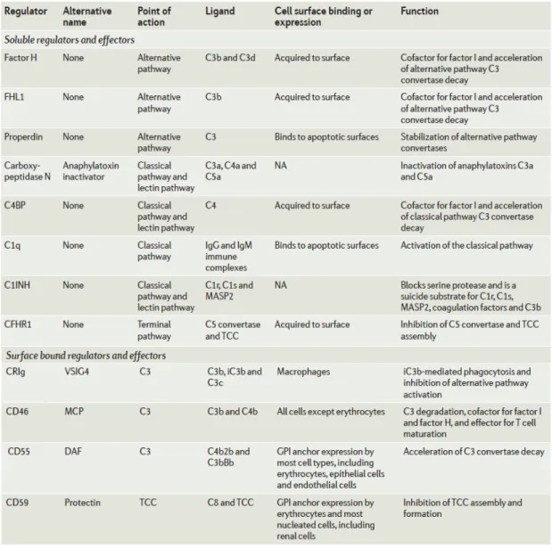
Even though complement proteins are specific to invading microorganism, it is possible to have activation on host membranes as well. Therefore, soluble, attached to surface of host and membrane integral proteins regulating activation of complement pathways were evolved in different levels (Table 1.1) [31]. To exemplify, C1 inhibitor (C1INH) and Factor H are crucial soluble inhibitors. C1NH displaces serine proteases (C1r/s, MASP2) to reduce activation of classical and lectin pathway. Factor H displace cleaved Bb from C3b of alternative pathway to inhibit the cascade. As a result of being important regulators, deficiency or loss-of-function of C1INH and Factor H leads to hereditary angioedema (HAE) and atypical hemolytic uremic syndrome (aHUS), respectively [32]. In addition to soluble factors, CD55 or decay-accelerating factor (DAF) and CD59 (or
7
protectin) are regulators that are attached to host cells via Glycosylphosphatidylinositol (GPI) anchor. While DAF binds to C3b to replace Bb from it, protectin inhibits C9 recruitment to C5b678 complex. Like deficiencies of C1NH and Factor H, mutation within the synthesis of GPI tails leads to Paroxysmal Nocturnal Hemoglobinuria (PNH) [33]. Similarly, deficiency of DAF has been identified to have distinct pathophysiology (will be mentioned in Section 1.2.1.3) [34–35]. Last but not least, there are inhibitory factors of complement that are produced by pathogens to avoid complement activation [36–37].
Table 1.1 Soluble and membrane bound regulators of complement cascade.
8
1.1.1.2 Pathogen Recognition Receptors (PRRs)
1.1.1.2.1 Toll-Like Receptors (TLRs)
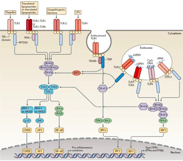
Long before the relationship between immune system and Toll gene was identified, it was described as a gene controlling dorso-ventral positioning of embryo throughout development of Drosophila melanogaster, fruit fly [38–39]. Then, strikingly, mutations in the product of this developmental gene predispose flies to suffer from gram-positive bacterial and fungal infections [40–42]. Furthermore, homologs of Toll proteins were identified and called as Toll-Like Receptors (TLRs) in humans [43].
Up to date, humans and mice do express 10 and 12 distinct TLRs, respectively [44]. TLRs can be grouped based on their location and adaptor molecules that are used within the intracellular signaling [45]. In the context of former, TLRs are divided into intracellular vesicles and cell surface TLRs. While TLR3, TLR7, TLR8, TLR9 and TLR13 are found within endosomes, TLR1, TLR2, TLR5, TLR6 and TLR11 are found on the surface membranes of cells. However, TLR4, first identified TLR, can be found on plasma membrane and in endosomes [3]. Whereas latter distinguishes TLRs via whether they use Myeloid differentiation primary response gene 88 (MyD88) or Toll/IL-1 receptor (TIR)-domain-containing adapter-inducing IFN-b (TRIF) adaptor proteins. In that essence, except TLR3, all TLRs use MyD88 adaptor protein. Surprisingly, TLR4 was identified as TLR using both MyD88 and TRIF proteins for its signaling [44].
1.1.1.2.2 TLR Signaling Pathway
Regardless of localization of receptors and recruited adaptor proteins, all TLRs are composed of three domains: Ectodomain, transmembrane region and cytoplasmic TIR domain [44]. Ectodomains contain leucine rich repeats (LRRs), which are crucial for ligand recognition. TIR domains recruit adaptor proteins such as MyD88 and TRIF and initiates signaling cascades.
These signaling cascades have being extensively studied and delineated (Figure 1.4). Upon dimerization of TLRs, they recruit c or/and TRIF adaptor proteins via their TIR domains. Death domains of recruited MyD88 protein further recruit IL-1-receptor associated kinase 4 (IRAK4) and IRAK1 to establish scaffold protein complex. The formed scaffold, recruits E3 ligase, called as tumor necrosis factor (TNF) receptor associated factor 6 (TRAF6) and activates it by phosphorylation. Activated TRAF6
9
along with accessory ubiquitin ligases, establish polyubiquitin scaffold to recruit transforming growth factor-β-activated kinase 1 (TAK1) and TAK binding protein1/2 (TAB1/2) adaptor proteins. As a consequence of being recruited, IRAK1 phosphorylates TAK1 and activates it. From activated TAK1, signaling divides into two distinct parts. On one hand, TAK1 activates IkB kinase (IKK) complex. As it names implies, activated IKK complex phosphorylates IkB leading to degradation of IkB and, consequently, enabling NFkB enter into nucleus for pro-inflammatory cytokine production (TNF-a, IL-6 and IL-1b). On the other propagation of signaling, TAK1 activates mitogen-activated protein kinase (MAPK) pathway contributing cytokine production [46].
In addition to pro-inflammatory cytokine production, TLR7 and TLR9 signaling pathway, depending on MyD88 adaptor protein, directly recruits and activates Interferon Regulatory Factor 7 (IRF7) via IRAK1. Phosphorylated and dimerized IRF7s enter nucleus and induce production of type I IFNs, especially IFN-a. [47].
In the contrast to MyD88-dependent signaling, TRIF pathway, that is utilized by TLR3 as well as TLR4, propagates via different proteins. Upon TLR dimerization, TRIF protein is recruited via their TIR domains. TRIF, in turn recruits TRAF3 having E3 ligase activity. This, enzymatic activity ensures formation of polyubiquitin complex. That scaffold activates TRAF-family member-associated NFkB activator (TANK) protein which, in turn, activates Tank-binding Kinase Protein 1 (TBK1). TBK1 phosphorylates transcriptional factor IRF3. Phosphorylated and dimerized IRF3 enters nucleus and initiates production of type I IFNs, especially IFN-b [48].
10
Figure 1.4 Mammalian TLRs (1-13) recognize different ligands and propagates through different pathways. (Adopted from [45])
1.1.1.2.2.1 Cell Surface Toll-Like Receptors
TLR1, TLR2, TLR4, TLR5 and TLR6, comprise Human Toll-like receptors confined to surface membranes. While TLR1, TLR2 and TLR6 forms two distinct heterodimers upon ligand binding such as TLR1&2 and TLR2&6, TLR4 and TLR5 homodimerize [18]. TLR1&2 and TLR2&6 recognizes lipopeptides: TLR1 and TLR6 enable TLR2 to sense diacyl- and triacyl-lipopeptides, respectively [49]. Moreover, TLR4 and TLR5, which are found on macrophages and dendritic cells, recognize lipopolysaccharide (LPS) and flagellin, respectively.
11
In addition to homodimerization, TLR4 further requires three accessory proteins such as LPS-binding protein (LPB), Myeloid Differentiation protein-2 (MD2) and CD14. LPB being a plasma soluble protein binds circulating LPS in blood and tissue; then, CD14 recruits LPS bound LBP to TLR4-MD2 complex. As a result of this, MD2 binds active part of LPS; consequently, TLR4 homodimerizes and signaling cascade is activated. TLR5, without using accessory proteins, recognizes flagellin, monomer of flagella [17]. Both LPS and Flagellin induces pro-inflammatory cytokine production via NFkB pathway.
1.1.1.2.2.2 Endosomal Toll-Like Receptors
Human TLRs residing within the endosomes are TLR3, TLR7, TLR8 and TLR9 [50]. Their common feature is that they are evolved to recognize nucleic acids found in extracellular environment such as RNAs and DNAs. They can bind their corresponding ligands after they fuse with phagocytosed and degraded pathogens or dying infected cells in endosomes.
While TLR3 is confined to macrophages, conventional dendritic cells and intestinal epithelial cells, it senses double stranded RNA (dsRNA) and its analog polyinosinic-polycytidylic acid (poly(I:C)) [51]. Even though TLR3 is specific for dsRNA, strikingly, it was reported that TLR3 is activated upon single stranded RNA (ssRNA) viruses such as West Nile virus, respiratory syncytial virus, and double stranded DNA (dsDNA) viruses (such as herpes simplex virus (HSV)) due to dsRNA production during viral gene transcription [52].
TLR7 (and TLR8 in humans [53]) recognize ssRNA entering endosomes and its synthetic analogs, derivatives of imidazoquinoline (R848, Imiquimod and Resiquimod), and guanine analogs; for example, loxoribine [54, 56]. Moreover, TLR7 has ability to sense some siRNAs as well [57]. While TLR7 is expressed by human plasmacytoid Dendritic Cells (pDCs) and B-cells, TLR8 is confined to DCs, macrophages and neutrophils [58–59].
In addition to be found in pDCs and B-cells, TLR9 is evolved to sense dsDNA, specifically CpG motif containing DNA without methylation since they are 20 times more common in viral or bacterial genome [60]. Therefore, bacteria, dsDNA viruses like Herpes Simplex Virus 1 and 2, protozoa parasites such as Trypanosoma cruzi are
12
sensed and demonstrated to elicit responses [61–63]. Strikingly, it was reported that malarial DNA along with hemozoin, a malarial pigment, induces strong activation [64]; however, in a previous report, hemozoin per se has been already shown to activate TLR9 signaling [65]. Nonetheless, ligand engagement with TLR9 induces MyD88-dependent immune signaling that eventually leading to B-cell proliferation and maturation, copious amount of type I IFN secretion from pDCs [66].
Besides natural DNAs, synthetic oligonucleotides (ODNs) were reported to induce TLR9 activation [67]. Although this effect was attributed to palindromic sequences, in 1995 and 1996, Krieg and Klinman demonstrated that ODN with unmethylated CpG dinucleotide, which is flanked by two 5’ purines and two 3’ pyrimidines, induces strong activation of innate immune cells [68–69]. Nowadays, three distinct classes of ODNs were determined [70–72]. First, when multiple CpG motifs reside on phosphorothioate backbone, ODN is called ‘K’ type (called ‘B’ type as well). Their recognition by TLR9 is achieved within late endosomes and leads to activation of NFkB transcriptional factor. They stimulate B-cells to differentiate and secrete pro-inflammatory cytokines, whereas pDCs were reported to secrete TNF-a. Another class of ODNs were named as D-ODNs (or ‘A’ type). In contrast to K-ODNs, D-ODNs (also known as A-Type ODN) bear one CpG motif on phosphodiester/phosphorothioate mixed backbone and possess poly-G tail on both 5’ and 3’ ends. Poly-G tails on both ends of ODN, enable them to form nanoparticle-like structures. As a consequence of their complicated structures, they are recognized in early endosomes. This feature enables them to induce pDCs to secrete massive amounts of type I IFNs. Lastly, mixed type (C-type) ODNs were recently described [72]. As the name implies, it can trigger pDCs to secrete type I IFNs and at the same time leads to maturation of B-cells.
1.1.1.2.3 Cytosolic Nucleic Acid Sensors
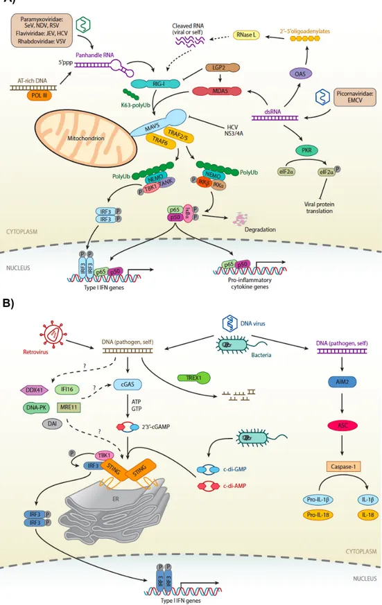
During an infection, it is crucial to detect microorganisms by presence of their nucleic acids in cytosol. As a consequence of this, immune system evolved to bear cytosolic nucleic acid sensors. There are three major sensors of nucleic acids in cytosol: i) RLRs, ii) DNA-dependent activator of interferon regulatory genes and iii) ALRs. While RLRs detects RNA in cytosol (Figure 1.5A), other two detects DNA (Figure 1.5B) [73].
13
Figure 1.5 Cytosolic nucleic acid sensors of innate immunity. A) RIG-I, MDA-5, B)
STING pathway. (Adopted from [74])
A)
14
1.1.1.2.4 RIG-I Like Receptors
There are three major distinct sensors for RNA in cytosol. They are RIG-I, Melanoma differentiation associated 5 (MDA-5) and LGP2 (laboratory of genetics and physiology 2 and a homolog of mouse D11lgp2) [75]. For the scope of this thesis, RIG-I and MDA-5 are merely discussed. Unlike most of the PRRs, their expression is ubiquitous. However, immune signaling are varies between cells; to illustrate, even though myeloid cells can respond to cytosolic RNAs, pDCs cannot initiate immune response via RLRs [75].
In order to sense ssRNA, RIG-I differentiates pathogenic RNA from eukaryotic is via sensing modification at 5’ end of host ssRNAs [76]. However, there are several microorganisms such as polio virus and hepatitis A, evade this differentiation by transcribing their RNAs within nucleus of host [4]. MDA-5 senses dsRNA, specifically long dsRNAs [77]. Moreover, they can be activated by synthetic compounds such as transfection of p(I:C) and poly(deoxyadenylic-deoxythymidylic) Although former can be recognized directly, latter is recognized by RNA intermediate that is formed by RNA polymerase III due to DNA rich in AT sequence [78].
Upon recognition of corresponding RNAs, autoinhibited conformation is altered; consequently, RIG-I and MDA-5 can bind to mitochondrial antiviral signaling protein (MAVS) on mitochondria. After binding, receptors and MAVS, aggregate and recruit TRAF which propagates signaling towards to activation of NFkB and IRF3. As a result of this, pro-inflammatory cytokines and type I IFNs are produced [79].
1.1.1.2.5 DNA-dependent activator of interferon regulatory genes
In healthy eukaryotic cells, DNA is strictly confined to nucleus. Therefore, either stress or presence of pathogen can lead to presence of DNA in nucleus. In order to sense and response against them, mammalian immune system is evolved to bear several cytosolic DNA sensors: AIM2, DAI (or ZBP1), RNA polymerase III, IFI16, DDX41 and most recently discovered cGAS [64, 71–72]. However, until the discovery of cGAS [81], none of these sensors were documented to be exclusive; that is, either they are confined to specific cell types or they have redundancy. For example, although IFI16, a protein confined to nucleus, and DAI are reported to sense cytosolic DNA, upon their depletion, cells were still observed to have DNA-dependent immune response [82–84].
15
As cyclic guanosine monophosphate-adenosine monophosphate (cGAMP) synthase (cGAS), binds to DNA in cytosol and produces a secondary messenger, cGAMP, from ATP and GTP in cytosol, cGAMP is sensed by an adaptor protein called Stimulator of IFN genes (STING). In addition to natural course of DNA sensing, DNA-dependent activation of STING can be studied by transfecting several product such as 2’3’ or 3’3’ cGAMP [85] , DNA of Herpes Simplex Virus (HSV) [82] and p(dA:dT) [86].
Except AIM2 [87], all cytosolic DNA sensors converge into same Endoplasmic Reticulum localized signaling adaptor, STING. Upon activation, STING proteins dimerize and recruit TBK1 to phosphorylate IRF3. Phosphorylated IRF3 homodimers enter into nucleus and produces type I IFNs. Meanwhile, homodimers of STING activate NFkB signaling to initiate production of pro-inflammatory cytokines as well [84– 88]. AIM2 as a member of PYHIN family protein, induces inflammasome formation which eventually lead to maturation and secretion of IL-1b and IL-18 rather than type I IFNs [89].
1.1.1.3 Neutrophils, Low-Density Granulocytes (LDGs) and Neutrophil
Extracellular Traps (NETs)
Being the most abundant immune cell type in peripheral blood circulation puts neutrophils on the frontier barricades of innate immunity [90]; in other words, they are the first cell type that are recruited to inflammatory area [91]. Neutrophils are terminally differentiated cells that are released to circulation from bone marrow; therefore, they have a short-life time in circulation (8-12 hours), however, it has been recently demonstrated that they can live up to 7 days during ongoing inflammatory conditions within tissues [92]. So as to limit the invasion, they have been equipped with three distinct powerful immune mechanisms: i) degranulation of antimicrobial proteins (such as neutrophil elastase (NE), myeloperoxidase (MPO)) in pre-formed granules, ii) phagocytose pathogens into reactive oxygen species (ROS) containing phagosomes and iii) propelling their chromosomes to extracellular environment to form a meshwork of DNA which is called as Neutrophil Extracellular Traps (NETs). [93–95].
16
Figure 1.6 Molecular signaling of NETosis. (Adopted from [95])
Formation of NETs (NETosis), is a unique subset of regulated necrosis in which invading pathogens can be immobilized, inactivated and killed [96]. Even though it is known that NETs are formed and propelled against certain sterile stimuli (Urate/Cholesterol crystals, autoantibody complexes [97–98]) and microbes (fungi [99], parasites [100], viruses[101]) especially ones that cannot be phagocytosed by neutrophils [102], neither ‘trap or phagocytose’ decision phenomena nor exact mechanism behind NET formation has been elucidated yet [91]. Meanwhile, there are more than one type of NETosis. In addition to regulated death of neutrophils upon stimuli, NETs and proteins in granules can be rapidly propelled against S. aureus while neutrophils are still viable and carry on functioning such as phagocytosis [103–104]. Besides non-lytic NETosis, it was shown that there are more than one signaling cascades leading to classical NETosis and, NADPH oxidase-dependent pathway is a well-studied one (Figure 1.6). Upon microbial or sterile stimulation (or mitogenic stimuli: PMA, Concavalin A and Ionomycin [105]) through receptors increases intracellular levels of Ca2+. Elevated calcium ion level
17
activates protein kinase C (PKC) and initiates assembly of nicotinamide adenine dinucleotide phosphate (NADPH) oxidase complex. PKC and NADPH generate ROS, especially hydrogen peroxide. Hydrogen peroxide activates special proteins such as NE, MPO within azurophilic granules. Contents of azurophilic granule is spilled over cytoplasm due to activity of MPO. They gather into nucleus to decondense chromatins via histone processing. While DNA is being released as a meshwork, proteins from granules such as MPO, NE, LL-37, HMGB1 are decorated on them [94–95–102–105– 106].
Although NETs are crucial for dissemination of invaders, aberrant NET formation or ineffective NET removal are reported to lead coagulation [107–108], sterile inflammation [109–110], organ/tissue damage [111] and contribute allergy [112], autoimmunity [113– 116].
In addition to diverse NETosis mechanisms, several novel subsets of neutrophils have been recently identified [116–120]. They have been named as low-density granulocytes (LDGs) or low-density neutrophils (LDNs) due to fact that they reside within Peripheral Blood Mononuclear Cell (PBMC) fractions after whole bloods were separated by density gradient centrifugation (Figure 1.7) [119–121]. Yet, there is a dilemma on whether LDGs are indeed subsets of neutrophils or modified versions of mature circulating neutrophils in diseased individuals [91]. However, it is certain that these ‘subsets’ of neutrophils contribute to progression and severity of diseases in which they are harbored. For example, cancer patients, apart from their tumor associated neutrophils (TANs) [122– 124], had LGDs with immunosuppressive effects inhibiting T-cell proliferation and functioning [125]. Circulating LDGs in autoimmune diseases like SLE, Psoriasis, Rheumatoid Arthritis [126] are recorded to have increased inflammatory cytokine secretion and NET formation to further exacerbating disease progression.
18
Figure 1.7 Subsets of neutrophils and their distribution through whole blood layering during health and disease. (Adopted from [91])
Moreover, it has been demonstrated that they can enhance activity and effector functions of adaptive immune cells [119–121, 127]. Strikingly, it has been demonstrated that presence of LDGs in HIV infected individuals, correlated strongly with the LDG numbers and HIV severity [128].
1.1.2. IMMUNE DEFICIENCIES
In the course of normal immune responses, pathogenic agents, that have breached the first line of defense barriers, provoke innate immune response, so that it limits the spread of the insult to the surrounding tissues. Professional antigen presenting cells that migrate to the nearest lymph node instruct naive B- and T-cells; consequently, adaptive arm is induced to establish long-lasting and antigen-specific immune response against threats. Alongside with innate immune cells, adaptive immune cells
19
eliminate threats; moreover, at the end, protective immunity is established as well. Nonetheless, in rare cases, immune responses may fail due to several reasons. These failures of defense mechanisms, are called as Immunodeficiencies [4]. In other words, immunodeficiencies arise when at least one element of or part of immune system cannot function as it is required to be mainly due to mutation(s) in the genes(s). As a consequence of this, infectious microorganisms or internal threats cannot be eliminated or cleared completely.
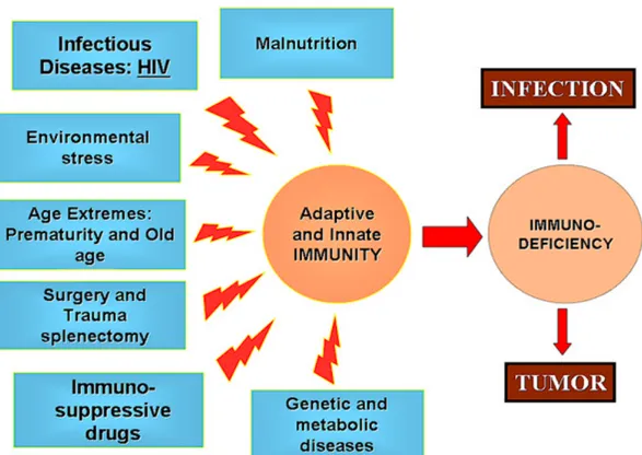
Defects of immune system are classified into two groups: Primary Immunodeficiency Disorders (hereafter PIDs) and Secondary (or Acquired) Immune Deficiencies [129]. Acquired Immune Deficiencies are consequences of environmental parameters instead of genetic mutations such as but not limited to malnutrition, metabolic diseases, medical therapies or interventions and infectious diseases are considered as general causes of acquired immunodeficiency disorders (Figure 1.8) [130].
Major cause of secondary immune deficiency is malnutrition [131]. Malnutrition is a term to define restricted access to food sources. Such as hypoproteinemia or lankness of micronutrients cause immune system to dysfunction and increase host’s susceptibility to internal and external threats [132]. Even though malnutrition is the most common factor leading to secondary immune deficiencies, the most studied one is Human Immunodeficiency Virus (HIV). Although HIV infects cells bearing CD4+ and
CCR5, CXCR4 co-receptors, major replication site of virus is CD4+ T-cells [133]. As a
consequence of this, CD4+ T-cell levels in blood circulation is reduced to a critical level
20
Figure 1.8 Major factors causing Secondary Immune Deficiencies. Secondary Immune
Deficiencies arise from extrinsic and/or environmental factors. (Adopted from [130])
1.1.2.1 Primary Immune Deficiency Disorders
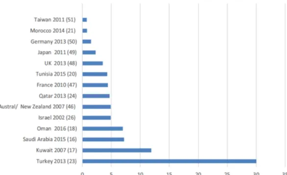
PIDs are congenital disorders in which inherited or de novo mutation(s) disrupts immune system functioning or proper development, or both of them. In contrast to being a group of rare disorders, as it is depicted in Figure 1.9, due to genetic basis, it is no surprise that prevalence of PIDs is reported to be high within inbred, consanguineous populations and countries [135].
21
Figure 1.9 Prevalence (in 100,000) of PIDs in different countries across the world.
(Adopted from [135])
Clinical manifestations of PIDs vary in a broad spectrum; to illustrate, even though many deficiencies lead to predisposition to infections, autoimmunity, malignancy as well as excessive inflammation and anatomical abnormalities can be manifested [129, 136–137].
More than 350 deficiencies have been characterized since the first identification of the PID which was agammaglobulinemia [138–139]. Although there is no consensus on classification, identified disorders are generally grouped based on functional part of immune system that is compromised. However, it is important to emphasize that malfunction of a certain part of the immune system may lead to perturbation of others. PIDs can be classified into two immense categories as: disorder of innate and adaptive immune system (Figure 1.10). Meanwhile, a group of deficiencies, called as Immune Dysregulations, leading to autoimmunity, autoinflammation is included in the PID categorization as well [140].
22
Figure 1.10 Classifications and examples of PIDs. Diseases that are written in bold and
indicated with asterisk are topics of this thesis (Compiled from [129–139–143])
Deficiencies within innate immune system can be further grouped into disorders of i) phagocytes and ii) complement system. In the case of phagocytes deficiencies, numbers, adhesion, activation, and killing capabilities of innate immune cells could be reduced or completely malfunctioning. For example, deficiency in neutrophil production, called as neutropenia, can be caused from mutations within ELA2 gene encoding NE. As a result of this, myelocytes undergo apoptosis during development from promyelocyte to myelocytes [144]. Defects in phagocytes reduce host’s capability to eradicate pus-forming bacteria; consequently, granulomas can be presented [142]. Non-functional complement proteins induced deficiencies can be divided into three as defects in early, late and regulatory components of predispose hosts to recurrent infections of encapsulated bacteria and Neisseria species; in addition, Systemic Lupus Erythematosus (SLE)-like syndrome can be manifested in certain complement deficiencies [140].
On the contrary, mutations interfering development or functionality of adaptive immune system can be grouped as i) T-cell deficiencies, ii) antibody-mediated deficiencies and iii) combined immune deficiencies (CIDs) [140]. T-cell deficiencies are composed of
23
defects in receptors or signaling pathways in which T-cell mediated antibody production is not compromised [4]. That is, subsets of T-cells have impaired responses. For example, defects in IL-12/IFN-g cytokines/receptors and IL17 cytokine/receptor manifest as recurrent intracellular bacterial infections and mucocutaneous candidiasis, respectively [140]. In the cases of antibody-mediated deficiencies where it can be due to defects in induction of B- and T-cell responses such as Bruton’s X-Linked agammaglobulinemia, hyper-IgM syndrome. Meanwhile, certain impaired genes leading to developmental defects in T- and/or B-cells are termed as CIDs [129]. Most prominent subtype of it is Severe Combined Immunodeficiencies (SCIDs) in which cell-mediated immune responses and T-cell dependent immunoglobulin presence are impaired. Individuals with SCIDs such as RAG1/2, JAK3, IL2RG deficiencies suffer from wide array of pathogens [143].
In the case of third group of deficiencies, immune dysregulations may manifest themselves with autoimmune and inflammatory symptoms without having increased predisposition to pathogenic infections [140]. In those diseases, even though lymphocytes and subsets are present, they are generally dysfunctional which may lead to autoreactivity as well as dysregulation [140]. For instance, Familial Mediterranean Fever (FMF) is an autosomal recessive disease that is resulted from mutations within the gene called Mediterranean Fever (MEFV). MEFV encodes a crucial regulatory protein called pyrin or marenostrin. Even though protein’s exact structure and function have not been elucidated yet, it was demonstrated that it inhibits caspase-1 associated inflammasome formation in cells [145]. FMF individuals have mutations leading to overactivation of inflammasome and consequently, sera of patients harbor elevated levels of IL-1b and IL-18 cytokines. Due to role of IL-1b in thermoregulation, patients manifests recurrent fever episodes, renal and dermal complications [146].
As it can be deduced, each classes and sub-classes of deficiencies discretely manifests themselves; thus, by conducting studies on these ‘experiments of nature’, one can delineate, comprehend normally-functioning immune pathways as well as their redundancy and interactions between other components of the immune system [4, 143–147].
Although certain PIDs can be managed through supportive treatments such as administration of antibiotics, antifungals, intravenous immunoglobulin (IVIG) and
24
cytokine replacements, others, especially SCIDs, are required to be corrected via hematopoietic-stem cells transplantation (HSCT) due to lethality [140–148].
1.1.2.2 Cytidine Triphosphate Synthase 1 (CTPS1) Deficiency
Cytidine Triphosphate Synthase 1 (CTPS1) deficiency has been identified as extremely rare deficiency of immune system in 2014 and it has been further documented in few numbers of articles [149–152]. Common clinical symptoms of CTPS1 deficiency are mild gastrointestinal symptoms, early onset of recurrent and severe infections of Herpes Simplex virus (HSV), Varicella Zoster virus (VZV), Epstein-Barr virus (EBV) and encapsulated bacteria. Moreover, due to EBV infections, individuals predispose to have malignancies, reduced memory B-cell and CD8+ T-cell numbers [149–152].
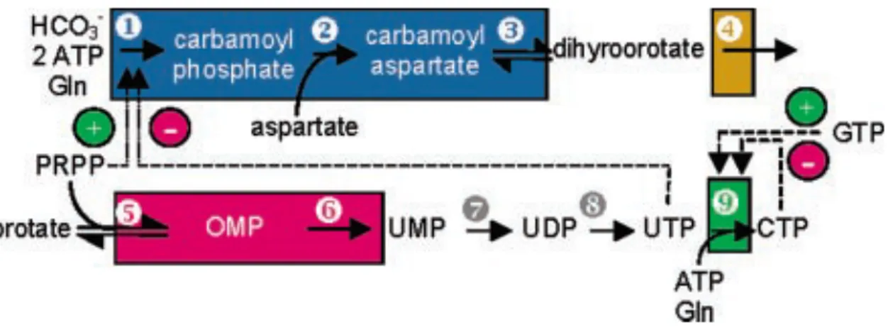
As its name implies, disease results from mutation within a gene encoding CTP synthase 1 enzyme which is one of the key enzymes in de novo pyrimidine biosynthesis. In order to synthesize CTP, CTPS1 and CTPS2 (another form of CTPS1) transfer an amide group from Glutamine to Uracil Triphosphate in presence of ATP (Figure 1.11) [153]. However, exact roles of CTPS1 and CTPS2 in nucleic acid biosynthesis pathway have not been elucidated yet [154].
Figure 1.11 Illustration of mammalian de novo pyrimidine synthesis pathway. The
exact positioning of CTPS1 in pathway was indicated by green box number as 9. (Adopted from [153])
25
As a result of pivotal role in nucleic acid synthesis, proliferation and consequently development of CTPS1 deficient cells even tissues are expected to be impaired. Nevertheless, patients do not manifest physical abnormalities or non-immune symptoms, therefore, it might be interpreted that in some tissues, CTPS2 enzyme is able to compensate deficiency of CTPS1 enzyme [149].
Having CID symptoms lead researchers to reveal consequences of CTPS1 deficiency in adaptive immune system [149]. First and foremost, patients do not have invariant
Figure 1.12 CTPS1 deficiency results in defective T-cell and B-cell proliferation.
(Adopted from [155])
Natural Killer Cells (iNKT) and Mucosal Associated invariant T-cells (MAIT). In contrast to CTPS2 which is highly expressed in non-activated T-cells, expression of CTPS1 enzyme was determined as low. However, upon CD3 and CD28 co-receptor cross-linking, healthy T-cells immediately express CTPS1 enzyme. CTPS1 deficient T-cells, however, are unable to induce production of CTPS1 enzyme; consequently, they are unable to produce CTP eventually leading to cell cycle arrest in G1 phase. Not only T-cells but also, activation of B-T-cells upon BCR and TLR9, CD40 engagement in the presence of IL-4 induced resulted in CTPS1 enzyme expression. All in all, proliferation capabilities of T-cells and B-cells were severely impaired upon TCR and BCR
![Figure 1.3 Activation of systemic complement cascade. (Adopted from [21])](https://thumb-eu.123doks.com/thumbv2/9libnet/5627476.111609/26.892.170.759.399.701/figure-activation-of-systemic-complement-cascade-adopted-from.webp)
![Figure 1.6 Molecular signaling of NETosis. (Adopted from [95])](https://thumb-eu.123doks.com/thumbv2/9libnet/5627476.111609/36.892.153.784.187.609/figure-molecular-signaling-of-netosis-adopted-from.webp)
![Bazı 2-(4h-[1,2,4]triazol-3-il-sulfanil)-asetamit türevlerinin antioksidan ve antitümör özelliklerinin araştırılması / The investigation of antitumuor and antioxidant properties of some 2-(4h-[1,2,4]triazole-3-yl-sulfanyl)-acetamide derivative](data:image/gif;base64,R0lGODlhAQABAIAAAP///wAAACH5BAEAAAAALAAAAAABAAEAAAICRAEAOw==)