INTRODUCTION
Cramp–fasciculation syndrome (CFS) is a rare peripheral nerve hyperexcitability (PNH) syndrome, characterized by disabling muscle cramps and twitches. CFS presents with a lower rate of clinical and electrophysiological signs of PNH as compared to the more severe member of the same spectrum-neuromyotonia (Isaacs’ syndrome) (1,2,3,4). Voltage-gated potassium channel (VGKC)-complex antibodies are often found in CFS patients. While these antibodies are usually directed against leucine-rich glioma-inactivated 1 (LGI1) and contactin-associated protein-like 2 (CASPR2) in other PNH syndromes, they are mostly against uncharacterized VGKC-complex antigens in CFS patients (2). In this study, we
Arch Neuropsychiatry 2017; 54: 183-186 • DOI: 10.5152/npa.2016.14816
Clinical, Electrophysiological, and Serological Evaluation of Patients with
Cramp–Fasciculation Syndrome
Kramp Fasikülasyon Sendromlu Hastaların Klinik, Elektrofizyolojik ve Serolojik
Olarak Değerlendirilmesi
Mürüvvet POYRAZ
1, Zeliha MATUR
2, Fikret AYSAL
1, Erdem TÜZÜN
3, Lütfü HANOĞLU
1, A. Emre ÖGE
3 1Department of Neurology, İstanbul Medipol University School of Medicine, İstanbul, Turkey2Department of Neurology, İstanbul Bilim University School of Medicine, İstanbul, Turkey 3Department of Neurology, İstanbul University İstanbul School of Medicine, İstanbul, Turkey
Short Research Report / Kısa Araştırma Raporu
Correspondence Address / Yazışma Adresi: Zeliha Matur, İstanbul Bilim Üniversitesi Tıp Fakültesi, Nöroloji Anabilim Dalı, İstanbul, Türkiye E-mail / E-posta: [email protected]
Received / Geliş tarihi: 19.01.2016 Accepted / Kabul tarihi: 18.02.2016 Available Online Date / Çevrimiçi Yayın Tarihi: 28.03.2016 ©Copyright 2017 by Turkish Association of Neuropsychiatry - Available online at www.noropskiyatriarsivi.com
©Telif Hakkı 2017 Türk Nöropsikiyatri Derneği Makale metnine www.noropskiyatriarsivi.com web sayfasından ulaşılabilir. Amaç: Kramp-fasikülasyon sendromu (KFS) nadir görülen bir
peri-ferik sinir hipereksitabilite sendromudur. Tek bir merkezden takip edilen KFS hastalarının klinik ve serolojik özelliklerinin bildirildiği az sayıda yazı vardır.
Yöntem: Altı KFS (5 erkek/1 kadın, yaş aralığı: 27-65 yıl) hastasının klinik, elektrofizyolojik ve serolojik özellikleri incelendi.
Bulgular: Bütün hastalarda kramp, fasikülasyon, kas ağrısı ve otonom belirtiler görülürken ikisinde ayrıca nöropatik ağrıyı düşündüren ekstremitelerde uyuşma ve yanma hissi mevcuttu. İki hastada ta-nımlanmamış voltaja duyarlı potasyum kanalı kompleks proteinlerine [voltage-gated potassium channel (VGKC)-complex proteins] karşı oluşmuş antikorlar, bir hastada “contactin-associated protein-like 2 (CASPR2)” antikoru bulundu. Hastaların hiçbirinde tümör yoktu.
Tibial sinir stimülasyonu sonucunda hastaların çoğunda uzamış ard boşalımlar görüldü. Sinir iletim çalışmaları ve R-R interval değişkenliği testleri normalken seronegatif olan 3 hastada sempatik deri yanıtı amplitüdleri artmıştı. Beş hasta karbamazepin ve pregabalin tedavisi-ne iyi cevap verirken VGKC-antikoru pozitif olan bir hasta karbama-zepin veya immünsüpresyon tedavisine dirençli idi.
Sonuç: Nöropatik ağrı ve VGKC-kompleks antikor birlikteliği KFS hastalarında görülebilir. Otonom belirtiler KFS’li hastalarda sık bu-lunmasına rağmen elektrofizyoloji laboratuvarlarında uygulanan rutin otonom sistem test sonuçları normal kalabilir.
Anahtar kelimeler: Kramp-fasikülasyon sendromu (KFS), periferik sinir hipereksitabilitesi, voltaja duyarlı potasyum kanal-kompleks pro-teinleri, nöropatik ağrı
ÖZ
Introduction: Cramp–fasciculation syndrome (CFS) is a rare peripheral nerve hyperexcitability syndrome. There are only a few reports on clinical and serological profile of a CFS cohort that was followed up by a single outpatient clinic.
Methods: Clinical, electrophysiological, and serological features of 6 CFS patients (5 men, 1 woman; 27–65 years old) were investigated. Results: All patients presented with cramps, fasciculations, muscle pain, and autonomic symptoms, and 2 also reported numbness and burning sensation in limbs, suggestive of neuropathic pain. Antibodies to uncharacterized voltage-gated potassium channel (VGKC)-complex proteins were found in 2 patients and to contactin-associated protein-like 2 (CASPR2) in 1 patient. None of the patients had a tumor. Most of the patients revealed prolonged after-discharges following tibial nerve stimulation. Nerve
conduction studies and R-R interval variability tests were normal, whereas sympathetic skin responses were increased in amplitude in 3 seronegative patients. Five patients showed favorable response to carbamazepine or pregabalin treatment, whereas 1 VGKC-antibody-positive patient was resistant to carbamazepine and immunosuppressant treatment.
Conclusion: Neuropathic pain and VGKC-complex antibodies may be encountered in CFS patients. Although autonomic symptoms are commonly found in CFS, routine autonomic system tests which are done in electrophysiology laboratories might yield normal results.
Keywords: Cramp–fasciculation syndrome (CFS), peripheral nerve hyperexcitability, voltage-gated potassium channel (VGKC)-complex proteins, neuropathic pain
ABSTRACT
report clinical and electrophysiological features of 6 CFS patients together with their detailed antibody screening and autonomic test findings.
METHODS
Patients
All consecutive CFS patients (27–65 years old; 5 men, 1 woman) fol-lowed up in our outpatient clinic were recruited, and their demographic, clinical, and laboratory data were recorded (Table 1). CFS was defined by the presence of muscle cramps and fasciculations in the upper and lower limbs on the neurological exam. None of the patients had any electrolyte disturbance, another neuromuscular disorder, or clinical and electrophysiological evidence for anterior horn disease or another auto-immune disease. Cranial magnetic resonance imaging (MRI) was normal in all patients. They were screened for occult malignancy with thorax and abdominal computerized tomography (CT), tumor markers, and flude-oxyglucose positron emission tomography (Cases 1 and 5), and none of the patients were found to have a tumor. The study was approved by the Institutional Review Board, and all patients gave their consents.
Electrophysiological Studies
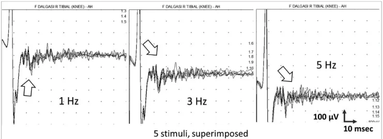
In all patients, routine sensory and motor nerve conduction studies (NCSs; at least 2 nerves in 1 upper and 1 lower extremity), F wave studies (recorded from at least 1 hand and 1 foot muscle), and needle electromy-ography (at least 1 distal and 1 proximal muscle in 1 upper and 1 lower extremity) were performed. Presence of after-discharges was evaluated by stimulation of the right posterior tibial nerve by trains of 5 stimuli given at 0.5-, 1-, 3-, and 5-Hz intra-train frequencies. Discharges which began during or immediately following the trains that were clearly distinct from the baseline without fluctuations, implying voluntary motor activity were accepted as after-discharges. Discharge intensities were graded on a scale of 0–3, as described in a previous report (5).
Sympathetic skin responses (SSRs) recorded simultaneously from the right hand and foot following electrical stimulation (0.1-s duration, 20–40 mA in intensity) of the left median nerve at the wrist level. Re-cordings were made by disk electrodes placed as palm/sole (active) and the dorsum of the hand/foot (reference) arrangement. Band pass was 0.1–1000 Hz, sensitivity was 1–2 mV/division, and sweep speed was 1 s/
division (6). SSR responses >6.3 mV in amplitude for hand and >3.0 mV for foot were considered as to be increased (7). For R-R interval varia-tion (RRIV), 1-min-long electrocardiograms of the supine patient during rest and hyperventilation were recorded with the oscilloscope program using triggering mode. The recording disk electrodes were placed on the wrist and 5th intercostal space at the midclavicular line. Then, RRIV
was calculated manually by the percentage of difference between the earliest and the latest R waves (range of the R-R intervals) /mean of R-R intervals*100 (8).
Immunologic Studies
Antibodies to N-methyl-D-aspartate receptor (NMDAR), alpha-ami-no-3-hydroxy-5-methyl-4-isoxazolepropionic acid receptor (AMPAR), LGI1, CASPR2, and gamma-aminobutyric acid B receptor (GABABR) were investigated by a cell-based immunofluorescence assay (Euroimmun, Luebeck, Germany). For uncharacterized VGKC-complex antibodies, ra-dioimmunoassay (RIA) using brain extracts labeled with 125I-dendrotoxin
(normal value <100 pM) were used (9). Paraneoplastic antibodies (Hu, Yo, CV2, Ri, Ma2, and amphiphysin) were evaluated by a commercial im-munoblot kit (Euroimmun).
RESULTS
All patients presented with cramps, fasciculations, muscle pain, and auto-nomic symptoms, and 2 of them (Cases 4 and 5) also reported numbness and burning sensation in the limbs, which were suggestive of neuropathic pain. Three patients had insomnia (Case 2, 4 and 5), and 1 of them had fatigue. All patients complained of hyperhidrosis, and all except Case 2 exhibited additional autonomic symptoms, such as erectile dysfunction, lightheadedness, constipation, and palpitation. Their follow-up durations were between 1 and 10 years (Table 1). They were diagnosed as having CFS 1 month to 1.5 years after the beginning of their complaints. Neuro-logical examination was normal in 2 patients, while increased deep tendon reflexes (Cases 1 and 6) and reduced vibration sensation in the lower limbs (Cases 3 and 4) were found in some patients. Cases 2–6 showed significant response to carbamazepine or pregabalin treatment, whereas symptoms of Case 1 was only moderately decreased following carbamaz-epine, gabapentin, pregabalin, pulse steroid, intravenous immunoglobulin, azathioprine, and cyclophosphamide treatments (Table 1).
Poyraz et al. Cramp–Fasciculation Syndrome Arch Neuropsychiatry 2017; 54: 183-186
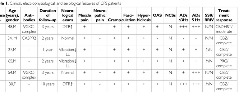
Table 1. Clinical, electrophysiological, and serological features of CFS patients
Age Duration Neuro- Neuro-
Treat-Case (years), Anti- of logical Muscle pathic Fasci- Hyper- OAS NCSs ADs ADs SSR/ ment
no. gender bodies follow-up exam pain pain Cramp culation hidrosis ≤3Hz 5 Hz RRIV response
1 48,M VGKC- 3 years DTR↑ + - + + + + N +++ +++ N/N CBZ+IST/
complex moderate
2 34, M CASPR2 2 years Normal + - + + + - N - - N/N CBZ/
complete 3 27,M - 1 year Vibration↓ + - + + + + N + + ↑/N CBZ/ LL complete 4 65,M - 2 years Vibration↓ + + + + + + N + + ↑/N PRG/ LL complete 5 54,M VGKC- 3 years Normal + + + + + + N + +++ N/N CBZ/ complex complete 6 30,F - 10 years DTR↑ + - + + + + N +++ +++ ↑/N CBZ/ complete
M: male; F: female; CBZ: carbamazepine; PRG: pregabalin; DTR: deep tendon reflexes; ↑: increased, vibration; ↓ LL: reduced vibration sense in the lower limbs; OAS: other autonomic symptoms; VGKC: uncharacterized voltage gated potassium channel antigens; CASPR2: contactin-associated protein-like 2; IST: immunosuppressive treatment; SSR: sympathetic skin response; RRIV: R-R interval variability; NCSs: nerve conduction studies; ADs: after-discharges; N: normal
Sensory and motor NCSs were normal in all patients. After-discharges were obtained in all patients except Case 2 (Figure 1). Needle electro-myography showed abnormal spontaneous activity (random or semirhyt-mically discharging fasciculations and myokymic discharges) in all patients. R-R interval variations were normal in all the cases, whereas SSR was increased in amplitude in Cases 3, 4, and 6 (Table 1).
None of the patients had evidence of other tested paraneoplastic anti-bodies, including Hu, Yo, CV2, Ri, Ma2, and amphiphysin. Patients were not screened for the presence of unclassified antibodies. Two patients (Cases 1 and 5) displayed antibodies to uncharacterized VGKC-complex antigens (both >500 pM), and 1 (Case 2) had CASPR2 antibody. None of the other investigated ion channel antibodies could be detected.
DISCUSSION
As a rare syndrome, CFS has mostly been reported in the form of case reports, and there are only a few reports on CFS cohorts followed up by single outpatient clinics (2,4). Overall, in line with previously reported cohorts, our CFS patients showed male predominance, no tumor associ-ation, VGKC-complex antibody positivity, high prevalence of autonomic symptoms, and presence of chronic pain. While the sensory complaints and reduced vibratory sensation in the distal extremities in our patients can easily be attributed to the mild peripheral nerve involvement found in the idiopathic/autoimmune peripheral nerve hyperexcitability syndromes, the mechanisms leading to the increased tendon reflexes in 2 cases are more difficult to be interpreted (1). It might be speculated that the same central mechanisms as those causing insomnia in 3 patients can be ef-fective in CFS, thereby reminding that this syndrome is a member of a larger coalition of syndromes with peripheral and central nervous system involvement (1).
Similar to previously reported cases, most of our patients, including sero-positive ones, responded to membrane-stabilizing agents with no immu-nosuppression requirement (2). Moreover, our antibody results corrob-orate the notion that CFS patients do not necessarily display antibodies to uncharacterized VGKC-complex antigens and might also present with antibodies to well-characterized antigens such as CASPR2 (2).
However, in contrast with a recent report (2), we failed to find an asso-ciation between VGKC-complex antibody positivity and autonomic test findings. Although autonomic symptoms are frequently observed in CFS
patients (1,2,3,10), autonomic studies might often give negative results. In previous studies, autonomic reflex screen and thermoregulatory sweat tests have given positive results only in a small fraction of CFS patients (2,4). Likewise, R-R interval variability test was normal in our patients, and SSR measurements gave large amplitude results in 3 patients. It is more likely that the large SSR responses were due to the already sweaty patients at the baseline. Nevertheless, none of the patients with increased SSR had VGKC-complex antibodies, indicating the presence of different pathogen-ic mechanisms in seronegative CFS. It was reported that after-discharges in seropositive patients were significantly longer in duration (5). We found similar after-discharge characteristics in seropositive and seronegative pa-tients.
In conclusion, VGKC-complex antibodies were found in most of our pa-tients. A parallelism between the antibody profile and some clinical char-acteristics may be found in larger series. Identification of novel anti-neu-ronal antibodies and non-VGKC-related disease mechanisms might be required for better identification of CFS cases.
Ethics Committee Approval: Ethics committee approval was received for this
study from the ethics committee of İstanbul University İstanbul School of Medicine.
Informed Consent: Written informed consent was obtained from patients who
participated in this study.
Peer-review: Externally peer-reviewed.
Author Contributions: Concept - F.A., A.E.O.; Design - A.E.O., Z.M.;
Super-vision - A.E.O.; Resource - M.P., Z.M., E.T.; Materials - F.A., M.P., Z.M., A.E.O.; Data Collection and/or Processing - Z.M., M.P., F.A.; Analysis and/or Interpretation - Z.M., A.E.O.; Literature Search - M.P., E.T.; Writing - M.P., E.T., Z.M.; Critical Reviews - A.E.O., L.H.
Conflict of Interest: No conflict of interest was declared by the authors. Financial Disclosure: The authors declared that this study has received no
fi-nancial support.
Etik Komite Onayı: Bu çalışma için etik komite onayı İstanbul Üniversitesi
İstan-bul Tıp Fakültesi Etik Kurulu’ndan alınmıştır.
Hasta Onamı: Yazılı hasta onamı bu çalışmaya katılan hastalardan alınmıştır.
Arch Neuropsychiatry 2017; 54: 183-186 Poyraz et al. Cramp–Fasciculation Syndrome
Figure 1. After-discharges occurred by repetitive nerve stimulation of the right posterior tibial nerve at 1, 3, and 5 Hz with surface electrodes over the right
ab-ductor hallucis muscle
Hakem Değerlendirmesi: Dış Bağımsız.
Yazar Katkıları: Fikir - F.A., A.E.O.; Tasarım - A.E.O., Z.M.; Denetleme - A.E.O.;
Kaynaklar - M.P., Z.M., E.T.; Malzemeler - F.A., M.P., Z.M., A.E.O.; Veri Toplanması ve/veya İşlemesi - Z.M., M.P., F.A.; Analiz ve/veya Yorum - Z.M., A.E.O.; Literatür Taraması - M.P., E.T.; Yazıyı Yazan - M.P., E.T., Z.M.; Eleştirel İnceleme - A.E.O, L.H.
Çıkar Çatışması: Yazarlar çıkar çatışması bildirmemişlerdir.
Finansal Destek: Yazarlar bu çalışma için finansal destek almadıklarını beyan
et-mişlerdir.
REFERENCES
1. Küçükali CI, Kürtüncü M, Akçay Hİ, Tüzün E, Öge AE. Peripheral nerve hyper-excitability syndromes. Rev Neurosci 2015; 26:239-251. [CrossRef]
2. Liewluck T, Klein CJ, Jones LK Jr. Cramp-fasciculation syndrome in patients with and without neural autoantibodies. Muscle Nerve 2014; 49:351-356.
[CrossRef]
3. Newsom-Davis J, Buckley C, Clover L, Hart I, Maddison P, Tüzün E, Vincent A. Autoimmune disorders of neuronal potassium channels. Ann N Y Acad Sci 2003; 998:202-210. [CrossRef]
4. Hart IK, Maddison P, Newsom-Davis J, Vincent A, Mills KR. Phenotypic variants of autoimmune peripheral nerve hyperexcitability. Brain 2002; 125:1887-1895.
[CrossRef]
5. Tahmoush AJ, Alonso RJ, Tahmoush GP, Heiman-Patterson TD. Cramp-fas-ciculation syndrome: a treatable hyperexcitable peripheral nerve disorder. Neurology 1991; 41:1021-1024. [CrossRef]
6. Uncini A, Pullman SL, Lovelace RE, Gambi D. The sympathetic skin response: normal values, elucidation of afferent components and application limits. J Neurol Sci 1988; 87:299-306. [CrossRef]
7. Elie B, Guiheneuc P. Sympathetic skin response: normal results in different ex-perimental conditions. Electroencephalogr Clin Neurophysiol 1990; 76:258-267.
[CrossRef]
8. Shahani BT, Day TJ, Cros D, Khalil N, Kneebone CS. RR interval variation and the sympathetic skin response in the assessment of autonomic function in peripheral neuropathy. Arch Neurol 1990; 47:659-664. [CrossRef]
9. Irani SR, Alexander S, Waters P, Kleopa KA, Pettingill P, Zuliani L, Peles E, Buckley C, Lang B, Vincent A. Antibodies to Kv1 potassium channel-complex proteins leucine-rich, glioma inactivated 1 protein and contactin-associated protein-2 in limbic encephalitis, Morvan’s syndrome and acquired neuromyo-tonia. Brain 2010; 133:2734-2748. [CrossRef]
10. de Entrambasaguas M, Ortega-Albás JJ, Martínez-Lozano MD, Díaz JR. Bron-chial involvement in the cramp-fasciculation syndrome. Eur Neurol 2006; 56:124-126. [CrossRef]
Poyraz et al. Cramp-Fasciculation Syndrome Arch Neuropsychiatry 2017; 54: 183-186