1 23
Lasers in Medical Science
ISSN 0268-8921
Volume 28
Number 5
Lasers Med Sci (2013) 28:1233-1239
DOI 10.1007/s10103-012-1201-5
Effects of different surface treatments
on shear bond strength in two different
ceramic systems
Tevfik Yavuz, Erhan Dilber, Haluk Baris
Kara, Ali Riza Tuncdemir & A Nilgun
Ozturk
1 23
Your article is protected by copyright and
all rights are held exclusively by
Springer-Verlag London Ltd. This e-offprint is for
personal use only and shall not be
self-archived in electronic repositories. If you wish
to self-archive your article, please use the
accepted manuscript version for posting on
your own website. You may further deposit
the accepted manuscript version in any
repository, provided it is only made publicly
available 12 months after official publication
or later and provided acknowledgement is
given to the original source of publication
and a link is inserted to the published article
on Springer's website. The link must be
accompanied by the following text: "The final
publication is available at link.springer.com”.
ORIGINAL ARTICLE
Effects of different surface treatments on shear bond strength
in two different ceramic systems
Tevfik Yavuz&Erhan Dilber&Haluk Baris Kara&
Ali Riza Tuncdemir&A Nilgun Ozturk
Received: 26 March 2012 / Accepted: 3 September 2012 / Published online: 13 September 2012 # Springer-Verlag London Ltd 2012
Abstract The purpose of this study was to evaluate the influence of different surface treatments (sandblasting, acid etching, and laser irradiation) on the shear bond strength of lithium disilicate-based core (IPS Empress 2) and feldspath-ic ceramfeldspath-ics (VITA VM 9). One hundred ceramfeldspath-ic discs were divided into two groups of 50 discs each for two ceramic systems: IPS Empress 2 (group I) and VITA VM 9 (group II). Each of the two groups was further divided into five surface treatment groups (ten each) as follows: group SB, sandblasting with alumina particles (50μm); group HF, 5 % hydrofluoric acid etching; group L, Er:YAG laser irradiation (distance, 1 mm; 500 mJ; 20 Hz; 10 W; manually, noncon-tact R14 handpiece); group SB–L, sandblasting + Er:YAG laser; and group HF–L, 5 % hydrofluoric acid + Er:YAG laser. Luting cement (Panavia 2.0) was bonded to the ce-ramic specimens using Teflon tubes. After 24 h of water storage, a shear bond strength test was performed using a universal testing machine at a crosshead speed of 0.5 mm/ min. The data were analyzed with a two-way analysis of variance (ANOVA) and Tukey's honestly significant differ-ence tests (α00.05). The two-way ANOVA indicated that the shear bond strength was significantly affected by the surface treatment methods (p<0.05), but there was no sig-nificant interaction between the ceramic systems. Group
SB–L had the highest mean values for each ceramic system. Sandblasting, followed by Er:YAG laser irradiation, en-hanced the bond strength, indicating its potential use as an alternative method. The atomic force microscopic evalua-tion revealed that group SB had the most distinct sharp peaks among the groups.
Keywords Bond strength . Luting cements . Surface treatment . Ceramic . Er:YAG
Introduction
Ceramics are still the most aesthetically pleasing materials available for prosthetic dentistry because of their high resis-tance to abrasion, compression, chemical stability, biocom-patibility, acceptable translucence, and fluorescence characteristics [1]. Feldspathic ceramics (silica-based ceram-ic) are the conventional ceramic materials for metal–ceramic restorations and consist of a mixture of feldspar and quartz [2]. They are frequently used to veneer metal frameworks and indirect restorations, such as inlays, onlays, and lami-nates. All-ceramic restorations have gained popularity among dentists and patients because of their favorable aes-thetics and liability in conservative tooth preparations [3].
A lithium disilicate glass–ceramic core veneered with a sintered glass–ceramic provides higher strength and allows for the fabrication of short-span fixed dentures [4].
Some clinical studies have indicated that an insufficient luting performance of restorations may result in clinical failure [5,6]. Obtaining desirable adhesion between cement and ceramic surfaces requires surface pretreatment to im-prove the retention, marginal adaptation, and fracture resis-tance of restorations [7, 8]. Surface pretreatment of porcelain increases surface area and creates microporosities on the surface, enhancing the potential for mechanical re-tention of the cement [9]. Different surface treatment
T. Yavuz (*)
:
E. Dilber:
A. N. OzturkDepartment of Prosthodontics, Faculty of Dentistry, Selcuk University,
Konya, Turkey
e-mail: [email protected] H. B. Kara
Department of Prosthodontics, Faculty of Dentistry, Medipol University,
Istanbul, Turkey A. R. Tuncdemir
Department of Prosthodontics, Faculty of Dentistry, Mustafa Kemal University,
Hatay, Turkey
Lasers Med Sci (2013) 28:1233–1239 DOI 10.1007/s10103-012-1201-5
methods (grinding, abrasion with diamond rotary instru-ments, airborne particle abrasion with Al2O3, acid etching,
sandblasting, and combinations of any of these methods) have been proposed to provide roughness and promote micromechanical retention [10].
Some authors declared that enhancing the roughness of a ceramic surface with hydrofluoric (HF) acid and then applying a silane coupling agent is the suggested surface treatment method for feldspathic ceramics [11, 12]. Conversely, for some conditions, such as when intraoral ceramic repair is needed, the use of acid etching is not recommended because of its harmful and irritating effects on oral tissues [13]. Other surface roughness methods can be assumed for this reason to induce similar or greater surface roughness on porcelains.
Another promising technique for the surface treatment of ceramic materials is laser irradiation [14]. Among the vari-ous laser types, the Er:YAG laser is highly recommended for use on the surfaces of dental materials, but there is no consensus in the literature regarding the best surface-conditioning method to produce optimal bond strength based on the cement and porcelain used [15].
Therefore, the purpose of this in vitro study was to investigate and compare the effects of Al2O3sandblasting,
HF acid etching, and their use in combination with ER:YAG laser irradiation on the surface roughness and shear bond strengths of lithium disilicate-based and feldspathic ceramics. The null hypothesis was that Er:YAG laser treat-ment would result in surface roughness and bond strength that is comparable with that of air abrasion and acid-etching surface treatment methods.
Materials and methods Specimen preparation
One hundred ceramic discs were divided into two groups of 50 each for two ceramic systems: IPS Empress 2 (group I; Ivoclar Vivadent, Amherst, NY, USA) and VITA VM 9 (group II; VITA VM 9, VITA Zahnfabrik H Rauter GmbH & Co. KG, Bad Säckingen, Germany).
For group I, to obtain 50 lithium disilicate-based core ceramic discs (diameter, 10 mm; thickness, 1 mm), IPS Empress 2 wax patterns were prepared and invested in the IPS Empress 2 Speed investment (Ivoclar Vivadent, Amherst, NY, USA). The wax was eliminated in a burnout furnace (EP 600; Ivoclar Vivadent, Amherst, NY, USA) preheated to 850 °C with an alumina plunger for 90 min. The IPS Empress 2 ingots were softened at 920 °C and automatically pressed into the mold in a furnace.
After pressing and cooling to room temperature, the specimens were divested with 125-μm glass beads at a pressure of 4 bars, ultrasonically cleaned in a special liquid
(Invex liquid, Ivoclar-Vivadent) for 10 min, washed in run-ning water, and dried. They were then subjected to airborne particle abrasion with 50-μm Al2O3particles at a pressure of
2 bars (Korox, Bego, Bremen, Germany).
For group II, to obtain 50 feldspathic ceramic discs (diameter, 10 mm; thickness, 1 mm; VITA VM 9), metal molds with disc-shaped holes were used, and an impression of the metal mold was made with silicon putty (Virtual vinyl polysiloxane, Ivoclar, Schaan, Liechtenstein). The refracto-ry die material (VITA Zahnfabrik H Rauter GmbH & Co. KG) was then poured into the silicon putty. Veneering porcelain powder (VITA VM 9 Powder, VITA Zahnfabrik H. Rauter GmbH & Co.) was mixed with the manufacturer-supplied condensing liquid and condensed using the vibra-tion blotting technique. The obtained slurry was blotted with tissue to eliminate excess water and then condensed into the mold. The prepared discs were fired in a programmable vacuum porcelain furnace (VITA Vacumat 4000 Premium T; VITA Zahnfabrik H Rauter GmbH & Co. KG) in accor-dance with the firing programs provided by the manufactur-er. No glaze was applied to the ceramic surface of the discs. The bonding surfaces of the specimens in the two groups were polished using silicon carbide paper (grit 800) under water cooling and then polished with OptraFine Assortment (Ivoclar) for standardization. The surfaces were cleaned with ethanol and dried carefully in air before surface treat-ment. After the finishing procedures, the discs were sub-jected to ultrasonic treatment (Biosonic JR, Coltene Whaledent) in 99.5 % acetone to remove any surface resi-dues and were dried. After these procedures, the specimens in both groups were further divided into five surface treat-ment groups of ten discs each.
Group SB (sandblasting)
For both groups, the ceramic surfaces were abraded with 50-μm Al2O3particles at a pressure of 2.8 bars from a distance
of 10 mm perpendicular to the treated surface for 20 s. Group HF (acid etching)
For group I, the ceramic surfaces were etched with 5 % HF acid (IPS Ceramic Etching Gel, Ivoclar Vivadent) for 20 s; the gel was rinsed off with water for 20 s, and then the surfaces were dried under oil-free compressed air for 20 s. Similar procedures were performed for group II at 60 s for each procedure.
Group L (Er:YAG laser irradiation)
An Er:YAG laser (Fotona, At Fidelis, Ljubljana, Slovenia) was used to irradiate the ceramic surfaces in both groups. A contact handpiece (R14; 1.3 mm in diameter) with an
1234 Lasers Med Sci (2013) 28:1233–1239
integrated spray nozzle was placed perpendicular to the ceramic surface at a 1-mm distance, and the entire ceramic area was manually scanned with water cooling for 20 s. The laser irradiation of all the specimens was performed by the same operator. The laser parameters were as follows: 500 mJ (pulse energy), 10 W (power), MSP mode (100-μs pulse length), 20 Hz (pulses per second), and 37.68 J/cm2(energy density).
Group SB–L (sandblasting + Er:YAG laser)
For both groups, the ceramic surfaces were abraded using the same parameters described for group SB. After the sand-blasting procedure, all the ceramic surfaces were irradiated with an Er:YAG laser using the same parameters described for group L.
Group HF–L (acid etching + Er:YAG laser)
For both groups, the ceramic surfaces were etched with 5 % HF acid using the same procedure described for group HF. The Er:YAG laser irradiation was performed using the same parameters described for group L.
After all the surface treatments, a silane-coupling agent (Clearfil Ceramic Primer, Kuraray, batch no. 00001A) was applied to the ceramic surface of all the specimens in both groups using a clean brush, and then the specimens were air dried using oil-free compressed air. To apply the resin cement, each treated ceramic was initially placed in its silicone mold. The resin cement (Panavia F 2.0, Kuraray Medical Inc., Osaka, Japan) was then mixed according to the manufacturer's instructions and injected into the Teflon tubes with an internal diameter and height of 4 and 3 mm, respectively. The bonding procedures were performed according to the manufacturer's recommendations. Curing light was applied to the top of the filled molds for 40 s (Bluephase, Ivoclar Vivadent). An oxygen-inhibiting gel (Oxyguard II, Kuraray) was applied to the exposed surfa-ces. After careful removal of the Teflon molds, every side of the cement cylinder was light cured for a total of 80 s. The bonded specimens were then stored in distilled water at room temperature for 24 h.
Evaluation of shear bond strength
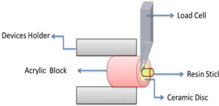
The samples were placed into the test device using special acrylic molds. The knife-edge-shaped apparatus was placed between the joint of the ceramic disc and the resin material. The shear bond strength of the ceramic discs was evaluated using a universal testing machine (TSTM 02500, Elista Ltd Sti, Istanbul, Turkey) with a crosshead speed of 0.5 mm/ min. The value at which the ceramic disc and the resin material ruptured was recorded (Fig.1).
After the shear bond strength test, ceramic bonding areas were observed with a stereomicroscope (Olympus CX41, ×40, Japan) to identify the mode of fracture. The fractured surface was classified according to one of three types: type 1, adhesive failure between the resin cement and the ceram-ic; type 2, cohesive failure in the ceramceram-ic; and type 3, cohesive failure in the resin cement. The data were analyzed by two-way analysis of variance (ANOVA) and Tukey's honestly significant difference (HSD) tests (SPSS/PC ver-sion 17.0, SPSS, Chicago, IL, USA) for pairwise compar-isons among the groups (p<0.05).
Atomic force microscopic evaluation
One additional specimen from each group was evaluated by atomic force microscopy (AFM; NTEGRA Solaris, NTMDT, Russia). The digital images were obtained in air. A 0.01–0.025-Ω-cm gold-doped silicon tip (40 μm) was used in the noncontact mode. The changes in the vertical position provided the height of the images, and these changes were registered as the bright and dark regions. A constant tip sample“tap” was maintained through a constant oscillation amplitude (set point amplitude). Five 25×25-μm digital images were acquired for each surface and recorded with a slow scan rate (1 Hz).
Results
Shear bond strength
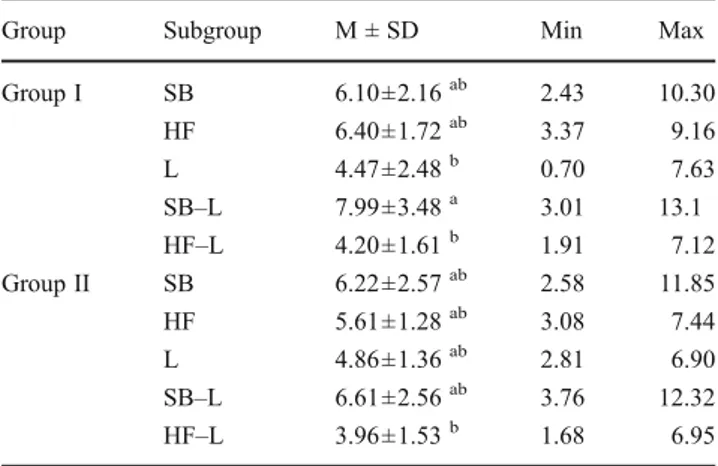
The mean and SD bond strength values for each ceramic material in the five surface treatment groups are given in Table 1. As shown in Table 1, the two-way ANOVA indicated that shear bond strength was significantly affect-ed by the surface treatment methods (p<0.05) and that there was no significant interaction between the two ce-ramic materials (p > 0.05; Table 2). Tukey's HSD test results on shear bond strength of the lithium disilicate-based ceramic revealed significant differences among the groups (p<0.05).
Fig. 1 Scheme of the test sample
Lasers Med Sci (2013) 28:1233–1239 1235
Tukey's HSD test results revealed that group SB–L had significantly higher shear bond strengths than groups HF–L and L for group I (p < 0.05). There were no significant differences among the groups for group II (p>0.05), but higher values were obtained from group SB–L.
Failure type analysis
The modes of failure for the specimens after the treatment methods are as follows: for the feldspathic ceramics, 31 specimens exhibited adhesive failure and 13 specimens exhibited cohesive failure; for the lithium disilicate-based ceramics, 44 specimens exhibited adhesive failure and no specimen exhibited cohesive failure. Six mixed failures were exhibited in both two ceramic systems.
AFM evaluation
The representative AFM images for the two ceramic groups treated with the surface treatment groups are presented in Figs.2
and3. The surface treatment groups exhibited similar topog-raphies, excluding the surfaces treated with the Er:YAG laser
(group L). The group L surfaces exhibited moderate irregular-ity, with peaks and valleys, and less roughness than what was achieved with sandblasting and HF acid. Group SB had the most distinct sharp peaks among the groups (Figs.2and3). Discussion
The properties of a luting agent and the surface treatments for ceramic surfaces before cement application play a major role in the clinical success of many indirect ceramic restorations [16]. The intent of this in vitro study was to investigate the influence of different surface treatments of feldspathic and lithium disilicate-based ceramics on the bond strength of resin cement. Of all the surface treatments for feldspathic ceramic, HF acid etching achieved the highest bond strength. The finding of the present study that sandblasting and laser application resulted in a strong shear bond between the resin cement and the feld-spathic or lithium disilicate-based ceramic was in agreement with the findings of other groups [14,17–27].
Airborne particle abrasion with Al2O3particles is effective
and practical for creating an activated and roughened surface on aluminum oxide ceramics [8, 28]. Changes in surface topography occurred after sandblasting procedures [29–31]. This technique was included in the present study, as it is a commonly used procedure in prosthodontic laboratories, and dental offices have miniaturized devices that facilitate its use [28]. Sandblasting produces a rough irregular surface with an increased surface area and enhances the wettability of the ceramic and composite resin. However, excessive sandblast-ing induces chippsandblast-ing or a significant loss of porcelain mate-rial, and it is not recommended for cementation of silica-based and feldspathic ceramic restorations [32–34].
Particle size, procedure duration, and pressure and dis-tance used in the procedure are important factors in the performance of a cement bond [26, 28, 35, 36]. It was indicated that a sandblasting procedure with very large particles or with a very high pressure does not increase the bond strength; nevertheless, it causes maladaptiveness in the restoration as a result of wear [35–37]. Kern and Thompson [32] reported that at the end of sandblasting procedures, material loss in the IPS Empress ceramics was 36-folds higher than that in the In-Ceram. Spohr et al. [37] used an electron microscope to analyze the IPS Empress 2 ceramics
Table 1 Median (min–max) shear bond strength (MPa) values accord-ing to surface treatments
Group Subgroup M ± SD Min Max
Group I SB 6.10±2.16ab 2.43 10.30 HF 6.40±1.72ab 3.37 9.16 L 4.47±2.48b 0.70 7.63 SB–L 7.99±3.48a 3.01 13.1 HF–L 4.20±1.61b 1.91 7.12 Group II SB 6.22±2.57ab 2.58 11.85 HF 5.61±1.28ab 3.08 7.44 L 4.86±1.36ab 2.81 6.90 SB–L 6.61±2.56ab 3.76 12.32 HF–L 3.96±1.53b 1.68 6.95
Values with same lowercase letter are not significantly different at p<0.05
SB sandblasting, HF etch, L laser, SB–L sandblasting + laser, HF–L etch + laser
Table 2 Results of two-way
ANOVA Source SS df MS F p
Ceramics 182.493 1 182.493 0.766 0.384
Surface treatments 6,538.319 4 1,634.580 6.863 0.000
Intercept 159,106.052 1 159,106.052 668.032 0.000
Ceramics* surface treatments 506.226 4 126.556 0.531 0.713
1236 Lasers Med Sci (2013) 28:1233–1239
to which 50- and 100-mm Al2O3 particles were applied.
They performed a shear bond test and indicated that surfaces on which 100-mm Al2O3 particles were applied did not
possess adequate retentive properties, whereas surfaces pre-pared using 50-mm Al2O3 particles were more highly
etched. They also found at the end of the test that there was a significant increase in the strength of the cement bond. Other researchers reported that chemical and mechan-ical etching methods can be used together [28,32,38,39]. In the present study, a statistical difference was found be-tween the bonding resistance of the group I and II samples conditioned with 50-mm Al2O3particles and the samples to
which 50-mm Al2O3particles were applied.
Among the chemical treatments of ceramic surfaces, etching with HF acid is one of the most commonly used methods [9,17,40–42]. In the procedure, the glassy matrix is selectively removed, and crystalline structures are ex-posed. Crystals influence the formation of microporosities [43]. Microporosity increases the surface area and makes micromechanical interlocking of resin possible [16]. Previ-ous studies [18,25,44] demonstrated that HF + silane was consistently the most effective ceramic surface treatment, although it was not always significantly better than the other
treatments that involved roughening and silane application [17]. In the present study, there were significant differences between the HF acid etching and the other techniques. HF acid is well recognized to have hazardous effects in vivo; it is harmful and irritating to soft tissues, [15] and this method cannot be considered for practical use in dentistry, particu-larly for intraoral ceramic repairs [17].
When Er:YAG laser irradiation is applied to a ceramic surface, it is expected that the laser irradiation will create a rough surface by removing the glass phase of the ceramic [27,
45] and increase the micromechanical retention of resin. How-ever, in this study, the Er:YAG laser irradiation actually pro-duced the lowest shear bond strength [27]. Erosion and melting were observed, but no fissures or cracks were observed on the surface after Er:YAG laser irradiation using AFM [27].
Similarly, Shiu et al. [17] observed that Er:YAG laser irra-diation of a feldspathic ceramic surface at an energy setting of 500 mJ/pulse and 4 Hz for 2 min was insufficient to cause adequate surface roughing. They also found that the surface treatment proposed with Er:YAG laser resulted in low bond strength [27].
In the present study, the methodology for Er:YAG laser application on ceramic surfaces (10 W, 20 Hz, 500 mJ, and
Fig. 2 AFM images of Group I. a SB. b HF. c L. d SB–L. e HF–L
Fig. 3 AFM images of Group II. a SB. b HF. c L. d SB–L. e HF–L
Lasers Med Sci (2013) 28:1233–1239 1237
37.68 J/cm2) was based on a study performed by Silveira et al. [16], who defined this protocol after a series of pilot studies on In-Ceram alumina and observed that the Er:YAG laser was the most effective surface treatment, followed by Al2O3
sandblasting.
The combination of Al2O3and Er:YAG laser (SB–L)
treat-ment resulted in a better performance than Er:YAG laser irradiation alone. The use of Er:YAG laser irradiation and acid etching (HF–L) for ceramic surface treatment resulted in lower bond strengths than those observed in other groups [17]. Laser irradiation after HF acid etching may remove crystalline components rather than glass components and may decrease the bonding to the porcelain surface. Converse-ly, laser irradiation after sandblasting may remove aluminum particles from the porcelain surfaces, thus enhancing the bonding. These mechanic alterations may affect the bond strength between resin cement and porcelain surfaces.
This study had limitations in its ability to simulate clin-ical loading forces on restorations and oral environmental changes. In the shear bond test, the loading was monotonic instead of representative of cyclic fatigue, and the tempera-ture and moistempera-ture of the oral cavity were not simulated; these factures should be included in investigations as many factors affect the bond strengths of the resin luting cements applied to the ceramics [28]. Future studies with a model that more closely resembles the oral environment and sim-ulates clinical loading conditions are required [17]. Conclusion
The present study demonstrated that the shear bond strengths of the resin cements tested on ceramics after sur-face treatments varied in accordance with the type of ceram-ic. For lithium disilicate-based ceramics, the shear bond strength was significantly different between the SB–L group and the HF–L and L groups. However, no significant dif-ference was observed for feldspathic ceramic.
Low bond strength was observed in the HF–L and L groups, indicating that these treatments are inadequate for clinical use within the parameters tested in this study. Laser irradiation used alone or combined with HF acid and SB within the parameters is not an alternative method for im-proving the bonding of resin cement to ceramic surface. References
1. Nakamura S, Yoshida K, Kamada K, Atsuta M (2004) Bonding between resin luting cement and glass-infiltrated alumina-reinforced ceramics with silane coupling agent. J Oral Rehabil 31 (8):785–789. doi:10.1111/j.1365-2842.2004.01304.x
2. Craig RG, Powers JM (2004) Materials for dental restorations. Livraria Editora Santos, São Paulo, p 704
3. Rosenstiel S, Land M, Fujimoto J (2006) Contemporary fixed pros-thodontics, 4th edn. Elsevier Health Sciences, St Louis, pp 909–925 4. Culp L (1999) Empress 2: first year clinical results. J Dent Technol
16(2):12–15
5. Ferrari M, Vichi A, Garcia-Godoy F (2000) Clinical evaluation of fiber-reinforced epoxy resin posts and cast post and cores. Am J Dent 13(Spec No):15B–18B
6. Monticelli F, Grandini S, Goracci C, Ferrari M (2003) Clinical behavior of translucent fiber posts: a 2-year prospective study. Int J Prosthodont 16(6):593–596
7. Burke FJ, Fleming GJ, Nathanson D, Marquis PM (2002) Are adhesive technologies needed to support ceramics? An assessment of the current evidence. J Adhes Dent 4(1):7–22
8. Ozcan M, Vallittu PK (2003) Effect of surface conditioning meth-ods on the bond strength of luting cement to ceramics. Dent Mater 19(8):725–731
9. Hayakawa T, Horie K, Aida M, Kanaya H, Kobayashi T, Murata Y (1992) The influence of surface conditions and silane agents on the bond of resin to dental porcelain. Dent Mater 8(4):238–240 10. Saracoglu ACC, Cotert HS (2004) Effect of various surface
treat-ment methods on the bond strength of the heat-pressed ceramic samples. J Oral Rehabil 31:790–797
11. Shen C, Oh WS, Williams JR (2004) Effect of post-silanization drying on the bond strength of composite to ceramic. J Prosthet Dent 91(5):453–458. doi:10.1016/S0022391304001301
12. Barghi N, Berry T, Chung K (2000) Effects of timing and heat treatment of silanated porcelain on the bond strength. J Oral Rehabil 27(5):407–412
13. Bertolini JC (1992) Hydrofluoric acid: a review of toxicity. J Emerg Med 10(2):163–168
14. Li R, Ren Y, Han J (2000) Effects of pulsed Nd:YAG laser irradiation on shear bond strength of composite resin bonded to porcelain. Trans Hua Xi Kou Qiang Yi Xue Za Zhi 18(6):377–379 15. Kamada K, Yoshida K, Atsuta M (1998) Effect of ceramic surface treatments on the bond of four resin luting agents to a ceramic material. J Prosthet Dent 79(5):508–513
16. da Silva Ferreira S, Hanashiro FS, de Souza-Zaroni WC, Turbino ML, Youssef MN (2010) Influence of aluminum oxide sandblast-ing associated with Nd:YAG or Er:YAG lasers on shear bond strength of a feldspathic ceramic to resin cements. Photomed Laser Surg 28(4):471–475. doi:10.1089/pho.2009.2528
17. Shiu P, De Souza-Zaroni WC, Eduardo Cde P, Youssef MN (2007) Effect of feldspathic ceramic surface treatments on bond strength to resin cement. Photomed Laser Surg 25(4):291–296. doi:10.1089/pho.2007.2018
18. Suliman AH, Swift EJ Jr, Perdigao J (1993) Effects of surface treatment and bonding agents on bond strength of composite resin to porcelain. J Prosthet Dent 70(2):118–120
19. Schmage P, Nergiz I, Herrmann W, Ozcan M (2003) Influence of various surface-conditioning methods on the bond strength of metal brackets to ceramic surfaces. Am J Orthod Dentofac Orthop 123(5):540–546. doi:10.1067/mod.2003.S0889540602569110
20. Phoenix RD, Shen C (1995) Characterization of treated porcelain surfaces via dynamic contact angle analysis. Int J Prosthodont 8 (2):187–194
21. Guler AU, Yilmaz F, Yenisey M, Guler E, Ural C (2006) Effect of acid etching time and a self-etching adhesive on the shear bond strength of composite resin to porcelain. J Adhes Dent 8(1):21–25 22. Guler AU, Yilmaz F, Ural C, Guler E (2005) Evaluation of 24-h s24-hear bond strengt24-h of resin composite to porcelain according to surface treatment. Int J Prosthodont 18(2):156–160
23. Brentel AS, Ozcan M, Valandro LF, Alarca LG, Amaral R, Bottino MA (2007) Microtensile bond strength of a resin cement to feldpathic ceramic after different etching and silanization regimens in dry and aged conditions. Dent Mater 23(11):1323–1331. doi:10.1016/ j.dental.2006.11.011
1238 Lasers Med Sci (2013) 28:1233–1239
24. Akova T, Yoldas O, Toroglu MS, Uysal H (2005) Porcelain surface treatment by laser for bracket–porcelain bonding. Am J Orthod Dentofac Orthop 128(5):630–637. doi:10.1016/j.ajodo.2004.02.021
25. Thurmond JW, Barkmeier WW, Wilwerding TM (1994) Effect of porcelain surface treatments on bond strengths of composite resin bonded to porcelain. J Prosthet Dent 72(4):355–359
26. Pisani-Proenca J, Erhardt MC, Valandro LF, Gutierrez-Aceves G, Bolanos-Carmona MV, Del Castillo-Salmeron R, Bottino MA (2006) Influence of ceramic surface conditioning and resin cements on microtensile bond strength to a glass ceramic. J Pros-thet Dent 96(6):412–417. doi:10.1016/j.prosdent.2006.09.023
27. Akyil MS, Yilmaz A, Bayindir F, Duymus ZY (2011) Microtensile bond strength of resin cement to a feldspathic ceramic. Photomed Laser Surg 29(3):197–203. doi:10.1089/pho.2009.2746
28. Kansu G, Gökdeniz B (2011) Effects of different surface treatment methods on the bond strengths of resin cements to full ceramic systems. J Dental Sci 6(3):134–139
29. Wood DJ, Bubb NL, Millar BJ, Dunne SM (1997) Preliminary investigation of a novel retentive system for hydrofluoric acid etch-resistant dental ceramics. J Prosthet Dent 78(3):275–280 30. Kato H, Matsumura H, Atsuta M (2000) Effect of etching and
sandblasting on bond strength to sintered porcelain of unfilled resin. J Oral Rehabil 27(2):103–110
31. Borges GA, Sophr AM, de Goes MF, Sobrinho LC, Chan DC (2003) Effect of etching and airborne particle abrasion on the microstructure of different dental ceramics. J Prosthet Dent 89 (5):479–488. doi:10.1016/S0022391302527049
32. Kern M, Thompson VP (1994) Sandblasting and silica coating of a glass-infiltrated alumina ceramic: volume loss, morphology, and changes in the surface composition. J Prosthet Dent 71(5):453–461 33. Calamia JR (1985) Etched porcelain veneers: the current state of
the art. Quintessence Int 16(1):5–12
34. Blatz MB, Sadan A, Kern M (2003) Resin–ceramic bonding: a review of the literature. J Prosthet Dent 89(3):268–274. doi:10.1067/mpr.2003.50
35. Retief DH (1991) Standardizing laboratory adhesion tests. Am J Dent 4(5):231–236
36. Della Bona A, Donassollo TA, Demarco FF, Barrett AA, Mecholsky JJ Jr (2007) Characterization and surface treatment effects on topography of a glass-infiltrated alumina/zirconia-reinforced ceramic. Dent Mater 23(6):769–775. doi:10.1016/ j.dental.2006.06.043
37. Spohr AM, Sobrinho LC, Consani S, Sinhoreti MA, Knowles JC (2003) Influence of surface conditions and silane agent on the bond of resin to IPS Empress 2 ceramic. Int J Prosthodont 16 (3):277–282
38. Peumans M, Van Meerbeek B, Lambrechts P, Vanherle G (2000) Porcelain veneers: a review of the literature. J Dent 28(3):163–177 39. Fradeani M (1998) Six-year follow-up with Empress veneers. Int J
Periodontics Restor Dent 18(3):216–225
40. Ozcan M, Alkumru HN, Gemalmaz D (2001) The effect of surface treatment on the shear bond strength of luting cement to a glass-infiltrated alumina ceramic. Int J Prosthodont 14(4):335–339 41. Ozcan M, Akkaya A (2002) New approach to bonding all-ceramic
adhesive fixed partial dentures: a clinical report. J Prosthet Dent 88 (3):252–254
42. Estafan D, Dussetschleger F, Estafan A, Jia W (2000) Effect of prebonding procedures on shear bond strength of resin composite to pressable ceramic. Gen Dent 48(4):412–416
43. Kara HB, Ozturk AN, Aykent F, Koc O, Ozturk B (2011) The effect of different surface treatments on roughness and bond strength in low-fusing ceramics. Lasers Med Sci 26(5):599–604. doi:10.1007/s10103-010-0806-9
44. Kupiec KA, Wuertz KM, Barkmeier WW, Wilwerding TM (1996) Evaluation of porcelain surface treatments and agents for composite-to-porcelain repair. J Prosthet Dent 76(2):119–124 45. Gokce B, Ozpinar B, Dundar M, Comlekoglu E, Sen BH, Gungor
MA (2007) Bond strengths of all-ceramics: acid vs laser etching. Oper Dent 32(2):173–178. doi:10.2341/06-52
Lasers Med Sci (2013) 28:1233–1239 1239
Author's personal copy
View publication stats View publication stats