AGE-DEPENDENT EFFECTS OF SHORT-TERM
INTERMITTENT FASTING AND RAPAMYCIN TREATMENT
IN ZEBRAFISH (DANIO RERIO) BRAIN
A DISSERTATION SUBMITTED TO
THE GRADUATE SCHOOL OF ENGINEERING AND SCIENCE OF BILKENT UNIVERSITY
IN PARTIAL FULFILLMENT OF THE REQUIREMENTS FOR THE DEGREE OF
DOCTOR OF PHILOSOPHY IN
NEUROSCIENCE
By
Ergül Dilan Çelebi Birand May 2020
ii
AGE-DEPENDENT EFFECTS OF SHORT-TERM INTERMITTENT FASTING AND RAPAMYCIN TREATMENT IN ZEBRAFISH (DANIO RERIO) BRAIN
By Ergül Dilan Çelebi Birand May, 2020
We certify that we have read this thesis and that in our opinion it is fully adequate, in scope and in quality, as a thesis for the degree of Doctor of Philosophy.
________________________ Michelle Marie Adams
(Advisor) ________________________ Ali Osmay Güre ________________________ Çağdaş Devrim Son ________________________ Özlen Konu Karakayalı ________________________ Erkan Kiriş
Approved for the Graduate School of Engineering and Science
__________________ Prof. Dr. Ezhan Karaşan
iii
ABSTRACT
AGE-DEPENDENT EFFECTS OF SHORT-TERM INTERMITTENT FASTING AND RAPAMYCIN TREATMENT IN ZEBRAFISH (DANIO
RERIO) BRAIN
Ergül Dilan Çelebi Birand Ph.D. in Neuroscience Advisor: Michelle M. Adams
May, 2020
World populations are rapidly aging, and there is an urgent need to develop interventions that prevent or reverse age-related deterioration of health. To date, several approaches have been developed to extend health span. Among these, non-genetic interventions have a higher potential to be utilized in translational studies. Caloric restriction (CR) and its pharmacological mimetic rapamycin, are two applications that have been shown to reliably extend life and health span across species. Despite a growing body of knowledge on how CR and rapamycin show their beneficial effects, their molecular mechanisms in the brain are not completely understood. Furthermore, most studies applied life-long CR, which is not suitable for translational research. To fill this gap, we investigated whether short-term durations of a CR approach intermittent fasting (IF) or rapamycin altered cellular and molecular markers of critical processes in the brain as well as metabolic parameters in the body. To assess how the age of the subjects affect the outcome of the
iv
treatments, we included young (6-10 months old) and old (26-31 months) zebrafish, which has recently emerged as a suitable model for gerontological research. Our results demonstrated that IF decreased whole-body glucose and cortisol levels, and increased neural progenitor marker DCAMKL1 in young and old animals. While this proliferation-promoting effect was preceded by suppression of mTOR activity in young, the upregulation of foxm1 and reduced autophagic flux as measured by LC3-II/LC3-I ratio were observed in old animals. Rapamycin, on the other hand, did not alter the metabolic parameters and induced entirely different molecular profiles at young and old ages. The most notable changes in young animals were reduced mTOR activity, LC3-II/LC3-I ratio and expression levels of a global proliferation marker PCNA. In old animals, the marker of activated astrocytes (i.e. GFAP) was decreased, indicating lower neuroinflammation, whereas excitatory-inhibitory balance as measured by PSD-95/Gephyrin ratio was shifted towards a more excitatory state. These results suggested that IF and rapamycin induced distinct metabolic profiles in young and old animals. Furthermore, there was an age-dependent reciprocal relationship between proliferation and autophagy, which might be partly due to differential regulation of mTOR activity. Interestingly, rapamycin treatment was more effective in suppressing mTOR activity in young animals, and compared to IF. Nevertheless, these results suggested that rapamycin crosses the blood-brain barrier in zebrafish, and that short-term durations of IF or rapamycin were sufficient to alter the expression levels of key proteins involved in critical mechanisms in the brain.
Keywords: Aging, brain, mTOR, intermittent fasting, rapamycin, zebrafish,
v
ÖZET
KISA SÜRELİ ARALI ORUÇ VE RAPAMİSİN UYGULAMALARININ ZEBRA BALIĞI (DANIO RERIO) BEYNİNDEKİ YAŞA BAĞLI ETKİLERİ
Ergül Dilan Çelebi Birand
Nörobilim Lisansüstü Programı, Doktora Tez Danışmanı: Michelle M. Adams
Mayıs, 2020
Dünya nüfusu hızla yaşlanmaktadır ve sağlık durumunda yaşa bağlı gerilemenin engellenmesini veya geriye çevrilmesini sağlayacak müdahalelerin geliştirilmesine ivedi ihtiyaç duyulmaktadır. Şimdiye kadar sağlıklı yaşam süresini uzatmaya yönelik çeşitli yaklaşımlar geliştirilmiştir. Bu yaklaşımlar arasında genetik tabanlı olmayan müdahaleler, insanlarda uygulanabilirlik açısından daha yüksek bir potansiyele sahiptir. Kalori kısıtlaması (KK) ve KK’nın farmakolojik mimetiği rapamisin, farklı canlı türlerinde yaşam ve sağlıklı yaşam süresini güvenilir bir şekilde uzattığı gösterilen uygulamalardır. KK ve rapamisinin yararlı etkilerini nasıl gösterdiğine dair artmakta olan bilgiye rağmen bunların beyindeki moleküler mekanizmaları henüz tam olarak anlaşılabilmiş değildir. Dahası birçok çalışma, insanlarda yapılacak çalışmalar için elverişli olmayan bir yaklaşım olan yaşam boyu KK uygulamasını incelemiştir. Bu boşluğu doldurmak için bir KK yaklaşımı olan aralı oruç (AO) ve rapamisinin kısa süreli uygulamalarının beyindeki kritik işlemlerin hücresel ve moleküler belirteçleri ile vücuttaki metabolik parametreleri değiştirme durumunu inceledik. Deneklerin yaşının tedavi sonucunu nasıl etkilediğini değerlendirmek için
vi
yakın zamanda gerontoloji çalışmalarında uygun bir model olarak ortaya çıkan zebra balıklarının genç (6-10 aylık) ve yaşlı (26-31 aylık) bireylerini çalışmamıza dahil ettik. Bulgularımız, AO uygulamasının genç ve yaşlı hayvanlarda tüm vücut glikoz ve kortizol seviyelerini düşürdüğünü, nöral progenitör hücre belirteci DCAMKL1’i ise artırdığını göstermiştir. Genç hayvanlarda bu çoğalma destekleyici etkinin önünde mTOR aktivitesinin baskılanması yer alırken yaşlı hayvanlarda foxm1 ifadesinin arttığı ve LC3-II/LC3-I oranı ile ölçülen otofajik akının azaldığı gözlemlenmiştir. Diğer tarafta rapamisin, metabolik parametreleri değiştirmemiş, genç ve yaşlı hayvanlarda tamamen farklı moleküler profillere sebep olmuştur. Genç hayvanlarda en kayda değer değişimler mTOR aktivitesi, LC3-II/LC3-I oranı ve geniş çaplı hücre çoğalması belirteci olan PCNA ifadesindeki azalmalar olmuştur. Yaşlı hayvanlarda ise etkinleştirilmiş astrosit belirteci (GFAP) seviyeleri düşmüş, bu durum nöroinflamasyonun azaldığını işaret etmiştir. Ayrıca PSD-95/Gephyrin oranı ile ölçülen eksitatör-inhibitör iletim dengesi eksitatör duruma kaymıştır. Bu bulgular, AO ve rapamisinin genç ve yaşlı hayvanlarda farklı metabolik profillere sebep olduğunu göstermiştir. Dahası hücre çoğalması ve otofaji arasında yaşa bağlı bir karşılıklı ilişki görülmüştür. Bu ilişkinin mTOR aktivitesinin ayrışık düzenlenmesine kısmen bağlı olması muhtemeldir. İlginç bir şekilde rapamisin muamelesi, mTOR aktivitesini baskılama konusunda genç hayvanlarda ve AO uygulamasına göre daha etkili olmuştur. Bununla birlikte elde edilen bulgular, rapamisinin zebra balığında kan-beyin bariyerini geçtiğini ve kısa süreli AO ve rapamisin uygulamalarının beyindeki kritik mekanizmalarda görevli anahtar proteinlerin ifade seviyelerini değiştirmeye yeterli olduğunu göstermiştir.
Anahtar Kelimeler: Yaşlanma, beyin, mTOR, aralıklı oruç, rapamisin, zebra balığı,
vii
viii
Acknowledgements
First and foremost, I would like to express my heart-felt gratitude to my advisor Professor Michelle Adams for providing a very productive environment along with her continuous support and guidance. It was a real privilege to be her PhD student. Her encouraging attitude helped me gain invaluable experience as a young scientist and I will always be grateful for everything I have learned from her.
I would like to thank my thesis committee members Associate Professor Ali Osmay Güre and Associate Professor Çağdaş Devrim Son for agreeing to be on my thesis progress committee and guiding me throughout the years. I am sincerely grateful for Associate Professor Özlen Konu and Assistant Professor Erkan Kiriş for kindly accepting to be on my thesis defense committee.
I thank the past and present members of the Adams Lab for their support, help and fruitful discussions that enriched our work environment. I thank Narin Ilgım Ardıç for patiently quantifying hundreds of Western blot images and helping with dissections, Elif Tuğçe Karoğlu Eravşar for helping me with the statistical analysis, Göksemin Fatma Şengül for her help during the feeding and drug treatment experiments, Melek Umay Tüz Şaşik for her guidance during the experimental procedures. I also thank Begün Erbaba, Hande Özge Aydoğan, Naz Mengi, Bilge Aşkın, Duygu Mutlu, Duygu Macaroğlu, Füsun Doldur Ballı and Ayşegül Dede. I would like to thank Bilkent University Molecular Biology and Genetics Department Zebrafish Facility manager Tülay Arayıcı and veterinarian Gamze Aykut for their excellent assistance with the animal care and experiments.
ix
This work was supported by The Scientific and Technological Research Council of Turkey (TUBITAK) 1001 Research Program (Grant number: 214S236) that was awarded to Professor Michelle Adams (2015-2016), and later led by Assistant Professor Hulusi Kafalıgönül (2016-2018). I would like to thank Dr. Hulusi Kafalıgönül for his continuous support and valuable feedback. His positive attitude gave me strength that I will never forget. I am sincerely grateful for having an opportunity to work with him.
I would like to share my greatest gratitude with my MBG family: Verda Bitirim, Begüm Han Horuluoğlu, Gözde Güçlüler, Deniz Cansen Kahraman, Tamer Kahraman, Merve Mutlu, Gülşah Dal Kılınç, Büşra Yağabasan, Şeyma Demirsoy. I also thank my then mentors, now friends Sinem Yılmaz Özcan, Gurbet Karahan and Nilüfer Sayar Atasoy, and my then intern, now friend Beril Simay Üner. We had an incredible journey together and these memories will remain for the rest of my life. I feel very blessed to have met them.
Last but not least, I would like to express my endless gratitude to my amazing family for their love, support and encouragement. My parents Zuhal Çelebi and Yaşar Çelebi bought me my first microscope when I was only nine years old, and provided everything to help me pursue my dream of becoming a scientist. I am thankful for my brother Çağatay Çelebi for teaching me responsibility and unconditional love. I thank Güher Birand, Ayşegül Birand Özsoy, Nar Özsoy and Kerem Özsoy for being a great family to me. Finally, I am profoundly thankful to my dear spouse Can Birand, for his never-ending patience, incredible support and love. This would not have been possible without you.
x
Contents
Chapter 1 Introduction ... 1
1.1. Aging ... 1
1.2. Age-related functional and structural changes in the brain ... 3
1.2.1. Cognitive changes during normal aging ... 4
1.2.2. Age-related structural changes in the brain ... 5
1.3. Age-related cellular and molecular changes in the brain ... 9
1.3.1. Changes in synaptic integrity and composition ... 10
1.3.2. A primary hallmark of aging: Loss of homeostasis ... 18
1.3.3. Deregulated nutrient sensing in aging ... 19
1.3.4. Impaired neurogenesis and cell senescence ... 22
1.4. Anti-aging interventions ... 23
1.4.1. Dietary interventions ... 24
1.4.2. Pharmacological interventions ... 25
1.5. Zebrafish as a gerontological model ... 26
1.6. Aims of this thesis ... 27
xi
2.1. Materials ... 30
2.1.1. Reagents for drug treatment and dietary restriction experiments ... 30
2.1.2. Reagents for protein isolation ... 30
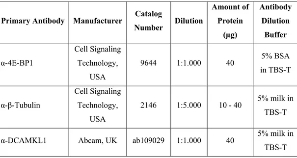
2.1.3. Western Blot reagents and antibodies ... 31
2.1.4. Quantitative Reverse Transcription Polymerase Chain Reaction (RT-qPCR) primers ... 34
2.1.5. Reagents for cortisol isolation and measurement of whole-body glucose ... 35
2.2. Methods ... 35
2.2.1 Animal Experiments ... 35
2.2.2. Protein Expression Experiments ... 42
2.2.4. Gene Expression Experiments ... 46
2.2.5. Body Cortisol Measurements ... 49
2.2.6. Whole-body Glucose Measurements ... 52
2.2.7. Statistical Analysis ... 52
Chapter 3 Results ... 55
3.1. Changes in the body weight and body mass index in response to different durations of treatment ... 55
xii
3.1.2. Intermittent fasting decreases the body mass index in old animals ... 57
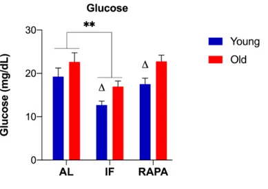
3.2. Changes in whole-body glucose and cortisol levels ... 58
3.2.1. Whole-body glucose levels decrease in animals subjected to intermittent fasting ... 58
3.2.2. Whole-body cortisol levels decrease in animals subjected to intermittent fasting ... 63
3.3. The suppression of mTOR activity through intermittent fasting and rapamycin treatment is age-dependent ... 65
3.3.1. Changes in the expression levels of total p-mTORSer2481, 4E-BP1, p-4E-BP1,
and p-4E-BP1/4E-BP1 in response to IF and RAPA ... 65
3.3.2. Changes in the expression levels of total S6, p-S6, and their ratio in response to IF and RAPA ... 68
3.4. The effects of intermittent fasting and rapamycin on the expression levels of key synaptic proteins, and markers of autophagy or proliferation ... 72
3.4.1. Changes in the expression levels of postsynaptic and presynaptic proteins involved in neurotransmission ... 72
3.4.2. Changes in the expression levels of autophagic flux markers ... 77
3.4.3. Changes in the markers of global proliferation, neurogenesis, and activated astrocytes ... 79
xiii
3.5. The expression levels of genes involved in the mTOR pathway, proliferation
and autophagy ... 83
3.5.1. Changes in the expression levels of genes in the nutrient signaling pathway . 84 3.5.2. Changes in the expression levels of rapamycin-responsive genes ... 87
3.5.3. Changes in the expression levels of autophagy-related genes ... 89
3.6. Principal component analysis for the proteins and genes of interest ... 91
3.6.1. PCA for the protein expression data ... 91
3.6.2. PCA for the gene expression data ... 97
Chapter 4 Discussion ... 101
Chapter 5 Conclusion and Future Directions ... 113
Bibliography ... 118
Curriculum Vitae and Publications ... 145
xiv
List of Figures
Figure 1.1. The World Health Organization’s 2050 projections of the percentage of
population over 60 years by country ... 1
Figure 1.2. Hallmarks of brain aging ... 10
Figure 2.1. Preparation of cortisol standard solutions through serial dilution ... 50
Figure 2.2. Workflow diagram for the statistical analysis. ... ..53
Figure 3.1. Changes in the mean body weight and BMI in response to IF and RAPA treatment throughout 8 weeks. ... 56
Figure 3.2. Changes in the whole-body glucose levels in response to IF and RAPA treatment. ... 60
Figure 3.3. Changes in the whole-body glucose levels in response to different durations of IF and RAPA treatment ... 61
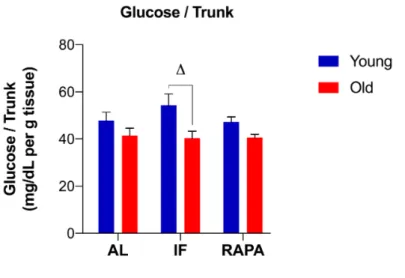
Figure 3.4. The levels of whole-body glucose normalized to trunk weight in response to IF and RAPA treatment ... 62
Figure 3.5. Changes in the whole-body cortisol levels in response to IF and RAPA treatment ... 64
Figure 3.6. Changes in the expression levels of p-mTORSer2481,4E-BP1, p-4E-BP1 and p-4E-BP1/4E-BP1 in response to IF and RAPA treatment in young and old animals. ... 66
Figure 3.7. Representative western blots for p-mTORSer2481, 4E-BP1, p-4E-BP1 and β-tubulin in old experiment groups at 4, 6 and 8 weeks.. ... 68
xv
Figure 3.8. Changes in the expression levels of S6, p-S6 and p-S6/S6 in response to IF and RAPA treatment ... 70 Figure 3.9. Changes in the p-S6/S6 ratio in response to different durations of IF and RAPA treatment. ... 71 Figure 3.10. Representative western blots for S6, p-S6 and β-tubulin in old
experiment groups at 4, 6 and 8 weeks ... 71 Figure 3.11. Changes in the expression levels of PSD-95, GEP and PSD-95/GEP, and SYP in response to IF and RAPA treatment ... 74 Figure 3.12. Changes in the expression levels of GEP in response to different
durations of IF and RAPA treatment ... 75 Figure 3.13. Representative western blots for PSD-95, GEP, β-tubulin and SYP in old experiment groups at 4, 6 and 8 weeks ... 76 Figure 3.14. Changes in the expression levels of LC3-I, LC3-II and LC3-II/LC3-I in response to IF and RAPA treatment ... 78 Figure 3.15. Representative western blots for LC3-I and LC3-II in old experiment groups at 4, 6 and 8 weeks ... 79 Figure 3.16. Changes in the expression levels of PCNA, DCAMKL1 and GFAP in response to IF and RAPA treatment ... 82 Figure 3.17. Representative western blots for DCAMKL1, GFAP, PCNA and β-tubulin in old experiment groups at 4, 6 and 8 weeks. ... 83 Figure 3.18. Log2 fold change values in the expression levels of ztor, akt1 and igf1
transcripts encoding proteins in the nutrient signaling pathway. ... 85 Figure 3.19. Log2 fold change values in the expression levels of ztor in response to
xvi
different durations of IF and RAPA ... 86 Figure 3.20. Log2 fold change values of rapamycin-responsive genes ddc and foxm1.
... 88 Figure 3.21. Log2 fold change values of autophagy-related genes atg5 and lc3b ... 90
Figure 3.22. Log2 fold change values of lc3b in response to different durations of IF
and RAPA ... 90 Figure 3.23. Visualization of the samples on scatterplot matrices for the principal components of the significantly affected proteins obtained from PCA ... 96 Figure 3.24. Visualization of the samples on scatterplots for the principal
components of the significantly affected genes obtained from PCA ... 99 Figure 3.25. Heatmaps for the gene expression data ... 100 Figure 4.1. Schematic representation of progenitor types identified by the
proliferative marker expression in the adult brain. ... 107 Figure A.1. Copyright permission for Figure 1.1. ... 151 Figure A.2. Copyright permission for reuse of figures published in Rejuvenation Research ... 152
xvii
List of Tables
Table 2.1. Western blot antibodies and their working conditions ... 32 Table 2.2. Sequences and melting temperatures (Tm) of forward and reverse primers
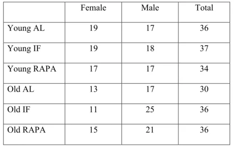
for RT-qPCR experiments ... 34 Table 2.3. Distribution of animals across experiment groups ... 37 Table 2.4. Feeding schedule for the experiment groups on odd weeks of the
experiment ... 38 Table 2.5. Feeding schedule for the experiment groups on even weeks of the
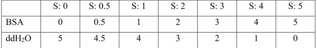
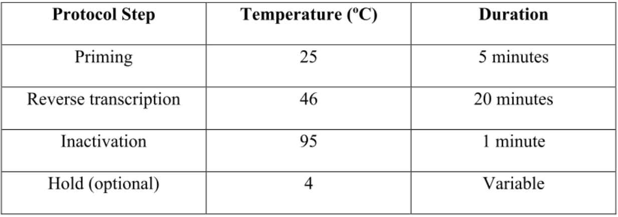
experiment ... 39 Table 2.6. BSA samples that were used as standards to measure protein
concentrations. ... 43 Table 2.7. Reaction protocol for cDNA synthesis ... 48 Table 3.1. Eigenvalues, explained variance and component loading scores for each principal component of protein expression data……….92 Table 3.2. Correlation coefficients and significance levels of the correlations for the protein expression data ... 93 Table 3.3. Letter coding for the proteins in Table 3.2 ... 94 Table 3.4. Eigenvalues, explained variance and component loading scores for each principal component of gene expression data ... 97 Table 3.5. Correlation coefficients and significance levels of the correlations for the gene expression data ... 98
xviii
Abbreviations
4E-BP1 ABP AD AI AKT1 AL eIF4E-binding protein 1 AMPA binding protein Alzheimer’s Disease Aged cognitively impaired AKT serine/threonine kinase 1ad libitum-fed AMP AMPA adenosine monophosphate alpha-amino-3-hydroxy-5-methyl-4-isoxazole propionic acid AMPAR AMPK ANOVA APS ATG5 AU BMI BrdU AMPA-type receptor
AMP-activated protein kinase Analysis of variance
Ammonium persulfate Autophagy-Related 5
Aged cognitively unimpaired Body mass index
Bromodeoxyuridine BSA
B0
Bovine serum albumin Zero standard
CA Cornu ammonis area
CaM kinase cDNA
Ca2+/calmodulin-dependent protein kinase
xix CI Cognitive impairment CNS CR Ct DCAMKL1 DDC ddH2O
Central nervous system Caloric restriction Cycle threshold Doublecortin-like kinase 1 Aromatic-L‐amino-acid-decarboxylase Double-distilled water DG DMSO Dentate gyrus Dimethyl sulfoxide DNA DPBS DR DTT EC E/I eIF4E Deoxynucleic acid
Dulbecco’s phosphate-buffered saline Dietary restriction
Dithiothreitol Entorhinal cortex Excitatory-inhibitory
Eukaryotic translation initiation factor 4E FDA FKBP12 FOXM1 GABA GEP GFAP GLUT GRIP IF
The Food and Drug Administration 12-kDa FK506-binding protein Forkhead Box M1
γ-aminobutyric acid Gephyrin
Glial fibrillary acidic protein Glucose transporter
Glutamate receptor-interacting protein Intermittent fasting
xx IGF-1 KMO MAP1LC3 mEPSC mIPSC MM
Insulin-like growth factor 1
Kaiser-Meyer-Olkin Measure of Sampling Adequacy
Microtubule-associated protein 1A/1B light chain 3
Miniature excitatory postsynaptic current Miniature inhibitory postsynaptic current Master mix mTOR mTORC1 mTORC2 NMDA NMDAR NO3 NPC NSB NSC O.D.
Mammalian target of rapamycin mTOR complex 1
mTOR complex 2
N-methyl-d-aspartate
NMDA-type receptor Nitrate
Neural progenitor cell Non-specific binding Neural stem cell Optical density PC PCA PCNA PFC PI3K pMM POI Principal component
Principal component analysis Proliferating cell nuclear antigen Prefrontal cortex
Phosphoinositide 3-kinase
Master mix containing primer pairs Protein of interest
xxi PSD PSD-95 PVDF RAPA Postsynaptic density
Postsynaptic density protein 95 Polyvinylidene fluoride Rapamycin-treated group RNA RT-qPCR S6 SDS SE SGL SVZ SYP TBS TBS-T TEMED Tm TOR WHO Ribonucleic acid
Quantitative reverse transcription polymerase chain reaction
Ribosomal protein S6 Sodium dodecyl sulfate Standard error
Subgranular layer Subventricular zone Synaptophysin Tris buffered saline TBS-Tween20
N,N,N',N'-tetramethylethane-1,2-diamine Melting temperature
Target of rapamycin
1
Chapter 1
Introduction
1.1. Aging
The populations are rapidly aging across the world. The accelerated rates of aging are due to reduction of childhood mortality in the low-income countries, and lower levels of mortality among older individuals in the high-income countries. This increase in average life expectancy is projected to lead to a doubling of the proportion of people aged 60 years or older by 2050 [1].
2
Figure 1.1. The World Health Organization’s 2050 projections of the percentage of population over 60 years by country. Reprinted from the World report on ageing and health, ISBN: 978 92 4 156504 2, WHO, Demographic and
epidemiological changes / Health in older age, Page 44, Copyright (2015), and with permission from WHO.
Living longer has been one of the greatest aspirations of humans for centuries, however, the life span increase is not without consequences. Aging has profound effects on health, societies, and global economy. The increased requirement for health care and inability to lead an independent lifestyle either due to age-related diseases or declines in certain abilities alter the society dynamics. Increased life expectancy without extending the healthy life span (i.e. health span) will lead to a larger proportion of people that is dependent on health care for a longer period of time. Furthermore, every individual ages differently, and the contribution of older people to the society is limited due to outdated stereotypes [2]. Therefore, there is an urgent need to develop strategies that will reduce the burden on healthcare providers and funding bodies and improve the quality of life in aging population.
The World Health Organization (WHO), launched an action plan on “Healthy
Ageing 2020-2030” in 2015 for preserving the physical and cognitive capacity in the
aged population, while providing a better and a safer environment for the elderly [2]. Good health and wellbeing through a healthier lifestyle (e.g. better nutrition, exercise) has been one of the steps that was listed in this multisectoral and international action plan. As a consequence, this necessitates a clear dissociation of healthy aging from pathological aging. Understanding the normal course of aging in
3
the absence of age-related diseases is therefore vital for managing the issues of older people. This approach may not only help us interpret the dysfunctional biological processes that lead to pathological aging, but also benefit from building an age-friendly environment that will facilitate the integration of older population in the society in a more effective manner.
1.2. Age-related functional and structural changes in the
brain
Advancing age is associated with many major changes in the body. These include but are not limited to: Gradual decline in cognitive function [3], changes in body composition and loss of muscle mass (i.e. sarcopenia) [4], deterioration of the collagen network and bone density [5], changes in cardiovascular structure and function [6], and immunosenescence [7]. While these changes have pathological implications such as cardiovascular and metabolic diseases, they are also observed during the course of normal aging. Understanding how advancing age affects this vast variety of processes will contribute to our search for finding the fine line between normal and pathological aging and facilitate development of palliative therapies.
Among these age-related changes, understanding how cognitive function deteriorates with age is particularly important since maintenance of cognitive skills is crucial for communication and independent lifestyle. Therefore, investigating the cellular and molecular correlates of the cognitive skills that are especially prone to age-related decline is very important. Identification of the mechanisms underlying critical
4
functional processes, and their alterations with respect to age will provide us with therapeutic targets.
1.2.1. Cognitive changes during normal aging
Cognitive impairment (CI) is among the most common conditions that are observed during aging. The prevalence of CI was reported as 37% in people >60 years in Iran [8], 19% in people >65 in Spain [9], 12.6% in people <60 years in the People’s Republic of China [10], and 25% in people aged >50 in India [11]. The level of CI ranges from mild to severe, and mild CI is often associated with normal aging. The manifestations of CI include deficits in short-term memory, difficulty in finding words, and decrease in processing speed [12]. A nascent form of dementia has been linked to CI, however, recent studies suggested that mild CI is not always proceeded by dementia [13]; and although mild CI might be regarded as a transition between normal aging and dementia, they are in fact different paradigms [12], [14].
Cognitive domains that measurably decline with age are attention, executive function, language, memory, and sensory perception [15]. Another cognitive skill that is affected by age is learning potential, also known as cognitive plasticity. Advancing age has been associated with lower performance in tasks related to verbal memory [16]. However, there are also domains and abilities that do not change with age. For example, general knowledge, vocabulary and sensory memory are stable with age [15]. Therefore, there is a need to dissociate these functional domains and understand the biological processes that stabilize them or cause their deterioration during aging.
5
1.2.2. Age-related structural changes in the brain
Aging is associated with numerous structural, cellular and molecular changes in the brain. While the debate over the ultimate consequences of these changes still continues, and it is not clear whether age-related deterioration in the brain is inevitable, the quest for finding remedies persists. One thing we have learned so far is that these changes are heterogeneous, and certain types of cells and brain areas are more prone to accumulation of damage than others [17]. The complexity of alterations, and their dramatically diverse effects on cognitive skills limit the potential to come up with a uniform approach. Nevertheless, targeting each contributor or a combination of them might prove useful in our fight against aging.
1.2.2.1. Neuron number
Neurons have been long considered as the primary target of age-related changes that underlie cognitive decline. Therefore, the effects of advancing age on the number of neurons, their structure (e.g. axons, dendrites, and even the cell body), and function have been extensively studied. The initial hypothesis was that aging causes significant neuron and synapse loss, which leads to the detrimental effects on cognitive abilities.
This conviction was in fact based on observations from as early as 1955, suggesting a significant amount of neuron death in aged humans [18]. These calculations relied on measuring neuron density in a given area. However, Coleman and Flood presented the limitations of this approach in a detailed review [19]. In this 1987 review, they listed a number of factors such as species, strains, as well as tissue
6
processing and counting methods that could potentially introduce a bias. This revelation was owing to the advances in technology and development of more sophisticated counting methods [20] involving morphometric procedures [21]. Later, stereological methods were introduced for more accurate and unbiased estimation of the neuronal changes during aging [22].
The studies using these techniques revealed species-, region- and cell-type-specific neuron loss during aging. In humans, a significant decrease in neuron number was observed in the subiculum (52%) and hilus (31%) of the dentate gyrus, respectively [23]. While the Cornu Ammonis area 1 (CA1) of the hippocampus showed a substantial neuron loss in the patients with Alzheimer’s Disease (AD), it was not affected by normal aging [24]. H. Haug reported that the number of human cerebral cortex neurons does not change or experience only a small decrease during aging. The only exception was extrapyramidal human cortex (area 6), which is implicated in motor activity and showed signs of aging earlier than other areas [25].
More recent studies supported the notion that there is no strong evidence for significant decline in the neuron number in the human entorhinal cortex and subregions of rat hippocampus during aging that could translate into a biologically meaningful effect on cognitive skills [26], [27]. Entorhinal cortex (EC) is a part of the hippocampal memory system, connecting the hippocampus and neocortex, and EC has been shown to be severely affected in the brains of AD patients, but not in normal aging groups [26]. Neuron loss was also absent in other areas related to learning and memory such as neocortex, superior prefrontal cortex and temporal cortex in normally aged humans and primates [28].
7
1.2.2.2. Dendrites
Structural changes in the brain during aging are not limited to the neuron number. Dendrites, the extensions of neurons that are assigned with the crucial task of receiving signals from neighboring cells and propagating them to the soma, are also prone to age-related changes. Dendrites are highly dynamic, particularly during development, and retain their plastic nature into adulthood. Dendritic branches, density, and patterns contribute to the neuron function since dendrites constitute up to 95% of the receptive surface, and distinct morphologies were observed in different types of neurons [19], [29].
Several studies reported increase in dendritic proliferation during normal aging in human and macaque monkey [30], [31]. The extent of dendritic branches were larger in nondemented old humans (average age, 79) compared to younger adults (average age, 51), and people with senile dementia (average age, 76) [30]. In the prefrontal cortex neurons of macaques, the number of branches and dendritic length increased significantly from young to middle age, followed by a highly significant decrease towards old age [31]. Since the maintenance of dendritic structure is critical for the survival of neurons, the increasing pattern has been suggested as a compensatory mechanism for the age-related decrease in neuron number that protect the integrity of the functional compartments in the brain [30].
On the contrary, later studies revealed opposite patterns in different regions. For instance, dendrites of the pyramidal neurons in the human motor cortex regressed with age [32]. In Sprague-Dawley rats, loss of dendrites was observed in auditory
8
cortex [33], further supporting that these patterns were both species/strain- and region-specific.
1.2.2.3. Synapses
Albeit the initial view on significant neuron and dendrite loss in the whole brain has been replaced with a refined hypothesis of asynchronous region-specific changes during aging, scientists continued to observe gray and white matter loss during normal human aging [34]–[36]. These findings drove the attention to structural changes in another important component of the neurons involved in connectivity: synapses. Some studies reported a decrease in synapse density with age in the neocortex of humans with AD [37] as well as the number of presynaptic terminals in the frontal cortex of people without dementia [38]. In rat dentate gyrus (DG), a subregion in the hippocampal formation that has been linked to learning and memory, a significant decrease in synapse number and density has been shown [39]. However, adding to the controversy, later studies with unbiased counting methods using electron microscopy instead of light microscopy concluded that the synapse number per neuron was not affected by age [40].
Taken together, the lack of major changes during normal aging in humans, nonhuman primates and rodents suggests that learning and memory deficits in old individuals of these species are likely to arise from more subtle changes rather than large scale structural rearrangements. Growing evidence indicates that cellular and molecular mechanisms might contribute to functional deteriorations in the aging
9
brain more than initially anticipated. Some of these mechanisms will be covered with regard to aging in section 1.3.
1.3. Age-related cellular and molecular changes in the brain
Recent advances in aging research provided evidence that the course of aging is controlled, at least in part, by evolutionarily conserved cellular, genetic and molecular processes. These include the changes in synaptic elements involved in neurotransmission, which are restricted to the nervous system, as well as global mechanisms that affect peripheral and nervous tissues. In their 2013 review, López-Otín et al. defined these changes as “the hallmarks of aging” and grouped them into three categories: Primary, antagonistic, and integrative hallmarks [41].
The primary hallmarks are regarded as the main contributors to aging phenotype. These are genomic instability, telomere attrition, epigenetic alterations, and loss of homeostasis. The antagonistic hallmarks are thought to initially protect against cellular damage; however, their chronic activation eventually becomes detrimental. Deregulated nutrient sensing, mitochondrial dysfunction, and cellular senescence fall into this category. Finally, integrative hallmarks are the phenotypical consequences of the first two categories. These are stem cell exhaustion and altered intercellular communication, which are the manifestations of age-related loss of function.
Most of these hallmarks are evident in the aging brain. However, since brain has a unique energy metabolism, its dysregulation is likely to be the major factor in determining brain health status. Taking this into account, Mattson et al. defined ten
10
hallmarks of brain aging as nine established hallmarks interacting with dysregulated energy metabolism in their 2018 review (Figure 1.2) [42]. Some of these hallmarks that are relevant to the scope of this thesis will be reviewed here.
Figure 1.2. Hallmarks of brain aging. The illustration is adapted from Mattson et
al.’s 2018 review [42], and created with BioRender.
1.3.1. Changes in synaptic integrity and composition
Synaptic transmission is a key factor in neuronal circuitry and function. Deficiencies in both the presynaptic and postsynaptic elements have severe consequences that impair the synaptic functioning. Therefore, understanding the molecular mechanisms that take place in both elements, and finding how these mechanisms change during
11
normal aging could provide us with appropriate targets to prevent or reverse age-related cognitive decline.
The intercellular communication at the synapses, by the simplest definition, are carried out by the synaptic vesicles carrying neurotransmitters at the presynaptic site, and their receptors at the postsynaptic site. Many processes occur at both sites to transmit, receive, and process the signal, and hundreds of proteins are required to work in harmony to ensure the efficiency of communication. Some of the key proteins, their functions, and how they change in response to advancing age will be reviewed in this section.
1.3.1.1. Presynaptic elements
One of the most critical components of the presynaptic site are the membranous organelles called synaptic vesicles. These vesicles have an essential role in transforming the electrical stimulus that is propagated in the cell body into chemical signals by storing the neurotransmitters that are produced in the presynaptic cell. When the action potential arrives at the nerve terminal, voltage-gated Ca2+ channels
allow Ca2+ influx. The change in membrane potential triggers the fusion of vesicles
with the plasma membrane, and neurotransmitters are released into the synaptic cleft through exocytosis. Empty vesicles are retrieved by endocytosis, recycled and refilled with neurotransmitters to be utilized in another round of exocytosis [43], [44].
12
The release of neurotransmitters from synaptic vesicles is mediated by different proteins that are either embedded in the lipid bilayer of vesicular membrane (e.g. synaptophysin, synaptotagmin and proteins with SNARE motifs such as synaptobrevin) or proteins associated with the cytoplasmic surface of vesicles (e.g. synapsins I-II and Ca2+/calmodulin-dependent protein kinase (CaM kinase) II) [43].
In this complex pool of proteins with distinct functions and binding partners, it is important to differentiate the constant factors from the ones with a rather supporting role. Studies approaching this process from a “vesicocentric” point of view revealed components that are present in majority, if not all, of the vesicles [44].
The SNARE proteins, Rab5, and synaptophysin are among the obligatory components [44]. Of these, synaptophysin is a major integral protein spanning the vesicle membrane and is involved in vesicle fusion and neurotransmitter release. As a substrate for protein kinases including CaM kinase II, its phosphorylation status is implicated in vesicle docking and recycling [43]. Furthermore, when in a complex with synaptobrevin, synaptophysin has been shown to provide a pool of available vesicles during development and synapse maturation [45]. Therefore, it is often regarded as a marker of synaptic vesicles and integrity.
Growing evidence link changes in synaptophysin expression to age-related cognitive decline [46]. For instance, cognitively impaired aged (AI) rats had reduced synaptophysin immunoreactivity in the CA3 region of hippocampus, relative to young or cognitively unimpaired aged (AU) animals [47]. Synaptophysin protein levels were significantly lower in people with dementia compared to nondemented controls, although there was no correlation between age and synaptophysin
13
expression [48]. Furthermore, in both humans with AD [49] and rodent models of AD [50], synaptophysin levels were reduced in hippocampus. The age-related decline in synaptophysin expression was also evident during normal aging in different subregions of rat hippocampus [51].
1.3.1.2. Postsynaptic elements
Spines are extensions of the neurons that may protrude from the soma, dendrites, or the axon hillock, and are specialized structures for synaptic transmission [52]. Receptors for the neurotransmitters are mostly localized to a cytoskeleton-bound matrix of receptor-associated proteins found in spines [53]. These matrices contain proteins that facilitate clustering of receptors and anchoring them to the plasma membrane, as well as signaling molecules that process and transduce the input to downstream cascades. The contents of a matrix therefore dictate its function.
Postsynaptic density (PSD), for instance, is predominantly a marker of excitatory synapses. As one of the most complex structures found in spines, PSD is estimated to harbor hundreds of receptor, adaptor, and signaling proteins [54]. The two types of ionotropic receptors for the major excitatory neurotransmitter in the central nervous system (CNS), glutamate, are N-methyl-d-aspartate (NMDA) and alpha-amino-3-hydroxy-5-methyl-4-isoxazole propionic acid (AMPA). Although both the NMDA and AMPA type receptors (NMDAR and AMPAR, respectively) have been observed within PSD, NMDARs are consistently found while AMPAR content varies highly, and some excitatory synapses completely lack AMPARs [55].
14
Both the NMDARs and AMPARs are composed of subunits with distinct ligand sensitivity profiles, kinetic properties and interaction partners [56], [57]. Assembly patterns of these subunits define the key features and functions of NMDARs and AMPARs. Not surprisingly, the expression levels of both NMDAR and AMPAR subunits have been shown to be affected by age. Age-related declines were observed in the expression levels of NR1, NR2A and NR2B subunits of NMDAR and the GluR1 and GluR2 subunits of AMPAR in the CA3 region of rat hippocampus [58], and of NR1 and GluR2 in the neocortex of macaques [59].
On the opposite side of the glutamatergic excitatory systems is the inhibitory neurotransmission. The primary inhibitory neurotransmitter in the mammalian CNS is γ-aminobutyric acid (GABA). There are three classes of GABA receptors: GABAA, GABAB and GABAC. The majority of fast inhibitory transmission is
mediated by GABAA, whose action is in a dynamic balance with fast excitatory
transmission [60].
GABAA channels form a pentameric structure that is assembled by different
subunits, and contains an intrinsic Cl- channel [60]. The types of subunits in a
GABAA receptor not only determine the receptor function and drug responses in
pharmacological studies, but also the cellular localization of the receptor. Although the GABAA receptors are predominantly postsynaptic, they have been also observed
in presynaptic and extrasynaptic locations as a consequence of their subunit composition [61].
15
There are contradicting reports on the age-related changes in GABAergic neurotransmission. Both GABA and GABAA receptor levels were reported to be
unchanged in the brains of aging rats [62]. Studies measuring receptor binding suggested different patterns for the binding affinity of GABA receptors: binding decreased in the brains of aging rats in one study [63], and was not affected in another [64]. In humans, GABA receptor number was increased while the GABAergic terminal density was decreased during aging [65]. These inconsistent observations compelled the researchers to try a different approach for investigating age-related changes in neurotransmitter dynamics. Since both the excitatory AMPA and NMDA, and inhibitory GABA receptors require clustering proteins for cellular localization, stability, and functional regulation, studies inclined towards measuring their levels [66]–[68].
The NMDA-type ionotropic glutamate receptors interact with the PDZ domain of postsynaptic density protein 95 (PSD-95) via the C-terminal of NR2 subunit. As mentioned earlier, PSD harbors many proteins, however PSD-95 has been shown to be the main component of these postsynaptic excitatory scaffolds, and the localization of NMDA receptors in postsynaptic site requires intact PSD-95 [69]. On the other hand, AMPA-type receptors, although colocalize with NMDA-type receptors in PSD, bind to other proteins containing PDZ domain. These include glutamate receptor-interacting protein (GRIP) and AMPA receptor-binding protein (ABP) [66].
16
The clustering of GABA receptors at postsynaptic sites are mainly mediated through gephyrin. Initially, gephyrin was thought to be in an interaction exclusively with the receptors for another inhibitory neurotransmitter: glycine [68]. More recent studies involving pharmacological agents or mutants lacking either gephyrin or GABAA
receptor subunits suggested that gephyrin is essential for the stabilization of GABAA
[70].
Advancing age has been associated with changes in postsynaptic clustering proteins. In humans, PSD-95 protein level was found to be positively correlated with age [48]. In the pathological cases of cognitive impairment in rats [71] or AD in humans [72], the expression of PSD-95 was reduced, yet other studies showed either increase or no change in PSD-95 protein levels [73], [74]. Although these opposing observations could be due to regional differences, contradictory data exists even for the same brain regions.
The reports on gephyrin are also conflicting, since there were species- and region-specific patterns during aging. For instance, gephyrin levels were stable across life span in zebrafish [75]. On the contrary, gephyrin clusters became more prominent in mature synapses as compared to young synapses during synaptogenesis in rat cerebellar cortex [76]. Furthermore, age-related increases in gephyrin protein levels and message were observed in dorsal cochlear nucleus, suggesting a mechanism for hearing loss [77], and in AI rats [71]. In addition to changes observed in normal aging, gephyrin have been shown to accumulate in the brains of humans with AD [78], although another study suggested the complete opposite pattern of a significant global decrease in postmortem brains [79].
17
1.3.1.3. Excitatory-inhibitory balance
Recent studies suggested that the balance between excitatory and inhibitory neurotransmission is as equally imperative as the expression levels of neurotransmitter receptors and clustering proteins in cognitive functioning. Maintenance of the excitatory-inhibitory (E/I) balance is an integral part of neuronal plasticity, learning, memory and behavior. The data obtained via electrophysiological and biochemical methods showed that E/I imbalance is associated with age-related cognitive decline [80].
In aged humans, increased prefrontal cortex (PFC) activity has been shown to correlate with memory impairments. This increase was attributed to lower E/I ratio due to higher inhibitory tone in old cohort as compared to young cohort, which had a stable E/I ratio [80]. In AI rats, electrophysiological recordings of miniature excitatory and inhibitory postsynaptic currents (mEPSCs and mIPSCs, respectively) showed a decrease in mEPSCs [81] or enhanced mIPSCs [82], suggesting an imbalance in E/I due to a shift towards inhibition. Furthermore, studies investigating the ratio of excitatory and inhibitory clustering proteins reported a significant decrease in PSD-95/gephyrin density in AI rats compared to young cohort, but not AU animals [83].
On the contrary, a declining trend in intrinsic inhibitory tone during aging has been observed in non-human primates and rats [77, 81]. This observation was also supported by the 30-60% reduction in synapse density in prefrontal layer I, whose major content is GABAergic neurons [80]. Other studies that reported a reduced
18
inhibition in aged animals indicate region- and species-specific changes, as well as the impact of individual differences in cognitive skills [85].
Nevertheless, E/I imbalance appears to be the common denominator regardless of the direction. Considering the part that E/I takes while responding to plastic changes driven by intrinsic and environmental factors, the imbalance pertains to cognitive decline. Therefore, the measures that are taken for slowing down or reversing the brain aging must take E/I balance into account.
1.3.2. A primary hallmark of aging: Loss of homeostasis
The hallmarks of aging as described by López-Otín et al. were principally based on the widely accepted idea that aging is accompanied, if not caused, by the accumulation of damage over time [86]. A chief mechanism that has been linked to age-related diseases through damage control is the protein homeostasis, also known as proteostasis. The proteome quality and functionality is ensured by the intrinsic control mechanisms that stabilize newly synthesized proteins, detect unfolded proteins, and target them for degradation through proteolytic pathways [87].
There are two main mechanisms for regulating the proteome quality: autophagy-lysosome and ubiquitin-proteasome pathways. Moreover, both have been shown to exhibit decreased activity in aged models [88], [89]. While mutant models with proteasome or autophagy inhibition showed phenotypes that resemble age-related pathologies, the life span extending interventions or genetic manipulations have been shown to initiate autophagic and proteolytic cascades [88].
19
Of these interventions, which will be covered in section 1.4., rapamycin treatment promotes longevity particularly through induction of autophagy [90]. Moreover, life span extension via rapamycin strictly requires autophagy induction in
Caenorhabditis elegans and Drosophila melanogaster, yet the mechanisms in
mammals are more complex and involve additional pathways such as inhibition of protein synthesis [88]. Autophagy is therefore a protective, rather than a destructive mechanism, and aberrant autophagy has been observed in accelerated aging models [91].
Removal of damaged/dysfunctional molecules is particularly important for neurons since they are post-mitotic, and generation of new neurons is limited and restricted to a few areas in the adult brain. Age-related impairments in waste management through autophagic and proteasomal mechanisms are evident in the brain. Several studies reported accumulation of autophagosomes and polyubiquitinated proteins, mitochondrial dysfunction in neurons [42]. Furthermore, aggregation of damaged molecules is established as an early step in the events that lead to neuronal death [92]. Thus, defective homeostatic mechanisms have been implicated in both normal and pathological aging.
1.3.3. Deregulated nutrient sensing in aging
Extracellular and intracellular nutrient levels are ever-fluctuating in response to environmental ques. The ability of the cells to sense these changes and initiate appropriate downstream cascades is a key step in survival. Elaborate mechanisms have thus evolved to detect levels of macronutrients such as amino acids, glucose and other sugars, and lipids. These could be summarized as anabolic reactions that
20
allow storage during nutrient abundance, and catabolic mechanisms that utilize internal reserves through autophagy or proteasome-mediated degradation when nutrient levels are scarce [93].
The brain has a considerably higher metabolism level and energy requirement than other organs, making it more sensitive to fluctuations in nutrient levels [94]. Different mechanisms are integrated through hormonal signals to fulfill the brain’s energy needs, especially under conditions of nutrient limitation. For instance, starvation leads to degradation of amino acids to synthesize glucose and ketone bodies, which are brain’s fuels [93].
Glucose sensing involves glucose transporters (GLUTs) at the plasma membrane and insulin/insulin-like growth factor 1 (IGF-1), adenosine monophosphate (AMP)-activated protein kinase (AMPK), and phosphoinositide 3-kinase (PI3K)-AKT pathways both at the membrane and intracellular domains [41], [93]. One of the significant proteins at the intersection of amino acid and glucose sensing, as well as autophagy is the target of rapamycin (TOR).
1.3.3.1. TOR signaling
TOR, as the name indicates, is the target of rapamycin: a macrolide compound. First isolated from a microorganism called Streptomyces hygroscopicus, which has been found in soil samples from Easter Island (Rapa Nui), rapamycin was initially identified as an antifungal compound [95]. Subsequent studies revealed that rapamycin is also an immunosuppressant and a potent inhibitor of cell growth, which
21
attracted attention as a valuable tool to investigate proliferation. These studies led to the identification of its molecular target (i.e. TOR) in yeast [96]. Shortly after, TOR homolog has been identified in mammals [97], and named mammalian target of rapamycin (also known as mechanistic target of rapamycin, mTOR). Interestingly, it is now known that mTOR is an evolutionarily conserved serine-threonine kinase belonging to the PI3K family [98], having homologs in Caenorhabditis elegans,
Drosophila melanogaster, and in zebrafish (Danio rerio) [99]–[101]. In addition to
its critical role in transitioning from anabolic to catabolic programs, TOR is involved in numerous cellular mechanisms including transcription, translation, proliferation, growth and survival [102].
mTOR is known to interact with many proteins in two multiprotein complexes, the mTOR complex 1 (mTORC1) and mTOR complex 2 (mTORC2) [103]. Both the amino acid and glucose sensing activities are suggested to be conveyed through mTORC1. For instance, mTORC1 promotes anabolic processes such as protein and lipid synthesis during nutrient abundance [104]. Insulin-IGF-1 pathway activation triggers the upstream effectors of mTORC1: PI3K-AKT.
Acute rapamycin treatment inhibits mTORC1 by interacting with the 12-kDa FK506-binding protein (FKBP12), a component in mTORC1, and disrupting the assembly of the protein complex. Although mTORC2 has been regarded as rapamycin-resistant, growing evidence suggest that its function is impaired by prolonged exposure to rapamycin [105]. The notion that deregulated nutrient sensing is a hallmark of aging originates from life and health span-extending effects of
22
mTOR inhibition through both genetic manipulations, and non-genetic interventions such as rapamycin and dietary restriction (DR).
With regards to brain aging, mTOR signaling has been implicated in regulation of synaptic protein synthesis through integrating signals relayed by neurotransmitter receptors at the postsynaptic sites [106]. Moreover, postsynaptic scaffolding proteins PSD-95 and gephyrin have been shown to be coupled with mTOR pathway, with gephyrin being an actual binding partner of mTOR [107], [108]. Noting the necessity for a balanced energy metabolism during healthy brain aging, mTOR is undeniably a key factor connecting nutrient signaling and autophagy to brain-specific programs such as synaptic protein synthesis and neurotransmission [109].
1.3.4. Impaired neurogenesis and cell senescence
While brain was once thought to not have a regenerative capacity, the evidence for neural stem cells (NSCs) in the adult brain suggest otherwise. Adult NSCs have the capacity for self-renewal and proliferation, and through the latter mechanism they give rise to neural progenitor cells (NPCs). Neuronally-committed NPCs undergo several cycles of cell division before they differentiate into neurons and glia. Although the neurogenic zones are restricted to a few areas in the adult brain such as the subventricular zone (SVZ) and subgranular layer (SGL) of DG, the potential they harbor for plasticity is promising [110].
The generation of new neurons in SVZ and SGL declines with age, however reports on cell proliferation rate suggest species-specific differences [111]. Whether the reductions in neurogenesis contribute to cognitive impairment is also not clear.
23
Hippocampus is one of the regions that is affected by age-related molecular changes which may lead to functional decline, yet it contains a well-defined neurogenic region. Some studies even suggested an inverse correlation between the maintenance of hippocampal neurogenesis and preservation of cognitive skills in rats [112], indicating a need for further investigations.
Still, understanding the mechanisms that cause age-related decline in the neurogenesis is necessary to assess to what extent the newly generated neurons are incorporated into functional circuits in the aging brain. Several mechanisms have been proposed as the basis for impaired neurogenesis. These are reduced mitochondrial metabolism, oxidative stress, accumulation of deoxyribonucleic acid (DNA) damage and impaired repair mechanisms, as well as inflammation [42]. In relation to these changes, increased number of senescent neural progenitor cells has been observed during aging. Interestingly, some NPCs might be destined to become senescent since isolated human NPCs have been observed to undergo senescence in vitro [113].
1.4. Anti-aging interventions
As we continue to uncover the changes that occur during aging, some mechanisms have emerged as potential therapeutic targets to reverse or slow down age-related functional deterioration. Several genetic, dietary, and pharmacological interventions have been shown to extend life and health span across species. Since the main concern with these approaches is the translational potential, and the genetic
24
manipulations that yielded long-lived mutants is not applicable in humans, many studies focused on dietary and pharmacological remedies.
1.4.1. Dietary interventions
The longevity-promoting effects of dietary restriction (DR), or caloric restriction (CR) have been reported as early as 1935 [114]. DR has been shown to reliably extend life span in a broad range of organisms, from yeast to primates, thus suggesting evolutionarily conserved responses [115]. These include improved glucose regulation, increased resistance to stressors, suppressed inflammation, activation of repair programs, and attenuation of cellular damage [116], [117]. At the cellular level, the master regulator of these responses is the nutrient sensing pathway which is seemingly centered around mTOR signaling.
The traditional DR approach follows a moderate reduction of daily calorie intake without causing malnutrition. When the ad libitum food intake of rodents was reduced by 30-60%, both the average and maximal life span increased. The onset of age-related diseases was delayed in calorically-restricted animals, and their prevalence decreased [117].
Later studies introduced intermittent fasting (IF) as an equally effective approach as reducing the daily calorie intake for extending life span. This is achieved through mTOR inhibition, and subsequent induction of autophagy which leads to removal of damaged molecules and overall cellular homeostasis. As a mechanism regulated by mTOR, IF reduces global protein synthesis [116]. One of the earliest reports of the IF-induced longevity showed 83% increase in life span of rats that have been
25
maintained on alternate-day feeding, an IF regimen [118]. Furthermore, IF promotes healthy aging by enhancing multiple cognitive domains such as learning and memory, and reversing effects of age-related diseases [119].
1.4.2. Pharmacological interventions
Experimental evidence from long- and short-lived mutants acknowledged the role of mTOR on life span. Lifespan extension by introducing mutations in mTOR gene was achieved in different organisms such as budding yeast (Saccharomyces cerevisiae), worm (Caenorhabditis elegans), fruit fly (Drosophila melanogaster), and mammals [109]. These data provided grounds that mTOR inhibition might slow down aging.
Rapamycin, which has been approved for use in humans by the U.S. Food and Drug Administration (FDA) [120] is a potent, well-characterized inhibitor of mTOR. It is now established that rapamycin extends life span, delay age-related impairments, and improve parameters of health [109]. As an autophagy-inducing agent, rapamycin has been suggested to prevent cognitive impairment by counteracting the accumulation of misfolded proteins in mouse models of AD. Furthermore, rapamycin seems to augment vascular function and hinder neuroinflammation in the CNS.
A major drawback of utilizing rapamycin for anti-aging therapies is its side effects. Although several rapamycin homologs (i.e. rapalogs) with milder side effects have been developed, there is still a need for finding the optimum dose and duration. This is particularly true for observing the beneficial effects in the brain since studies focusing on the brain are limited.
26
1.5. Zebrafish as a gerontological model
Various model organisms from different phylogenetic levels are used for aging research. Each organism, from yeast to worms, flies to rodents and nonhuman primates provide valuable information regarding the very complex process of aging. Yet until recently, a large gap between invertebrate and vertebrate models existed. This gap was filled with zebrafish (Danio rerio), a freshwater fish native to India [121].
Zebrafish first attracted attention as a model to study developmental biology due to its transparent embryos which develop ex utero. Their high fecundity and low-cost husbandry allow generation of large populations fairly quickly, easily and inexpensively. While the life span of zebrafish (3-4 years) is about 50% longer than mouse and this could be considered as a disadvantage for gerontology, it is considerably less than nonhuman primates. Moreover, the life span of zebrafish is a contributing factor to its suitability as a bridge model for aging research [121].
Zebrafish are diurnal vertebrates, and as humans, age gradually [122], [123]. Circadian clock is now recognized as an integral part of the molecular processes regulating aging. Similar to the observations from mammals, zebrafish aging involved selective alterations in circadian clock, reduced nighttime sleep, progressive decline in melatonin, and expression levels of circadian genes [122]. As part of the gradual aging, age-related changes such as accumulation of DNA damage, oxidative damage, stress-related markers, and senescence have been reported in zebrafish [121], [123], [124]. The zebrafish genome has been fully
27
sequenced, showing 70% homology with human genome, which granted further means to investigate molecular and genetic processes underlying age-related changes [125].
In the context of brain aging, zebrafish have recently emerged as a convenient model to study brain function. Zebrafish CNS is morphologically similar to mammalian CNS at both the macro and cellular levels. Major neurotransmitters, receptors, and proteins involved in cellular metabolism are similar in structure and function that have been observed in the mammals. Finally, zebrafish behavior spans multiple domains including anxiety, mood/depression, cognitive and social skills [125]. In spite of these advantages, zebrafish have not been employed fully in the aging research. Considering the ease of testing drugs and compounds, zebrafish offers a unique model for developing therapeutics [126].
1.6. Aims of this thesis
Despite the growing evidence for the benefits of IF, and DR in general, there are controversial reports on the magnitude of effects. The responses to dietary interventions are influenced by feeding paradigms, diet composition, duration, genetic background, gender, and age of the subjects at the onset of intervention [116]. Therefore, there is need for understanding how these factors determine the response to IF.
In terms of translation to humans, several impediments to the applicability of IF exist. Cultural norms have been dictating a diet of three meals per day as a way of
28
healthy living for so long that it is a challenge to change the common view. Another drawback is the initial side effects of IF such as irritability, feeling cold, hunger, and fatigue. However, these negative side effects have been reported in less than 15% of the participants, usually disappeared within a month, and followed by overall improvements in mood and physiological parameters [116], [127]. Nonetheless, researchers have been pursuing to identify compounds, either natural or synthetic, that could mimic the effects of IF to eliminate the aforementioned obstacles associated with a diet-based remedy.
As the most conventional DR mimetic, rapamycin has been used extensively in aging research. While the life and health span extending effects of rapamycin have been shown in a variety of organisms, its reported side effects limit the applications in humans. Similar to evidence from DR studies, the effects of rapamycin differ between tissues, ages, and genders [128]. Despite their inhibitory action on mTOR, to what degree rapamycin mimics DR is still not well-understood. Furthermore, most studies on longevity-promoting interventions are done with lifelong DR or drug treatment, which is not feasible in humans and there is need for understanding their short-term effects. The growing body of data on general health parameters and cognitive performance is not accompanied by brain-specific effects, which needs to be addressed separately due to unique brain metabolism and blood brain barrier. Finally, females are still underrepresented since most studies utilize male organisms only.
In this study, we aimed to apply different short-term durations of IF and rapamycin in wild type male and female zebrafish to investigate: (i) How young and old
29
animals respond to these interventions to understand whether the age at the onset of intervention affects the outcome (ii) the minimum short-term duration to observe significant effects, (iii) the efficacy of rapamycin as a CR mimetic, (iv) the mechanisms of action for IF and rapamycin irrespective of the gender of the subjects, which is essential for comprehension of the global aging patterns.
![Figure 1.2. Hallmarks of brain aging. The illustration is adapted from Mattson et al.’s 2018 review [42], and created with BioRender](https://thumb-eu.123doks.com/thumbv2/9libnet/5552619.108218/31.892.223.732.265.677/figure-hallmarks-illustration-adapted-mattson-review-created-biorender.webp)