ehçet Syndrome (BS) has been defined as a multisystemic disease af-fecting the skin, joints, peripheral vessels and nervous system that can also affect all other tissues, first defined by Hulusi Behçet, MD, a Turkish dermatologist. The current criteria for BS diagnosis were deter-mined by the principles that were defined and proposed by the “Interna-tional Study Group for Behçet Disease” in 1990. According to these criteria, BS diagnosis can be made in the presence of oral ulceration and two of the following; genital ulceration, typical eye lesions (uveitis), typical skin
le-Turkiye Klinikleri J Gynecol Obst 2015;25(3)
213
A Rare Presentation of Behçet Ulcer on the
Vaginal Cuff: Case Report
AABBSS TTRRAACCTT Behçet Syndrome (BS) is a rare chronic, inflammatory, recurrent and multisystemic dis-ease that is characterised by vasculitis throughout the body. BS is an autoimmune disdis-ease that af-fects the neurological and gastrointestinal systems that is characterized by reoccurring oral and genital aphthosis, arthritis, eye and cutaneous lesions. In BS, genital lesions have been reported at rates of 60-90% in different series. Lesions are observed most commonly on the vulva labium ma-jora, followed by the vagina and uterine cervix in descending order. However, no previous study has reported a lesion on the vaginal cuff of a patient who has undergone total hysterectomy. In this study, for the first time, we defined a rare vaginal cuff ulcer in a patient previously diagnosed with BS. This case study will be the first in literature and contribute to it by revealing the appearance of a genital ulcer on the vaginal cuff caused by BS. Moreover, we think that it should be considered in the differential diagnosis of vaginal lesions.
KKeeyy WWoorrddss:: Behçet syndrome; ulcer; vagina Ö
ÖZZEETT Behçet sendromu (BS) nadir görülen, vaskülit ile karakterize kronik, inflamatuar, tekrarla-yan, multisistemik bir hastalıktır. BS, tekrarlayan oral ve genital aftlar, artrit, göz lezyonları, kuta-nöz lezyonlar, gastrointestinal sistem ve merkezi sinir sistemi tutulumuyla karakterize otoimmün bir hastalıktır. Behçet sendromunda genital tutulum farklı serilerde %60-90 olarak rapor edilmiştir. Genital bölgede en sık tutulum vulvada labium majuslarda görülmektedir. Daha sonra azalan sıklıkta vajen ve servikste rapor edilmiştir. Fakat şu ana kadar histerektomize bir hastada vajinal cuffta her-hangi bir tutulum bildirilmemiştir. Biz bu makalede daha önceden BS olduğu bilinen hastada ilk defa görülen nadir bir vajinal cuff ülseri tanımladık. Bu olgu BS’ye bağlı genital ülserin vaginal cuff da görülmesi bakımından literatürde ilk olup bu anlamda literatüre katkı sağlayacağını ve vajinal lez-yonların ayırıcı tanısında göz önüne alınması gerektiğini düşünüyoruz.
AAnnaahh ttaarr KKee llii mmee lleerr:: Behçet sendromu; ülser; vajina TTuurrkkiiyyee KKlliinniikklleerrii JJ GGyynneeccooll OObbsstt 22001155;;2255((33))::221133--66
Betül YORGUNLAR,a Suna ÖZDEMİR,a
Lebriz Hale AKTÜN TAMER,a Mustafa ACETa
aDepartment of Gynaecology
and Obstetrics,
Medipol University Faculty of Medicine, İstanbul
Ge liş Ta ri hi/Re ce i ved: 24.05.2014 Ka bul Ta ri hi/Ac cep ted: 17.11.2014
This case report was presented as a poster in X. Turkish German Gynecology Congress, 30 April-04 May 2013, Antalya, Turkey.
Ya zış ma Ad re si/Cor res pon den ce: Betül YORGUNLAR
Medipol University Faculty of Medicine, Department of Gynaecology and Obstetrics, İstanbul, TÜRKİYE/TURKEY [email protected]
doi: 10.5336/gynobstet.2014-40567 Cop yright © 2015 by Tür ki ye Kli nik le ri
sions and a positive pathergy skin test.1,2Although BS is seen on all over the world, it is more common in countries along the historical silkroad including Turkey, the Mediterranean, Middle Asia, Asia and Japan.3-6
Genital ulcers, like oral ulcers, commonly ap-pear on the skin and mucosa of the genital tract. In women, these lesions commonly develop on the vulva and less frequently on the vagina or cervix.5 However, at present, no Behçet’s ulcer has been re-ported on the vaginal cuff.
CASE HISTORY
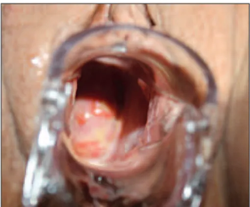
A 47 year old female patient applied to our clinic with the complaints of vaginal discharge and groin pain for the past one week. In her medical history, she had one normal vaginal delivery and total ab-dominal hysterectomy operation for myoma uteri 7 years ago. Moreover, the patient had been taking colchicine since diagnosed with BS 25 years ago. In addition, when oral and genital sores developed, she applied topical corticosteroid cream for treat-ment. The patient had not been suffering from gen-ital ulcers for 3 years. Furthermore, the patient had had an attack of uveitis 8 years ago and made a full recovery with medical treatment. Additionally, the patient was being followed by cardiovascular surgeons for thromboflebitis affecting the lower extremities. In the patients gynaecological exam-ination, the perineum and vulva were normal; with speculum examination, there were two ulcers of 1 cm and 0.5 cm in size observed on the vaginal cuff and smear was taken (Figure 1). The uterine cavity could not be observed by ultrasonography (USG) due to the hysterectomy and the ovaries were bilaterally atrophic. As topical treatment, be-tamethasone valerate cream was prescribed. The smear results were normal. The patient was called for a control examination 3 weeks after the first ad-mission. In her control examination, there was no genital ulcer or scar observed (Figure 2).
DISCUSSION
Mucosal ulcerations are among the main findings of BS and are characterized by recurrent oral
ul-cerations.7 Other criteria include genital ulcers, erythema nodosum, papulopustular rashes, eye le-sions and pathergy skin test positivity.4,5 Recent studies with large series have determined an equal incidence of BS in both males and females.6The same study reported that symptoms including ery-thema nodosum and genital aphts were more com-monly seen in females.6
Genital ulcers are the second most common finding in BS, and like the oral ulcers, they are usu-ally located on the mucosa and skin of the genital tract. The most common location of genital ulcers is the labia majora of the vulva; however they may be
Betül YORGUNLAR et al. A RARE PRESENTATION OF BEHÇET ULCER ON THE VAGINAL CUFF: CASE REPORT
Turkiye Klinikleri J Gynecol Obst 2015;25(3)
214
FIGURE 1: Two ulcers of 1 cm and 0,5 cm on the vaginal cuff.
located less often on the vagina or cervix.5In BS, genital lesions have been shown to occur at a rate of 60-90%.8The ulcer of the present case was detected on vaginal cuff. To our knowledge, there are no data in the literature about the Behçet’s ulcer of vaginal cuff. In a previous case report, vaginal ulceration was described in a patient with ulcerative colitis after abdominal hysterectomy.9Histopathology was found as nonspecific chronic granulamatous in-flammation on biopsy at the first time, the patient was diagnosed as pyoderma gangrenosum because recovery was determined after local and systemic steroid treatment. However, BS was diagnosed in the patient after recurrent vaginal ulceration.
In addition to pyoderma gangrenosum, granu-loma, cellulitis of the vaginal vault, BS should be considered in the differential diagnosis of the vagi-nal ulcerations. About half of the pyoderma gan-grenosum are associated with underlying systemic conditions, such as inflammatory bowel disease, arthritis, and haematological malignancies.10About 2% of patients with inflammatory bowel disease will develop pyoderma.11In vaginal cuff cellulitis, the vaginal surgical margin is erythematous, ede-matous, and hyperemic early following surgery, and there are purulent vaginal secretion.12
In another one of our studies, the incidence of genital lesions (scar + ulcer) in BS was found to be 74.3%, with the labia majora being the most com-monly affected region at 55.1%, followed by labia minora, pubic area and perineal area. The duration for ulcer recovery was 2.6±0.9 weeks.13
The genital lesions are usually circular or oval and look like they have been punctured with a sta-ple. In contrast to oral ulcers, genital lesions re-cover with scar formation that is specific to BS.3 The recovery time for these lesions has been re-ported as being 10-30 days in various studies.8,14 The size and depth of the lesion and the presence of bacterial infection are important in scar for-mation.15On the other hand, a recent study sug-gested that ulcers of the labia minora recover without scarring like oral lesions.16This sugges-tion could be due to the difficulties in differing mucosal scar tissue on the labia minora with the naked eye from scar-free normal mucosa.
Pub-lished data regarding vaginal or cervical lesion cover is insufficient. In our case, we noted that re-covery was without scarring like in oral lesions. This may be due to the structural characteristics of the vaginal mucosa.
Genital lesions are usually painful however they may rarely be asymptomatic and asymptomatic females have no symptoms.16Active ulcerated le-sions and scars may cause gynaecological complica-tions in female patients with BS. The present case admitted to our clinic with vaginal discharge. For instance, deep ulcers located in the vagina may re-sult in the formation of fistulas to the bladder and urethra.17In addition, genital lesions may cause uri-nary and walking difficulties.18Moreover, large gen-ital ulcers may recover with scar tissue that may cause dyspareunia and affect the quality of life. Al-though there are a large number of studies regarding oral and systemic symptoms in BS, studies relating to genital ulcers, the formation of scar tissue and other gynaecological symptoms are limited in literature.
Recurrent painful genital ulcers may decrease the quality of life of the patients. Topical anaes-thetics, dapsone, sucralfate, granulocyte colony-stimulating factor, cyclosporine, topical and intralesional corticosteroids have been used either alone or in combination, having varying degrees of supporting evidence in the treatment of genital ul-cers in BS.19-23For the topical treatment of genital ulcer and cutaneous lesions, corticosteroid and an-tiseptic creams can be applied for a short period of time (7 days). Topical sucralfate reduces the heal-ing duration and pain of genital ulcer.20Colchicine can be used as a secondline alternative. A recent study has shown that colchicine reduces the oc-currence of genital ulcer.23 Similary, we applied local steroid treatment to our case, and then no scar or ulcer formation occured.
In conclusion, the variety of lesions may be observed in the vaginal and vulvar regions in fe-males. However, the approach to these lesions dif-fers depending on the etiological agent. BS should be considered in the differential diagnosis of lesions located in the genital region of patients even though they may be from an area where the inci-dence of BS is low.
Betül YORGUNLAR et al. A RARE PRESENTATION OF BEHÇET ULCER ON THE VAGINAL CUFF: CASE REPORT
Turkiye Klinikleri J Gynecol Obst 2015;25(3)
Betül YORGUNLAR et al. A RARE PRESENTATION OF BEHÇET ULCER ON THE VAGINAL CUFF: CASE REPORT
Turkiye Klinikleri J Gynecol Obst 2015;25(3)
216
1. Criteria for diagnosis of Behçet’s disease. In-ternational Study Group for Behçet’s Disease. Lancet 1990;335(8697):1078-80.
2. O’Neill TW, Rigby AS, Silman AJ, Barnes C. Validation of the International Study Group cri-teria for Behcet’s disease. Br J Rheumatol 1994;33(2):115-7.
3. Evereklioglu C. Current concepts in the etiol-ogy and treatment of Behçet disease. Surv Ophthalmol 2005;50(4):297-350.
4. Kaklamani VG, Vaiopoulos G, Kaklamanis PG. Behçet’s disease. Semin Arthritis Rheum 1998;27(4):197-217.
5. Pickering M, Haskard DO. Behçet’s syn-drome. J R Col Physicians Lond 2000;34(2):169-77.
6. Tursen U, Gurler A, Boyvat A. Evaluation of clinical findings according to sex in 2313 Turk-ish patients with Behçet’s disease. Int J Der-matol 2003;42(5):346-51.
7. Evaluation of diagnostic (‘classification’) crite-ria in Behcet’s disease--towards internation-ally agreed criteria. The International Study Group for Behcet’s disease. Br J Rheumatol 1992;31(5):299-308.
8. Zouboulis CC. Epidemiology of Adamanti-ades-Behçet’s disease. Ann Med Interne (Paris) 1999;150(6):488-98.
9. Lilford RJ, Tindall VR, Batchelor AG.
Post-sur-gical pyoderma gangrenosum of the vaginal vault associated with ulcerative colitis and Be-hcet’s disease; a case report. Eur J Obstet Gy-necol Reprod Biol 1989;31(1):93-4. 10. 10-Bennett ML, Jackson JM, Jorizzo JL,
Fleis-cher AB Jr, White WL, Callen JP. Pyoderma gangrenosum. A comparison of typical and atypical forms with an emphasis on time to re-mission. Case review of 86 patients from 2 in-stitutions. Medicine (Baltimore) 2000;79(1): 37-46.
11. Callen JP. Pyoderma gangrenosum. Lancet 1998;351(9102):581-5.
12. Hemsell DL. Infection after hysterectomy. In-fect Dis Obstet Gynecol 1997;5(1):52-6. 13. Ozdemir S, Ozdemir M, Celik C, Balevi A, Toy
H, Kamış U. Evaluation of patients with Be-hçet’s disease by cervical cytology and colpo-scopic examination. Arch Gynecol Obstet 2012;285(5):1363-8.
14. Zouboulis CC. Genitoanal lesions in Adaman-tiades--Behçet’s disease. J Eur Acad Derma-tol Venereol 1997;9(Suppl 1):106-7. 15. Singer AJ, McClain SA. Persistent wound
fection delays epidermal maturation and in-creases scarring in thermal burns. Wound Repair Regen 2002;10(6):372-7.
16. Ghate JV, Jorizzo JL. Behçet’s disease and complex aphthosis. J Am Acad Dermatol 1999;40(1):1-18.
17. Al-Otaibi LM, Porter SR, Poate TW. Behçet’s disease: a review. J Dent Res 2005;84(3):209-22.
18. Kontogiannis V, Powell RJ. Behçet’s disease. Postgrad Med J 2000;76(900):629-37. 19. Lin P, Liang G. Behçet disease:
recommen-dation for clinical management of mucocuta-neous lesions. J Clin Rheumatol 2006;12(6): 282-6.
20. Alpsoy E, Er H, Durusoy C, Yilmaz E. The use of sucralfate suspension in the treatment of oral and genital ulceration of Behçet disease: a randomized, placebo-controlled, double-blind study. Arch Dermatol 1999;135(5):529-32.
21. Bacanli A, Yerebakan Dicle O, Parmaksizoglu B, Yilmaz E, Alpsoy E. Topical granulocyte colony-stimulated factor for the treatment of oral and genital ulcers of patients with Be-hçet’s disease. J Eur Acad Dermatol Venereol 2006;20(8):931-5.
22. Avci O, Gürler N, Güneş AT. Efficacy of cy-closporine on mucocutaneous manifestations of Behçet’s disease. J Am Acad Dermatol 1997;36(5 Pt 1):796-7.
23. Yurdakul S, Mat C, Tüzün Y, Ozyazgan Y, Hamuryudan V, Uysal O, et al. A double-blind trial of colchicine in Behçet’s syndrome. Arthri-tis Rheum 2001;44(11):2686-92.