Can double fluoroscopy in the oblique position reduce
surgical time and radiation exposure during
intertrochanteric femur fracture nailing?
Haluk Çelik, M.D.,1 Adnan Kara, M.D.,2 Yavuz Sağlam, M.D.,3 İsmail Türkmen, M.D.,1 Serkan Aykut, M.D.,4 Mehmet Erdil, M.D.2
1Department of Orthopaedics and Traumatology, Ümraniye Training and Research Hospital, İstanbul-Turkey 2Department of Orthopaedics and Traumatology, İstanbul Medipol University Faculty of Medicine, İstanbul-Turkey 3Department of Orthopaedics and Traumatology, Biruni University Faculty of Medicine, İstanbul-Turkey
4Department of Orthopaedics and Traumatology, Metin Sabancı Baltalimanı Bone Diseaeses Training and Research Hospital, İstanbul-Turkey
ABSTRACT
BACKGROUND: The purpose of this study was to determine the comparative effectiveness of the use of single fluoroscopy versus double fluoroscopy during intramedullary nailing in the oblique position for intertrochanteric femur fractures in terms of surgery and radiation time.
METHODS: Fifty-two patients (20 men, 32 women; average age: 78.2 years; range: 69-88 years) were included in the study. While double fluoroscopy was used for 25 patients, single fluoroscopy was used for the remaining 27 patients. Data of the preparation time between anesthesia and surgery, surgery time, radiation time, bleeding volume, postoperative collodiaphyseal angle between the frac-tures and intact parts, and the tip-apex distance (TAD) were compared.
RESULTS: The surgery time in the double and single fluoroscopy groups averaged 34.48±8.92 minutes and 50.37±16.63 minutes, respectively (p<0.01). The radiation time was 42.72±16.00 seconds for the double-fluoroscopy group and 68.22±21.53 seconds for the single-fluoroscopy group (p<0.01). The surgical preparation time, bleeding volume, collodiaphyseal angle and TAD distance did not vary significantly between groups (p>0.05).
CONCLUSION: The use of double fluoroscopy in the oblique position in the surgical treatment of intertrochanteric femur fractures reduced the surgical time and the anesthesia time for patients, as well as the exposure to radiation, thereby also reducing the risk of complications.
Keywords: Intertrochanteric femur fracture; proximal femur intramedullary nailing; radiation time; single versus double fluoroscopy.
fluoroscopy during IM nailing may result in high rates of ra-diation exposure regardless of patient position. In addition, during the surgery, the position of the fluoroscope has to be changed to provide anteroposterior (AP) and lateral images and this necessity can lead to unnecessary views. It might be possible to decrease the time of exposure to radiation with newly developed fluoroscopic techniques.
INTRODUCTION
It is estimated that around 6.3 million hip fractures will oc-cur worldwide by 2050.[1] As a result of the already growing
number of intertrochanteric femur fractures, intramedullary (IM) nailing is becoming an increasingly popular treatment technique among orthopedic surgeons.[2] IM nailing is
com-monly performed using a traction table or, in the supine or lateral decubitus positions, a radiolucent table.[3] The use of
Cite this article as: Çelik H, Kara A, Sağlam Y, Türkmen İ, Aykut S, Erdil M. Can double fluoroscopy in the oblique position reduce surgical time and radiation exposure during intertrochanteric femur fracture nailing? Ulus Travma Acil Cerrahi Derg 2018;24:581-586.
Address for correspondence: Haluk Çelik, M.D.
Ümraniye Eğitim ve Araştırma Hastanesi, Ortopedi ve Travmatoloji Kliniği, Ümraniye, 34766 İstanbul, Turkey Tel: +90 216 - 632 18 18 E-mail: [email protected]
Ulus Travma Acil Cerrahi Derg 2018;24(6):581-586 DOI: 10.5505/tjtes.2018.04048 Submitted: 14.11.2017 Accepted: 16.04.2018 Online: 12.09.2018 Copyright 2018 Turkish Association of Trauma and Emergency Surgery
In this study, IM nailing for an intertrochanteric fracture was performed in the oblique position, a modified lateral decubi-tus position. The objective was to examine the differences between single and double fluoroscopy in IM nailing per-formed in the oblique position in terms of the preparation time between anesthesia and surgery, surgery time, radi-ation time, bleeding volume and postoperative radiological results. The research hypothesis was that the use of double fluoroscopy in the oblique position would reduce the surgery time and radiation time.
MATERIALS AND METHODS
Patient Population-Treatment Groups
The sample group comprised patients who underwent IM nailing for intertrochanteric femur fractures at Medipol Mega Hospital between March 2013 and March 2015. This was a retrospective study that included patients whose fracture was type A1 or A2 according to the AO/OTA Fracture and Dislocation Classification, whose injury had been diagnosed as a non-pathological isolated intertrochanteric femur frac-ture, and those who underwent nailing with closed reduction in the oblique position. Of 84 patients who underwent IM nailing for an intertrochanteric femur fracture, the follow-ing patients were excluded from the study: 4 patients with a pathological fracture, 3 patients with multi-trauma, 2 pa-tients who underwent open reduction, 11 papa-tients with type A3 fractures according to the AO/OTA classification, and 12 patients who underwent operations in positions other than the oblique position (Table 1). In all, 52 patients (20 men, 32 women) were included in the study. The average age was 78.2 years (range: 69–88 years). The causes of the fracture included falling from a height in 2 cases, a traffic accident in 5, and a simple fall for 45 patients. The mean time interval between the trauma and the operation was 3.5 days (range: 1–7 days). Double fluoroscopy was used for 25 patients, and single fluoroscopy was used for 27 patients.
Written informed consent was obtained from each patient be-fore enrollment. All of the study procedures involving human participants were performed in accordance with the ethical standards of the institutional research committee and with the 1964 Helsinki Declaration and its later amendments or comparable ethical standards. Approval for the study was ob-tained from the local ethical committee.
Technique
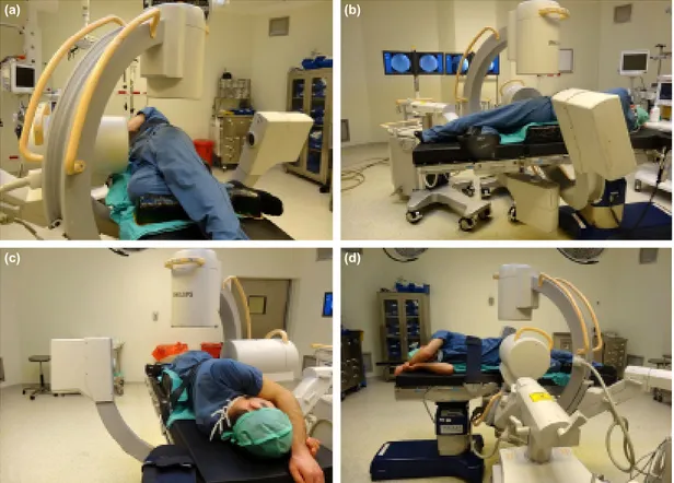
All of the surgeries were performed using a closed technique under spinal anesthesia. An anti-rotational screw antero-grade proximal femur intramedullary nail (Intertan; Smith & Nephew plc, London, UK) was used. After the induction of anesthesia, all of the patients were placed in the oblique po-sition on the radiolucent table at 70° to the floor in order to prevent two-hip superposition (Fig. 1). In order to maintain the position, silicon supports were placed behind the hips and under the feet.
Philips BV Endura fluoroscopes (Koninklijke Philips N.V., Am-sterdam, Netherlands) were used for all patients. The flu-oroscopes were positioned before sterile draping, and AP and lateral images of the proximal femur were taken. When double fluoroscopy was used, the fluoroscope used to take AP images was placed such that the X-ray tube was behind the hips and the image intensifier was in front of the patient. A 20° tilt was applied in the caudal direction in order to ease the entrance of the fluoroscope used to record the lateral im-ages. The fluoroscope used to take lateral images was placed at a 90° angle to the floor in the caudocranial direction such that the image intensifier was above. Once suitable images were obtained, the positions of fluoroscopes were marked on the floor with adhesive bandages. After sterile draping, the fluoroscopes were also draped and returned to the marked areas. Reduction of the fracture was provided by an assistant using manual longitudinal traction and internal rotation. Once a longitudinal skin incision was made proximal to the greater trochanter, a guide pin was inserted into the trochanteric re-gion under fluoroscopic guidance. An entry reamer was then inserted and reamed to the level of the lesser trochanter. The appropriate nail size was selected for each patient accord-ing to the preoperative plannaccord-ing. Once the nail was inserted, the guide pin was placed into the neck-head. Under manual traction, the quality of the reduction and the position of the guide pin were evaluated with AP and lateral images. After obtaining the appropriate position, a lag screw and interlock-ing screw were placed. A sinterlock-ingle distal lockinterlock-ing screw was then inserted through the guide of the nail (Figs. 2 and 3). It was determined that it was not necessary to change the position of the fluoroscope during surgery.
In all of the operations performed with single fluoroscopy, draping was performed after checking the preoperative AP and lateral proximal femur images. In the various operative phases, the fluoroscope’s position was changed by a single
ra-Table 1. Patient distribution according to AO fracture
classification
OTA/AO fracture Patients with hip fractures classification
Double fluoroscopy Single fluoroscopy (n=25) (n=27) A1-2 A1-3 3 2 4 2 A2-1 5 6 A2-2 11 13 A2-3 4 2
diology technician with 7 years of experience. All of the surg-eries were performed by 2 very experienced, senior trauma surgeons (AK and ME).
Variables
The surgical preparation time between anesthesia induction and the incision, the surgical time between the incision and
Figure 3. (a) Preoperative radiograph of a right intertrochanteric femur fracture; (b, c) Postoperative radiographs of
the patient after treatment with proximal femoral intramedullary nailing.
(a) (b) (c)
Figure 2. (a-d) The position of the fluoroscopes and anteroposterior-lateral images taken during surgery. (a)
(b)
(c) (d)
Figure 1. (a-d) To prevent the superposition of both hips, the patient is positioned on the operating table in a 70°
oblique position. (a)
(c)
(b)
closure, bleeding volume, and the total radiation time dur-ing surgery were recorded for both groups. Collodiaphyseal angles were evaluated in comparison with the intact hip on a postoperative graph. The apex distance between the implant end and the femoral head and the position of the screw within the head was then compared.
Statistical Analysis
NCSS 2007 and PASS 2008 statistical software programs (NCSS LLC, Kaysville, UT, USA) were used for the statistical analyses. While evaluating the study data, in addition to the definitive statistical methods (average, SD, median, frequency, and ratio), the conformity of the variables with normal distri-bution was evaluated using the Kolmogorov-Smirnov test. The Student’s t-test was used for inter-group comparisons of the variables with normal distribution. The Mann-Whitney U test was used to compare variables that did not have normal dis-tribution. The results were evaluated within a 95% confidence interval and at a significance level of p<0.05.
RESULTS
No complication was noted in either group. There were no significant differences between the 2 groups in terms of sur-gical preparation time (p>005). The sursur-gical time was 15.89 minutes shorter (p<0.01) and the radiation time was 25.5 seconds shorter (p<0.01) in the double-fluoroscopy group compared with the single-fluoroscopy group. Bleeding vol-ume, collodiaphyseal angle and type, and apex distance did not vary significantly between the groups (p>0.05) (Table 2).
DISCUSSION
As closed surgery has become more and more popular, flu-oroscopy has become the third eye of orthopedic surgeons. However, due to the ionizing radiation emitted during the fluoroscopy, it may have harmful effects on the surgical team and the patient.[4,5] If the time of fluoroscopy exceeds 1.7
min-utes during an operation, radiation exposure increases
signif-icantly.[6] A study of radiation-exposed and unexposed
per-sonnel found that the tumor risk in orthopedic surgeons had increased significantly after 24 years.[7] Another clinical study
demonstrated that the incidence of breast cancer among fe-male orthopedic surgeons is higher than that of the general population.[8] The radiation exposure represents a relatively
small increased risk of future cancer development in the pa-tients who undergo fluoroscopy-dependent surgeries.[9] In
light of this information, the principle of as low as reasonably achievable (ALARA) was developed in order to decrease ra-diation exposure.[10] Methods to decrease radiation exposure
were discussed and collected under the title of Distance, Exposure, Barriers and Techniques/Technologies (DEBT).[11]
The purpose of this study was to develop a new technique to decrease radiation exposure. Since type-A3 fractures may be accompanied by several complications during surgery, this type of fracture has not been included in the present study in order to be able to evaluate the effectiveness of double-fluoroscopy used with an oblique position. In addition, the experience of the surgeon is one of the important factors affecting radiation exposure, and senior surgeons can signifi-cantly reduce the radiation dose.[12] Thus, all of the surgeries
were performed by 2 experienced, senior trauma surgeons. In order to obtain 2-directional images during surgical at-tempts, the position of the C-arm fluoroscopy must be ad-justed. Extra and often unnecessary images can be taken in order to obtain the appropriate image and, as a result, the radiation exposure time increases. This can be prevented by using 2 fluoroscopes in fixed positions under appropriate conditions. There are a limited number of studies in the lit-erature pertaining to the use of double-fluoroscopy during orthopedic procedures. A study that researched the effec-tiveness of single- and double-fluoroscopy in intramedullary nailing for intertrochanteric fractures determined that the ra-diation exposure time decreased by 29% in the group under-going double fluoroscopy.[13] The study results indicated that
the surgical time decreased in statistically significant terms in the double fluoroscopy group. In that study, the surgeries
Table 2. Comparison of fluoroscopy groups
Double fluoroscopy (n=25) Single fluoroscopy (n=27) p
Surgical preparation time in the OR (min); mean±SD 19.36±7.35 17.26±6.83 a0.291 Surgical time (min); mean±SD 34.48±8.92 50.37±16.63 a0.001* Radiation time (sec); mean±SD (median) 42.72±16.00 (38) 68.22±21.53 (65) b0.001* Bleeding volume (mL); mean±SD 210.61±49.88 230.62±62.55 a0.212 Collo-diaphyseal angle (degrees); mean±SD 128.63±6.62 124.22±3.86 a0.069
TAD (mm); mean±SD 20.81±2.22 24.41±3.55 a0.301
TAD <25 mm; n (%) 21 (80.0) 22 (81.5) c1.000
TAD >25 mm; n (%) 4 (20.0) 5 (18.5) c1.000
were performed on traction tables, and the bleeding volume, collodiaphyseal angles and TAD distances were not evalu-ated. According to a similar study on traction tables, in the double fluoroscopy group, the duration of surgery was 15.9 min. shorter, and the radiation time was 25.7 seconds shorter than in the single fluoroscopy group.[14] To the best of our
knowledge, there are no other studies in the literature that evaluate the effectiveness of double fluoroscopy in IM nail-ing for proximal femur fractures; however, a study in which an evaluation of the effectiveness of single and double fluo-roscopy during slipped capital femoral percutaneous screw-ing determined that the radiation time in the double fluo-roscopy group decreased by 34%.[15] A similar study reported
that use of double fluoroscopy did not significantly extend the surgical preparation time and increased the accuracy of the screw position.[16] We found that double fluoroscopy
de-creased the radiation exposure time by 37.4% in the oblique position. This suggests that unnecessary images taken during single fluoroscopy in order to obtain the appropriate images occurred much more frequently than previously suspected. The use of double fluoroscopy appears to minimize the num-ber of unnecessary images and significantly decrease radiation exposure.
Shorter orthopedic surgical times provide many benefits. It has been reported that the infection risk is higher in ex-tended surgeries.[17,18] The techniques used to decrease
surgi-cal time are particularly important for major trauma centers, and adoption of these techniques can also decrease patients’ total hospitalization time as well as costs.[19] We observed
in our study that the time of operations using double roscopy were shorter by 31.5% compared with single fluo-roscopy. The preoperative preparation phase was longer in the double fluoroscopy group than in the single fluoroscopy group. However, the difference was not significant. There were also no significant differences between the 2 groups in the evaluation of the bleeding amount.
In the treatment of intertrochanteric femur fractures, re-duction is essential for surgical stability. When we compared the operated collodiaphyseal angles with those of intact hips on postoperative graphs and evaluated the TAD and screw positions, we did not detect any significant differences be-tween the 2 groups. We can conclude that use of double fluoroscopy did not have any positive effects on reduction and screw position.
There are certain limitations to this study. First, the number of patients was limited. Second, more powerful data could have been obtained by measuring the amount of the dose with a dosimeter (badge) as well as radiation time. However, the present study is the first study in the literature to evalu-ate the benefits of the double fluoroscopy technique in the oblique position. Further studies are necessary to evaluate larger patient samples and complicated fracture types.
Conclusion
The use of double fluoroscope while a patient is in the oblique position during surgical treatment of intertrochanteric femur fractures may decrease the potential complications of long surgical and anesthesia times for the patient and it may help prevent the side effects of radiation by lessening radiation exposure.
Conflict of interest: None declared.
REFERENCES
1. Kannus P, Parkkari J, Sievanen H, Heinonen A, Vuori I, Järvinen M. Epi-demiology of hip fractures. Bone 1996;18:57S–63S. [CrossRef ]
2. Anglen JO, Weinstein JN; American Board of Orthopaedic Surgery Research Committee. Nail or plate fixation of intertrochanteric hip frac-tures: changing pattern of practice. A review of the American Board of Orthopaedic Surgery Database. J Bone Joint Surg Am 2008;90:700–7. 3. Bishop JA, Rodriguez EK. Closed intramedullary nailing of the femur in
the lateral decubitus position. J Trauma 2010;68:231–5. [CrossRef ]
4. Blakely EA. Biological effects of cosmic radiation: deterministic and sto-chastic. Health Phys 2000;79:495–506. [CrossRef ]
5. Dewey P, George S, Gray A. Ionising radiation and orthopaedics. Curr Orthop 2005;19:1–12. [CrossRef ]
6. Sanders R, Koval KJ, DiPasquale T, Schmelling G, Stenzler S, Ross E. Exposure of the orthopaedic surgeon to radiation. J Bone Joint Surg Am 1993;75:326–30. [CrossRef ]
7. Mastrangelo G, Fedeli U, Fadda E, Giovanazzi A, Scoizzato L, Saia B. Increased cancer risk among surgeons in an orthopaedic hospital. Occup Med (Lond) 2005;55:498–500. [CrossRef ]
8. Chou LB, Chandran S, Harris AH, Tung J, Butler LM. Increased breast cancer prevalence among female orthopedic surgeons. J Womens Health (Larchmt) 2012;21: 683–9. [CrossRef ]
9. Beebe MJ, Jenkins P, Rothberg DL, Kubiak EN, Higgins TF. Prospective Assessment of the Oncogenic Risk to Patients From Fluoroscopy During Trauma Surgery. J Orthop Trauma 2016;30:e223–9. [CrossRef ]
10. Giordano BD, Grauer JN, Miller CP, Morgan TL, Rechtine GR 2nd. Radiation exposure issues in orthopaedics. J Bone Joint Surg Am 2011;93:e69. [CrossRef ]
11. Agarwal A. Radiation Risk in Orthopedic Surgery: Ways to Protect Yourself and the Patient. Operative Techniques in Sports Medicine 2011;19:220–3. [CrossRef ]
12. Botchu R, Ravikumar K. Radiation exposure from fluoroscopy during fixation of hip fracture and fracture of ankle: Effect of surgical experience. Indian J Orthop 2008;42:471–3. [CrossRef ]
13. Brin YS, Palmanovich E, Aliev E, Laver L, Yaacobi E, Nyska M, et al. Closed reduction and internal fixation for intertrochanteric femoral frac-tures is safer and more efficient using two fluoroscopes simultaneously. Injury 2014;45:1071–5. [CrossRef ]
14. Kara A, Celik H, Seker A, Uzun M, Sonmez MM, Erdil M. Procedural outcomes of double vs. single fluoroscopy for fixing intertrochanteric fe-mur fractures. Arch Orthop Trauma Surg 2016;136:929–34. [CrossRef ]
15. Klug R, McCarthy JJ, Eilert RE. The use of a two C-arm technique in the treatment of slipped capital femoral epiphysis. Orthopedics 2004;27:1041–2.
16. Westberry DE, Davids JR, Cross A, Tanner SL,Blackhurst DW. Simul-taneous biplanar fluoroscopy for the surgical treatment of slipped capital femoral epiphysis. J Pediatr Orthop 2008;28:43–8. [CrossRef ]
OLGU SUNUMU
İntertrokanterik femur kırıklarında oblik pozisyonda uygulanan femur intramedüller
çivilemesi sırasında çift skopi kullanımı cerrahi süre ve skopi süresini azaltabilir mi?
Dr. Haluk Çelik,1 Dr. Adnan Kara,2 Dr. Yavuz Sağlam,3 Dr. İsmail Türkmen,1 Dr. Serkan Aykut,4 Dr. Mehmet Erdil2 1Ümraniye Eğitim ve Araştırma Hastanesi, Ortopedi ve Travmatoloji Kliniği, İstanbul2İstanbul Medipol Üniversitesi Tıp Fakültesi, Ortopedi ve Travmatoloji Anabilim Dalı, İstanbul 3Biruni Üniversitesi Tıp Fakültesi, Ortopedi ve Travmatoloji Anabilim Dalı, İstanbul
4Metin Sabancı Baltalimanı Kemik Hastalıkları Eğitim ve Araştırma Hastanesi, Ortopedi ve Travmatoloji Kliniği, İstanbul
AMAÇ: Bu çalışmada intertrokanterik femur kırıklarında, oblik pozisyonda uygulanan intramedüller çivileme esnasında çift skopi kullanımının tek skopi kullanımına üstün olup olmadığı araştırılmıştır.
GEREÇ VE YÖNTEM: İntertrokanterik femur kırığı sonrası oblik pozisyon kullanılarak intramedüller çivileme yapılan 52 hasta çalışmaya dahil edildi. Hastaların 20’si erkek, 32’si kadın ve ortalama yaş: 78.2 (dağılım: 69–88) idi. Kırıklar AO/OTA sınıflamasına göre tip A1 ve tip A2 kırıklardan oluş-makta idi. Yirmi beş hastada çift skopi kullanılırken, 27 hastada tek skopi kullanıldı. Anestezi ile cerrahi arasındaki hazırlık süresi, cerrahi süresi, skopi kullanım süresi, kanama miktarı, cerrahi sonrası kollodiafiziyal açı ve tip-apeks mesafesi iki grup arasında karşılaştırıldı.
BULGULAR: Cerrahi süresi çift ve tek skopili gruplarda sırası ile 34.48±8.92 dakika ve 50.37±16.63 dakika idi (p<0.01). Skopi kullanım süresi çift skopili grupta 42.72±16.00 iken, tek skopili grupta 68.22±21.53 idi (p<0.01). Cerrahi hazırlık süreleri, kanama miktarı, kollodiafiziyel açı ve tip-apeks mesafeleri karşılaştırmalarında iki grup arasında istatistiksel fark saptanmadı (p>0.05).
TARTIŞMA: İntertrokanterik femur kırıkları cerrahi tedavisinde oblik pozisyonda çift skopi kullanılarak yapılan intramedüller çivilemede tek skopi kullanımına göre daha az radyasyon maruziyeti ile daha kısa sürelerde cerrahiyi tamamlamak mümkündür.
Anahtar sözcükler: Çift skopi kullanımı; intertrokanterik femur kırığı; proksimal femur intramedüller çivileme; radyasyon zamanı.
Ulus Travma Acil Cerrahi Derg 2018;24(6):581-586 doi: 10.5505/tjtes.2018.04048
ORİJİNAL ÇALIŞMA - ÖZET
17. Edwards C, Counsell A, Boulton C, Moran CG. Early infection after hip fracture surgery: risk factors, costs and outcome. J Bone Joint Surg Br 2008;90:770–7. [CrossRef ]
18. Lee J, Singletary R, Schmader K, Anderson DJ, Bolognesi M, Kaye KS.
Surgical site infection in the elderly following orthopaedic surgery. Risk factors and outcomes. J Bone Joint Surg Am 2006;88:1705–12. 19. Lawrence TM, White CT, Wenn R, Moran CG. The current hospital