Herz 2015 · 40:549–551 DOI 10.1007/s00059-013-4023-8 Received: 3 October 2013 Accepted: 2 November 2013 Published online: 18 December 2013 © Urban & Vogel 2013
I. Rencuzogullari1 · I.T. Ozcan2 · A. Cirit2 · S. Ayhan2
1 Division of Cardiology, Mugla Sitki Kocman University Education and Research Hospital, Mugla 2 Division of Cardiology, Department of Medicine, Mersin University School of Medicine, Mersin
Isolated interrupted
aortic arch in adulthood
A case report
Case report
A 5-year-old man presented to our hos-pital with the complaints of dyspnea and chest pain during exercise over months. He had a history of hypertension for sev-eral years, which had been kept under control with nifedipine plus valsartan. In addition, he had a hemorrhagic stroke 2 years earlier but he had no physical lim-itations. However, he had not undergone a complete evaluation at that time. His brother died at 41 years of age of myocar-dial infarction.
On physical examination, peripheral pulses were palpable over the carotid ar-teries and in the upper limbs, with a ra-dial pulse stronger in the right arm than in the left arm. Bounding pulses in the neck were also detected. The blood pres-sure was 135/70 mmHg in the right arm
and 95/0 mmHg in the left arm. Low-er limb pulses wLow-ere not palpable. ThLow-ere was 2/ systolic ejection murmur on the second intercostal area at the left. A chest x-ray demonstrated cardiomegaly and in-creased vascularity. Transthoracic echo-cardiography from parasternal and api-cal views showed concentric hypertrophy and moderate aortic regurgitation, but a satisfactory image could not be obtained from the suprasternal view because of poor image quality.
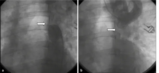
During cardiac catheterization, the guidewire did not pass beyond the prox-imal part of the descending aorta. Aor-tography via the femoral artery showed a complete interruption of the aortic arch just distal to the origin of the left subcla-vian artery (. Fig. 1a). We inserted a pig-tail catheter distal to the subclavian artery via the right brachial artery and we could
e-Herz: Case study
Fig. 1 9 Aortography showing the distal portion of the interruption (a,
ar-row) and the total
occlu-sion of aorta distal to the left subclavian artery (b,
arrow)
e-Herz
549
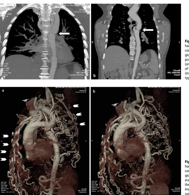
clearly demonstrate the interruption and collaterals (. Fig. 1b). Computed tomog-raphy angiogtomog-raphy revealed severe hypo-plasia of the transverse aortic arch proxi-mal to the origin of the left subclavian ar-tery (. Fig. 2). The bilateral common ca-rotid arteries were dilated. The descend-ing thoracic aorta was supplied by exten-sive collateral vessels from the vertebro-basilar system down to the posterior chest wall and the spine (. Fig. 3).
In accordance with the results of the cardiac catheterization, retrograde flow could be seen in the proximal left sub-clavian artery and the left vertebral ar-tery. A single-stage extra-anatomic re-pair was made by placing a 1-mm Da-cron graft between the ascending and de-scending portions of the thoracic aorta.
The patient recovered uneventfully and was doing well at the first follow-up vis-it 1 month later.
Discussion
Interrupted aortic arch (IAA) is a rare congenital malformation that occurs in three cases per million live births [1]. It is defined as the absence of luminal con-tinuity between the ascending and de-scending portions of the aorta [2]. In most cases, IAA is associated with intra-cardiac malformations such as ventric-ular septal defect, patent ductus arterio-sus, bicuspid aortic valve, left ventricular outflow tract obstruction, or aortopulmo-nary window. In this anomaly, the prog-nosis, which depends on the associated
congenital anomalies, is very poor un-less there is surgical treatment [1, 2]. In infants, its clinical presentation involves severe congestive heart failure and 90% of affected infants die at a median age of 4 days if not treated [3]. In a few docu-mented cases in adults, the presentation ranges from a lack of symptoms to limb swelling with blood pressure difference between the arms and legs. Substantial collateral circulation must be present to maintain flow, enabling survival. How-ever, collateral vessels are prone to atro-phy and atherosclerosis, which can lead to other challenging problems [4].
IAA was first described by Steidele in 1778 [5]. The first classification sys-tem, introduced by Celoria and Patton in 1959 [], is still used today almost
uni-Fig. 2 9 a, b Contrast-en-hanced, three-dimensional computed tomography an-giogram reveals severe hy-poplasia of the aortic arch (arrow) distal to the origin of the left subclavian artery (interrupted aortic arch, type A)
Fig. 3 9 a, b Contrast-en-hanced, three-dimensional computed tomography an-giogram. Extensive collat-eral vessels are evident in the paraspinal region and involve the vertebrobasilar system (arrows)
550 |
Herz 3 · 2015versally. This system describes and clas-sifies the site of aortic arch discontinui-ty, which may be: distal to the left subcla-vian artery (type A); between the left ca-rotid and left subclavian arteries (type B); or between the innominate and left carot-id arteries (type C). The most common type is B (53%), followed by type A (43%), and C (4%).
Several methods can be used for the diagnosis of IAA. Two-dimensional echo-cardiography plays an important role in the delineation of IAA. This technique is also necessary for ruling out associat-ed intracardiac anomalies that were not detailed at cardiac catheterization [7]. In general, cardiac catheterization is war-ranted for definitive anatomical evalu-ation in patients with IAA. However, it may be difficult to perform without pri-or knowledge of the vascular anatomy to ensure visualization of both proximal and distal segments [8]. Thoracic three-dimensional magnetic resonance angiog-raphy is a reliable, noninvasive diagnos-tic modality for the correct diagnosis of aortic coarctation and aortic arch anom-alies as well as visualization of collateral vessels [9, 10].
Treatment of the IAA is definitely sur-gical correction [1, 2, 3, 4]. IAA is rarely encountered in an adult patient and the malformation may be repaired in a sin-gle-stage procedure by means of an ex-tra-anatomic approach with a low risk of morbidity and mortality [4].
Corresponding address
I. Rencuzogullari Division of Cardiology,
Mugla Sitki Kocman University Education and Research Hospital
Orhaniye Mahallesi, İsmet Çatak caddesiMugla Turkey
Compliance with ethical
guidelines
Conflict of interest. I. Rencuzogullari, I.T. Ozcan, A.
Cirit, and S. Ayhan state that there are no conflicts of interest.
Consent was obtained from all patients identifiable from images or other information within the manu-script. In the case of underage patients, consent was obtained from a parent or legal guardian.
References
1. Canova CR, Carrel T, Dubach P et al (1995) In-terrupted aortic arch: fortuitous diagnosis in a 72-yearold female patient with severe aortic in-sufficiency [in German]. Schweiz Med Wochenschr 125(1–2):26–30
2. Backer CL, Mavroudis C (2000) Congenital Heart Surgery Nomenclature and Database Project: patent ductus arteriosus, coarctation of the aor-ta, interrupted aortic arch. Ann Thorac Surg 69(4 Suppl):S298–S307
3. Collins-Nakai RL, Dick M, Parisi-Buckley L et al (1976) Interrupted aortic arch in infancy. J Pediatr 88:959–962
4. Messner G, Reul GJ, Flamm SD et al (2002) Inter-rupted aortic arch in an adult, single-stage ex-traanatomic repair. Tex Heart Inst J 29:118–121 5. Steidele RJ (1778) Samml Chir u Med Beob
(Vien-na) 2:114
6. Celoria GC, Patton RB (1959) Congenital absence of the aortic arch. Am Heart J 58:407–413 7. Collins-Nakai RL, Dick M, Parisi-Buckley L et al
(1976) Interrupted aortic arch in infancy. J Pediatr 88:959–962
8. Kosucu P, Kosucu M, Dinc H, Korkmaz L (2006) In-terrupted aortic arch in an adult: diagnosis with MSCT. Int J Cardiovasc Imaging 22(5):735–739 9. Varghese A, Gatzoulis M, Mohiaddin RH (2002)
Images in cardiovascular medicine: magnetic res-onance angiography of a congenitally interrupted aortic arch. Circulation 106(3):E9–E10
10. Yucel EK, Anderson CM, Edelman RR et al (1999) Magnetic resonance angiography: update on ap-plications for extracranial arteries. Circulation 100(22):2284–2301