exploration of general acid function by site-directed mutagenesis
☆
Mustafa Köksal
a,1, Kevin Potter
b, Reuben J. Peters
b,⁎
, David W. Christianson
a,⁎
aRoy and Diana Vagelos Laboratories, Department of Chemistry, University of Pennsylvania, Philadelphia, PA 19104-6323, USA
bDepartment of Biochemistry, Biophysics and Molecular Biology, Iowa State University, Ames, IA 50011, USA
a b s t r a c t
a r t i c l e i n f o
Article history: Received 12 July 2013
Received in revised form 30 August 2013 Accepted 4 September 2013
Available online 12 September 2013 Keywords:
Protein crystallography Terpene cyclase Enzyme mechanism Gibberellin biosynthesis
Background: The diterpene cyclase ent-copalyl diphosphate synthase (CPS) catalyzes thefirst committed step in the biosynthesis of gibberellins. The previously reported 2.25 Å resolution crystal structure of CPS complexed with (S)-15-aza-14,15-dihydrogeranylgeranyl thiolodiphosphate (1) established theαβγ domain architecture, but ambiguities regarding substrate analog binding remained.
Method: Use of crystallization additives yielded CPS crystals diffracting to 1.55 Å resolution. Additionally, active site residues that hydrogen bond with D379, either directly or through hydrogen bonded water molecules, were probed by mutagenesis.
Results: This work clarifies structure–function relationships that were ambiguous in the lower resolution struc-ture. Well-defined positions for the diphosphate group and tertiary ammonium cation of 1, as well as extensive solvent structure, are observed.
Conclusions: Two channels involving hydrogen bonded solvent and protein residues lead to the active site, forming hydrogen bonded“proton wires” that link general acid D379 with bulk solvent. These proton wires may facilitate proton transfer with the general acid during catalysis. Activity measurements made with mutant enzymes indicate that N425, which donates a hydrogen bond directly to D379, and T421, which hydrogen bonds with D379 through an intervening solvent molecule, help orient D379 for catalysis. Residues involved in hydrogen bonds with the proton wire, R340 and D503, are also important. Finally, conserved residue E211, which is located near the diphosphate group of 1, is proposed to be a ligand to Mg2+required for optimal cata-lytic activity.
General significance: This work establishes structure–function relationships for class II terpenoid cyclases. © 2013 Elsevier B.V. All rights reserved.
1. Introduction
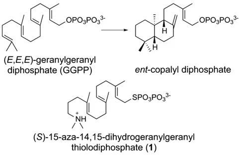
The diterpene cyclase ent-copalyl diphosphate synthase (CPS) from Arabidopsis thaliana is a class II terpenoid cyclase that initiates the cycli-zation of the substrate, (E,E,E)-geranylgeranyl diphosphate (GGPP), by protonation of the C14,C15 double bond to form a tertiary carbocation, thereby triggering a cascade of carbon–carbon bond forming reactions that ultimately yield the bicyclic product ent-copalyl diphosphate (Fig. 1)[1–3]. This comprises thefirst committed step in the biosynthesis of gibberellins, ubiquitous phytohormones that
function in plant growth and development[4–6]. The active site of CPS plays a critical role as a template in this reaction, since the highly flexible C20-isoprenoid substrate must befixed in a specific
conforma-tion to direct carbon–carbon bond formation with structural and stereo-chemical precision. While Mg2+ is required for optimal catalytic activity, this metal ion does not participate directly in the chemistry of catalysis. Instead, Mg2+is believed to anchor the position of the
sub-strate diphosphate group to help chaperone the subsub-strate binding con-formation[2].
The general acid responsible for substrate protonation is the “mid-dle” aspartic acid residue in the DXDD amino acid sequence motif that characterizes a class II terpenoid cyclase[7–10]. This motif has diverged to VXDC in the related oxidosqualene cyclases[11–13], in which the aspartic acid protonates the oxirane ring of the substrate to generate the initial carbocation. Neither general acid motif is to be confused with the“DDXXD” motif in which the first and sometimes the last as-partate residue coordinate to metal ions in the active site of a class I ter-penoid cyclase[8,14]. While carbon–carbon bond forming chemistry similarly proceeds through multiple carbocation intermediates in class
☆ This work was supported by US National Institutes of Health Grant GM56838 to D.W.C. and GM076324 to R.J.P.
⁎ Corresponding authors.
E-mail addresses:[email protected](R.J. Peters),[email protected] (D.W. Christianson).
1Current Address: Department of Molecular Biology and Genetics, Izmir Institute of
Technology, Urla, Izmir, 35430 Turkey.
0304-4165/$– see front matter © 2013 Elsevier B.V. All rights reserved. http://dx.doi.org/10.1016/j.bbagen.2013.09.004
I cyclase reactions, initial carbocation formation is achieved by the metal-dependent ionization of the substrate diphosphate group to gen-erate an allylic carbocation. Thus, class I and class II diterpene cyclases utilize the same substrate, GGPP, but they initiate carbocation formation at opposite ends of this substrate. The evolution of alternative cyclase classes reflects the evolution of alternative regiochemical strategies for carbocation generation in a common substrate.
Four crystal structures of class II terpenoid cyclases have been re-ported to date: the C30-triterpene cyclases squalene–hopene cyclase
and oxidosqualene cyclase at 2.0 Å and 2.1 Å resolution, respectively, and the C20-diterpene cyclases abietadiene synthase (a bifunctional
class I and class II cyclase) and CPS at 2.3 Å and 2.25 Å resolution, re-spectively[15–19]. Each of these cyclases exhibits the characteristic αβγ domain architecture predicted in an elegant bioinformatics study
[20]. Common structure–function relationships include the DXDD (or VXDC) general acid motif positioned at the“bottom” of a hydrophobic active site cleft at the interface of theβ and γ domains. This cleft is complementary to the nonpolar portion of the isoprenoid substrate; additional polar channels leading to the general acid motif could be im-plicated in proton transfer during catalysis. Given that the protonation of a carbon–carbon double bond (C = C) typically requires a strong acid such as H3O+(pKa =−1.7), it is curious as to how the aspartic
acid general acid in the active site of a class II terpenoid cyclase can pro-vide a sufficiently acidic proton to efficiently protonate a C = C bond. The environment of the aspartic acid presumably modulates its pKa from ~4 measured for the free aspartic acid side chain to a value compa-rable to that measured for H3O+to ensure an appropriately strong acid
function in the active site.
Here, we report the 1.55 Å-resolution crystal structure of CPS com-plexed with (S)-15-aza-14,15-dihydrogeranylgeranyl thiolodiphosphate (1) (Fig. 1), which mimics the tertiary carbocation formed upon proton-ation of the C14,C15 double bond of substrate GGPP. The structure of this complex prepared in the presence of Mg2+ion clarifies various
as-pects of the previously-reported structure at 2.25 Å-resolution, includ-ing a well-defined position for the diphosphate group and tertiary ammonium cation of 1. Additionally, the 1.55 Å-resolution structure provides an unparalleled view of solvent structure in the active site of a class II terpenoid cyclase. Intriguingly, hydrogen bonded water mole-cules appear to form“proton wires” that link general acid D379 with bulk solvent through secondary channels distinct from the main en-trance to the active site cavity. These proton wires define possible tra-jectories for regenerating the active form of the general acid during
catalysis. The results of site-directed mutagenesis support the proposed roles for active site residues in substrate binding and catalysis. 2. Materials and methods
2.1. Crystallization and structure refinement.
The crystallization of CPS has been previously described[19]. Briefly, a 2-μL drop of protein solution [5 mg/mL CPS, 25 mM 3-morpholino-2-hydroxypropanesulfonic acid (pH 6.8), 2.0 mM 1, 2.0 mM MgCl2, 10%
glycerol, 300 mM NaCl, 1.0 mM dithiothreitol (DTT) (incubated at 4 °C for 6 h and centrifuged to clear the solution)] was added to a 1.6-μL drop of precipitant solution [100 mM sodium citrate (pH 5.4), 30% polyethylene glycol 400, 200 mM KH2PO4]. Subsequently, 0.4-μL
drop of 40% (v/v) 1,4-butanediol was added to this mixture and equilibrated against a 100-μL reservoir of precipitant solution. Rectan-gular prism-shaped plates appeared in 2–3 weeks and grew to maximal dimensions of 50μm × 100 μm × 200 μm. Crystals were flash-cooled after transfer to a cryoprotectant solution consisting of the mother liquor augmented with 10% glycerol.
Crystals of the CPS-1 complex diffracted X-rays to 1.55 Å-resolution at the National Synchrotron Light Source (NSLS), Brookhaven National
Laboratory, beamline X29A, using incident radiation with λ =
0.9795 Å. All diffraction data were processed with HKL2000[21]. Crys-tals of the CPS-1 complex belonged to space group P212121with unit
cell parameters a = 130.2 Å, b = 51.4 Å, and c = 114.3 Å. Data collec-tion and reduccollec-tion statistics are recorded inTable 1. The atomic coordi-nates from the previously reported for the CPS-1 complex determined at 2.25 Å-resolution (PDB ID: 3PYA)[19]were used as the starting point for refinement with PHENIX[22]. Manual model rebuilding was performed with COOT[23]. In thefinal model of the CPS-1 complex, 678 of 727 residues are present; disordered segments excluded
from thefinal model include N-terminal residues M84-S89 (M84 is
the N-terminus of the construct), the C-terminal hexahistidine tag and one of its associated linker residues (S804-H810), and surface loops I617-G638, G687-E689, and R730-E740. The refined structure also in-cludes one S-hydroxy-L-cysteine residue at position 411, presumably the result of radiation damage to C411. Refinement statistics are recorded inTable 1. Ramachandran plot statistics were calculated with
PROCHECK[24] and simulated-annealing omit maps were calculated
with CNS[25]. Protein structurefigures were prepared with the graphics program PyMol (http://www.pymol.org). Atomic coordinates of the
1.55 Å-resolution structure of the CPS-1 complex have been deposited in the Protein Data Bank (www.rcsb.org) with accession code 4LIX. 2.2. Mutagenesis
The following forward and reverse primer sets were used for site di-rected mutagenesis of selected residues by using the QuickChange pro-tocol (Agilent, USA): CPS_T421A_F, CAATCAAACCAAGCAGTAGCCGGTA TGTTCAACCTATAC; CPS_T421A_R, GTATAGGTTGAACATACCGGCTACTG CTTGGTTTGATTG; CPS_T421S_FN, GTGGGGCAATCAAACCAAGCAGTA TCCGGTATGTTCAACCTATACCGGGC; CPS_T421S_RN, GCCCGGTATAG GTTGAACATACCGGATACTGCTTGGTTTGATTGCCCCAC; CPS_F424A_FN, CAATCAAACCAAGCAGTAACCGGTATGGCCAACCTATACCGGGCATCACAA TTG; CPS_F424A_RN, CAATTGTGATGCCCGGTATAGGTTGGCCATACCGG TTACTGCTTGGTTTGATTG; CPS_N425A_FN, CCAAGCAGTAACCGGTATG TTCGCCCTATACCGGGCATCACAATTGG; CPS_N425A_RN, CCAATTGTG ATGCCCGGTATAGGGCGAACATACCGGTTACTGCTTGG; CPS_N425H_FN, CCAAGCAGTAACCGGTATGTTCCACCTATACCGGGCATCACAATTGG; CPS_ N425H_RN, CCAATTGTGATGCCCGGTATAGGTGGAACATACCGGTTACTG CTTGG; CPS_E211A_FN, GATGAGCATATGCCAATCGGATTCGCAGTAGC ATTCCCATCGTTGCTTGAG; CPS_E211A_RN, CTCAAGCAACGATGGGAA TGCTACTGCGAATCCGATTGGCATATGCTCATC; CPS_R340A_FN, CACAT ATGGATAGTGGATCGGTTACAAGCTTTAGGGATATCGAGATACTTTGAAG;
CPS_R340A_RN, CTTCAAAGTATCTCGATATCCCTAAAGCTTGTAACCGAT
CCACTATCCATATGTG; CPS_D503A_FN, GATCAATATGGTGGAGAAAAC GCCGTTTGGATTGGCAAGACTCTTTATAG; CPS_D503A_RN, CTATAAAG AGTCTTGCCAATCCAAACGGCGTTTTCTCCACCATATTGATC.
2.3. Enzyme purification and assay
The various CPS constructs described here were expressed from pET22bCV (a custom-made variant of pET22b) as 6xHis tagged fusion
NaCl, and 10 mM MgCl2, with 10 mM (buffer A) or 250 mM (buffer B)
imidazole. Cell lysate was passed over a 5-mL column equilibrated with buffer A; this was then washed with 30 mL of buffer A, and the tagged CPS eluted with a 0–100% gradient of buffer B over 20 mL. Frac-tions were checked by SDS–PAGE, with those containing N90% pure CPS pooled (≥5 mL total) and dialyzed overnight against 1 L of storage buff-er (10 mM Bis-Tris, pH 6.8, 150 mM KCl, 10 mM MgCl2, 1 mM DTT, 10%
glycerol). Enzymatic kinetic assays were conducted as previously de-scribed[3]. Briefly, assays were performed at 30 °C in 1 mL of assay buffer (50 mM HEPES, pH 7.75, 100 mM KCl, 0.1 mM MgCl2, 10%
glyc-erol). Reactions were terminated by the addition of 110μL of
20 mM N-ethylmaleimide in 0.5 M glycine (pH 11), and incubation at 75 °C for 5 min. Excess N-ethylmaleimide was deactivated by the addi-tion of 20μL 1 M DTT and incubation for 15 min at room temperature. The samples were then neutralized with 60μL of 1 M HCl. ZnCl2and
MgCl2 were added to final concentrations of 10 mM and 100 mM
(12μL) before the addition of 4 units of bovine alkaline phosphatase (Invitrogen) to dephosphorylate both substrate and products. The sam-ples were either incubated at room temperature, overnight, or at 37 °C for 3 h. The resulting alcohols (geranylgeraniol and copalol) were extracted thrice with 1 mL hexanes; extractions were pooled, dried under N2, and resuspended in 50μL hexanes for GC analysis. The
pro-duction of CPP was verified by GC–MS, with subsequent quantification of the relative catalytic turnover via GC-FID, as previously described
[3]. For the reported kinetics analyses, enzyme concentration and incu-bation time were varied to achieve 2–8% turnover for GGPP (Isoprenoid Co). To correct for handling errors, catalytic rates were derived from the observed fractional turnover (i.e. the ratio of product to the sum of product and substrate), along with the known starting concentration of GGPP. Kinetic parameters were calculated byfitting the resulting re-action rate data to the substrate inhibition equation for wild-type, E211A, R340A, T421S, T421A, N425A and D503A CPS enzymes, or the Michaelis–Menten equation for N425H CPS (KaleidaGraph 4.0; Synergy Software, Reading, PA, USA). For all curvefits, R2≥ 0.90, except N425H
where R2= 0.8.
3. Results and discussion
3.1. Structure of the CPS-1 complex at 1.55 Å-resolution
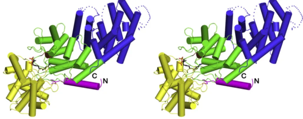
The overall fold of CPS in the 1.55 Å-resolution structure of the CPS-1 complex is essentially identical to that observed in the previously-reported 2.25 Å-resolution structure[19]. The enzyme adoptsαβγ do-main architecture with the active site located at the interface of theβ andγ domains (Fig. 2). However, there are important differences at the atomic level that now clarify structure–mechanism relationships.
The key difference between the previously reported 2.25 Å-resolu-tion structure of CPS[19]and the 1.55 Å-resolution structure reported herein is the presence of 1.0 mM MgCl2in the mother liquor of the
crys-tal. Although this magnesium salt was added with the intention of locat-ing the catalytically-important Mg2+binding site[2], no bound Mg2+
ions are interpretable in the electron density map, even at the higher resolution of the current crystal structure determination. The lack of Mg2+ binding is attributed to the relatively low pH of the crystal
Ligand atomsd 40
R.m.s. deviations
Bonds (Å) 0.015
Angles (°) 1.6
Dihedral angles (°) 21.8
Improper dihedral angles (°) 1.6 Average B factors (Å2) Main chain 22 Side chain 28 Ligand 44 Solvent 37 Ramachandran plot Allowed (%) 93.5 Additionally allowed (%) 6.3 Generously allowed (%) 0.2 Disallowed (%) 0 a
Number in parentheses refer to the outer shell of data.
b
Rmerge=Σ|I − bIN |/ΣI, where I is the observed intensity and bIN is the average
intensity calculated from replicate data.
c R
work=Σ| |Fo|− |Fc| |/Σ|Fo| for reflections contained in the working set, and
Rfree=Σ| |Fo|− |Fc| |/Σ|Fo| for reflections contained in the test set held aside during
refinement (1% of the total number of reflections). |Fo| and |Fc| are the observed and
calculated structure factor amplitudes, respectively.
d
structure determination (the pH of the crystallization drop was mea-sured to be approximately 5.4 [19]), which would weaken metal– ligand coordination interactions. We were unable to prepare crystals at higher pH values. However, the presence of Mg2+in the
crystalliza-tion buffer apparently affects the conformacrystalliza-tion of the diphosphate group of 1, which was previously disordered between two positions in the absence of Mg2+and is now ordered in a single position in the
pres-ence of Mg2+(Fig. 3a).
Since 1 binds with an extended conformation, as previously noted in the 2.25 Å-resolution structure of the CPS-1 complex[19], it cannot mimic the overall conformation or intermolecular interactions of GGPP required for cyclization to ent-copalyl diphosphate. However, the binding conformation of 1 may reflect the penultimate precatalytic substrate binding conformation, such that the substrate diphosphate
group is not likely to shift too far away from the conserved basic seg-ment K241–R248 upon assuming a cyclization-competent precatalytic conformation. The diphosphate group of 1 accepts direct hydrogen bonds from conserved residues K245 and K463, and the backbone NH group of G209. Since electron density defining the side chains of K245 and K463 is very weak, hydrogen bonds with these residues may not be strong. The carboxamide group of N417 is close to the diphosphate group of 1, but with a separation of 3.5 Å this interaction is too long to be considered a hydrogen bond. The diphosphate group of 1 also accepts a hydrogen bond from a water molecule that also donates a hydrogen bond to E211 (Figs. 3a and b). A second water molecule is 3.5 Å away from the diphosphate group of 1, but this interaction is too long to be considered a hydrogen bond. The bound conformation of 1 may reflect an incompletely-desolvated substrate binding mode that precedes
Fig. 2. Overall structure of the CPS-1 complex color-coded as follows:α domain = blue; β domain = green; γ domain = yellow; N-terminal helix = magenta; DXDD general acid motif = brown. The N- and C-termini are indicated by“N” and “C,” respectively. The γ domain is inserted between the N-terminal helix and the second helix of the β domain. Inhibitor 1 is shown as a stickfigure bound in the active site at the interface of the β and γ domains.
Fig. 3. (a) Simulated annealing omit map (contoured at 4.0σ) of the CPS-1 complex at 1.55 Å resolution. Atoms are color-coded as follows: C = green (β domain), brown (DXDD general acid motif in theβ domain), or yellow (γ domain), N = blue, O = red, P = orange, S = yellow; selected water molecules are shown as small red spheres, and hydrogen bond interactions are indicated by dash red lines. (b) Superposition of the 1.55 Å-resolution structure of the CPS-1 complex color-coded as in (a) with the 2.25 Å-resolution structure (all atoms cyan).
would correspond to the C15 atom of substrate GGPP) makes no other hydrogen bond interactions and is 4.2 Å from general acid D379. The C14 atom of 1, which corresponds to the C14 atom of GGPP that would be protonated by the general acid, is 5.7 Å from D379. A
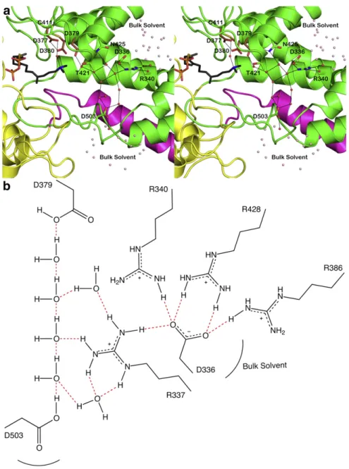
theα and β domains and in the active site at the interface of the β andγ domains. Additionally, two hydrogen-bonded solvent channels lead away from general acid D379 (Fig. 4). These channels are distinct from the main active site cleft, i.e., the substrate entry and product
Fig. 4. (a) Cut-away view of the CPS active site showing solvent channels leading to general acid D379. One channel leads to D503 and the solvent-exposed surface of theβ domain (green) near the interface with theγ domain (yellow); the other channel leads to D336–R340, which are exposed to bulk solvent at the interface of the α (blue) and β domains. These channels are distinct from the main active site cleft, which opens to the left in this view. (b) Molecular scheme showing hydrogen bond interactions linking general acid D379 with bulk solvent through the two polar solvent channels shown in (a). For clarity, R337, R386, and R428 are omitted from the view in (a) but are shown here to emphasize the highly polar nature of the hydrogen bond networks with D379.
egress route, and may influence proton transfer to and from D379 dur-ing catalysis. The hydrogen bond geometry around D379 suggests that this residue serves its general acid function through the more acidic
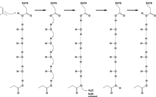
[27] anti-oriented proton; the resulting carboxylate conjugate base form of D379 can subsequently be reprotonated through the more basic syn geometry by Grotthuss diffusion through one of the hydrogen bonded solvent channels, as illustrated inFig. 5.
3.2. Catalytic activity of CPS mutants
In order to probe the function of residues that could influence the general acid function of D379, we prepared site-specific mutants of CPS in which adjacent residues T421 and N425 were substituted by al-ternative amino acids. Additionally, we prepared exploratory mutants in which selected polar residues lining the solvent channels, D336 and R340, were substituted. Finally, we studied a mutant of the proposed Mg2+ ligand, E211. Steady-state kinetic parameters measured from
these mutants are recorded inTable 2.
The side chain hydroxyl group of T421 hydrogen bonds with a water molecule, which in turn donates a syn-oriented hydrogen bond to the side chain Oδ2 atom of D379 (Fig. 4). The T421A mutant exhibits a 163-fold reduction in kcat (KM is increased only 2-fold), suggesting
that this residue and its hydrogen bonded water molecule play an im-portant role in catalysis. That kcatis reduced by less than 3-fold and KM
is essentially invariant in T421S CPS further supports an important role for the γ-hydroxyl group of residue 421. Possibly, the water-bridged hydrogen bond between T421 and D379 is important for
orienting the carboxylic acid side chain of D379 for its general acid function.
The side chain carboxamide group of N425 donates a syn-oriented hydrogen bond directly to the side chain Oδ1 atom of D379 (Fig. 4), so it is likely that this residue similarly plays an important role in orienting the general acid catalyst. In accord with this expectation, the catalytic activity of N425A CPS exhibits a 13-fold reduction in kcat. The
corre-sponding residue in squalene–hopene cyclase is a histidine[15,16], and the substitution of a histidine residue in N425H CPS exhibits a 75-fold reduction in kcat.
The substitution of selected polar residues lining solvent channels leading to general acid D379 influences kcat, while KMremains relatively
invariant. Although D336A CPS is not sufficiently stable to allow for kinet-ic measurements, D503A CPS exhibits a 7-fold reduction in kcat; this
res-idue, through hydrogen bonding, connects the active site solvent network directly to the surface of theβ domain. In contrast, R340A CPS exhibits an 850-fold reduction in kcat; this residue hydrogen bonds with
D336 and blocks the solvent channel leading to the interface between theα and β domains. However, since R340 hydrogen bonds with solvent in the channel as well as bulk solvent, R340 and/or its hydrogen bond partner D336 could mediate proton transfer to D379. While the structural consequences of these mutations must be fully evaluated in future X-ray crystal structure determinations to confirm that no unanticipated struc-tural changes are triggered by the mutations, our working hypothesis is that the solvent channel leading to the interface between theα and β do-mains is important for catalytic function. This channel corresponds to that first identified in the X-ray crystal structure determination of oxidosqualene cyclase[17], which exhibitsβγ domain architecture.
Finally, deletion of the putative Mg2+ligand in E211A CPS results in
a nearly 500-fold reduction in kcatbut only a slight reduction in KM. In
the high resolution structure, a water molecule bridges the diphosphate group of 1 and the E211 side chain. While this confirms the catalytic im-portance of E211, the kinetic results suggest that the postulated
coordi-nation of the substrate diphosphate group to Mg2+ may be more
important for stabilizing the cyclization-competent conformation in the transition state[28].
3.3. General acid function of D379
It is unusual tofind general acid catalysis function within a cluster of carboxylic acid-containing amino acids in a protein structure, such as
Fig. 5. General acid D379 could be reprotonated by Grotthuss diffusion of a proton across a 4-solvent“proton wire” in a polar channel leading to another aspartic acid serving as a proton shuttle to bulk solvent, such as D503 or D336; in order for D336 to serve in this capacity, its hydrogen bond with R340 must be displaced by a solvent molecule.
Table 2
Steady-state kinetic parameters for CPS mutants.
kcat(s−1) KM(μM) kcat/KM(M−1s−1) Wild-type 0.9 ± 0.2 3 ± 1 3 × 10⁵ T421S 0.3 ± 0.1 3 ± 2 1 × 10⁵ T421A 0.006 ± 0.002 6 ± 3 1 × 10³ N425H 0.012 ± 0.001 0.8 ± 0.4 2 × 10⁴ N425A 0.071 ± 0.007 1.8 ± 0.5 4 × 10⁴ E211A 0.0019 ± 0.0003 1.7 ± 0.6 1.1 × 10³ D503A 0.13 ± 0.02 2.9 ± 0.7 4.5 × 10⁴ R340A 0.0011 ± 0.0001 2.0 ± 0.5 5.5 × 10²
ation by D379, the carboxylic acid cluster bears a net charge of−2. It is proposed that this additional negative charge provides electrostatic sta-bilization for carbocation intermediates in the cyclization cascade[11]. As noted upon thefirst crystal structure determination of a class II terpenoid cyclase, squalene–hopene cyclase [15], the general acid aspartic acid is located such that the more acidic anti-oriented proton is oriented toward the substrate [11]. This is also the case for CPS (Fig. 3)[19]and abietadiene synthase[18]. Interestingly, this is not the case for oxidosqualene cyclase[17], perhaps due to the fact that the moi-ety being protonated is not an isoprenoid C = C bond, but instead an ep-oxide ring; the epep-oxide is more reactive and may not require as powerful a Brønsted acid for efficient protonation. Additionally, oxidosqualene cy-clase does not have a residue comparable to N425 of CPS or H451 of squalene–hopene cyclase to orient the general acid aspartic acid side chain. Instead, these residues appear as A536 in oxidosqualene cyclase.
Since the anti-oriented conformer of a carboxylic acid is less stable than the syn-oriented conformer by 6–8 kcal/mol[30–32], the enzyme active site must provide an environment that stabilizes the anti-oriented conformer of D379 so that it is poised for general acid catalysis. The hydrogen bond network involving D379, N425, and T421 ensures that only the anti-oriented position of the side chain Oδ2 atom of D379 is oriented toward the substrate binding site. Mutagenesis results suggest that N425 helps to orient D379 for catalysis; T421 plays a similar role, and may also orient the bridging solvent molecule to enable proton transfer with D379 to regenerate the active form of the general acid at the conclusion of the cyclization cascade.
Finally, regeneration of the anti-oriented general acid D379 requires reprotonation after each catalytic turnover, and this may occur through either one or both of the solvent channels identified in the structure of the CPS-1 complex (Fig. 5). Proton transfer across hydrogen bonded sol-vent networks in such channels through Grotthuss diffusion is more ef fi-cient than the hydrodynamic diffusion of H3O+[33]and may account for
mutagenesis results with our exploratory site-specific mutants D503A and R340A (Table 2). Future studies will probe structure–function rela-tionships in these active site solvent channels in greater depth. Acknowledgements
We thank the National Synchrotron Light Source at Brookhaven National Laboratory for access to beamline X29A.
References
[1] T.-P. Sun, Y. Kamiya, The Arabidopsis GA1 locus encodes the cyclase ent-kaurene syn-thetase A of gibberellin biosynthesis, Plant Cell 6 (1994) 1509–1518.
[2] S. Prisic, R.J. Peters, Synergistic substrate inhibition of ent-copalyl diphosphate synthase: a potential feed-forward inhibition mechanism limiting gibberellin me-tabolism, Plant Physiol. 144 (2007) 445–454.
[11] K.U. Wendt, G.E. Schulz, E.J. Corey, D.R. Liu, Enzyme mechanisms for polycyclic triterpene formation, Angew. Chem. Int. Ed. 39 (2000) 2812–2833.
[12] K.U. Wendt, Enzyme mechanisms for triterpene cyclization: new pieces of the puz-zle, Angew. Chem. Int. Ed. 44 (2005) 3966–3971.
[13] I. Abe, Enzymatic synthesis of cyclic triterpenes, Nat. Prod. Rep. 24 (2007) 1311–1331.
[14] J.A. Aaron, D.W. Christianson, Trinuclear metal clusters in catalysis by terpenoid synthases, Pure Appl. Chem. 82 (2010) 1585–1597.
[15] K.U. Wendt, K. Poralla, G.E. Schulz, Structure and function of a squalene cyclase, Sci-ence 277 (1997) 1811–1815.
[16] K.U. Wendt, A. Lenhart, G.E. Schulz, The structure of the membrane protein squalene–hopene cyclase at 2.0 Å resolution, J. Mol. Biol. 286 (1999) 175–187. [17] R. Thoma, T. Schulz-Gasch, B. D'Arcy, J. Benz, J. Aebi, H. Dehmlow, M. Hennig, M.
Stihle, A. Ruf, Insight into steroid scaffold formation from the structure of human oxidosqualene cyclase, Nature 432 (2004) 118–122.
[18] K. Zhou, Y. Gao, J.A. Hoy, F.M. Mann, R.B. Honzatko, R.J. Peters, Insights into diterpene cyclization from structure of bifunctional abietadiene synthase from Abies grandis, J. Biol. Chem. 287 (2012) 6840–6850.
[19] M. Köksal, H. Hu, R.M. Coates, R.J. Peters, D.W. Christianson, Structure and mecha-nism of the diterpene cyclase ent-copalyl diphosphate synthase, Nat. Chem. Biol. 7 (2011) 431–433.
[20] R. Cao, Y. Zhang, F.M. Mann, C. Huang, D. Mukkamala, M.P. Hudock, M.E. Mead, S. Prisic, K. Wang, F.Y. Lin, T.K. Chang, R.J. Peters, E. Oldfield, Diterpene cyclases and the nature of the isoprene fold, Proteins: Struct. Funct. Bioinform. 78 (2010) 2417–2432.
[21] Z. Otwinowski, W. Minor, Processing of X-ray diffraction data collected in oscillation mode, Methods Enzymol. 276 (1997) 307–326.
[22] P.D. Adams, P.V. Afonine, G. Bunkóczi, V.B. Chen, I.W. Davis, N. Echols, J.J. Headd, L.-W. Hung, G.J. Kapral, R.W. Grosse-Kunstleve, A.J. McCoy, N.W. Moriarty, R. Oeffner, R.J. Read, D.C. Richardson, J.S. Richardson, T.C. Terwilliger, P.H. Zwart, PHENIX: a comprehensive Python-based system for macromolecular structure solu-tion, Acta Crystallogr. D66 (2010) 213–221.
[23] P. Emsley, B. Lohkamp, W.G. Scott, K. Cowtan, Features and development of Coot, Acta Crystallogr. D66 (2010) 486–501.
[24] R.A. Laskowski, M.W. MacArthur, D.S. Moss, J.M. Thornton, PROCHECK: a program to check the stereochemical quality of protein structures, J. Appl. Crystallogr. 26 (1993) 283–291.
[25] A.T. Brünger, P.D. Adams, G.M. Clore, W.L. DeLano, P. Gros, R.W. Grosse-Kunstleve, J.-S. Jiang, J. Kuszewski, M. Nilges, N.S. Pannu, R.J. Read, L.M. Rice, T. Simonson, G.L. Warren, Crystallography & NMR System: a new software suite for macromolecular structure determination, Acta Crystallogr. D54 (1998) 905–921.
[26] K. Zhou, R.J. Peters, Investigating the conservation pattern of a putative second ter-pene synthase divalent metal binding motif in plants, Phytochemistry 70 (2009) 366–369.
[27] R.D. Gandour, On the importance of orientation in general base catalysis by carbox-ylate, Bioorg. Chem. 10 (1981) 169–176.
[28] R.B. Peters, R.B. Croteau, Abietadiene synthase catalysis: conserved residues in-volved in protonation-initiated cyclization of geranylgeranyl diphosphate to (+)-copalyl diphosphate, Biochemistry 41 (2002) 1836–1842.
[29] J.V. Needham, T.Y. Chen, J.J. Falke, Novel ion specificity of a carboxylate cluster Mg(II) binding site: strong charge selectivity and weak size selectivity, Biochemistry 32 (1993) 3363–3367.
[30] X. Wang, K.N. Houk, Theoretical elucidation of the origin of the anomalously high acidity of Meldrum's acid, J. Am. Chem. Soc. 110 (1988) 1870–1872.
[31] K.B. Wiberg, K.E. Laidig, Barriers to rotation adjacent to double bonds. 3. The carbon–oxygen barrier in formic acid, methyl formate, acetic acid, and methyl acetate. The origin of ester and amide resonance, J. Am. Chem. Soc. 109 (1987) 5935–5943.
[32] K.B. Wiberg, K.E. Laidig, Acidity of (Z)- and (E)-methyl acetates: relationship to Meldrum's acid, J. Am. Chem. Soc. 110 (1988) 1872–1874.
[33] C. Knight, G.A. Voth, The curious case of the hydrated proton, Acc. Chem. Res. 45 (2012) 101–109.