Full Terms & Conditions of access and use can be found at
https://www.tandfonline.com/action/journalInformation?journalCode=icey20
Current Eye Research
ISSN: (Print) (Online) Journal homepage: https://www.tandfonline.com/loi/icey20
The Effect of Sildenafil on Selenite-Induced
Cataract in Rats
Hatice Tuba Atalay , Ahmet Yucel Ucgul , Ummuhani Ozel Turkcu , Mehmet
Cuneyt Ozmen , Samet Yilmaz & Ayse Bilgihan
To cite this article: Hatice Tuba Atalay , Ahmet Yucel Ucgul , Ummuhani Ozel Turkcu , Mehmet Cuneyt Ozmen , Samet Yilmaz & Ayse Bilgihan (2020) The Effect of Sildenafil on Selenite-Induced Cataract in Rats, Current Eye Research, 45:9, 1082-1088, DOI: 10.1080/02713683.2020.1726405
To link to this article: https://doi.org/10.1080/02713683.2020.1726405
Published online: 11 Feb 2020.
Submit your article to this journal
Article views: 111
View related articles
View Crossmark data
D
Taylor & Francis~ Tll~lorf,J,;ti,caC:rlklj,
The Effect of Sildenafil on Selenite-Induced Cataract in Rats
Hatice Tuba Atalaya, Ahmet Yucel Ucgulb, Ummuhani Ozel Turkcuc, Mehmet Cuneyt Ozmena, Samet Yilmazd, and Ayse Bilgihand
aDepartment of Ophthalmology, Gazi University Medical School, Ankara, Turkey;bDepartment of Ophthalmology, Izzet Baysal Training and
Research Hospital, Abant Izzet Baysal University, Bolu, Turkey;cDepartment of Medical Biochemistry, Training and Research Hospital, Mugla Sıtkı
Kocman University, Mugla, Turkey;dDepartment of Medical Biochemistry, Gazi University Medical School, Ankara, Turkey
ABSTRACT
Purpose: To investigate the effect of sildenafil on an experimental sodium selenite-induced cataract model in rats.
Materials and Methods: Twenty-six young Wistar rats were separated into four groups. On postpartum day 10, six rats received only selenite (group 1, selenite-induced cataract), seven rats received selenite and high dose oral sildenafil (group 2, high-dose sildenafil-treated), seven rats received selenite and low dose oral sildenafil (group 3, low-dose sildenafil-treated), and six rats received only saline (group 4, controls). On postpartum day 30, cataract formation was graded and recorded using an operating microscope. The rats were sacrificed, lens tissues were isolated, and serum samples were collected. Nitrite oxide metabolites (NOx), advanced oxidative protein products (AOPP), and total sulfhydryl (TSH) levels were assessed in both serum and lenticular samples.
Results: The rats treated with low-dose sildenafil showed lower levels of AOPP and NOx, and the higher levels of TSH than the rats in other experimental groups. Otherwise, the rats treated with high-dose sildenafil, similar to the selenite-induced cataract group, showed higher levels of AOPP and serum NOx than rats in the low-dose sildenafil-treated group. The rats treated with low-dose sildenafil also showed less cataract development than rats in the other experimental groups.
Conclusion: Low doses (0.7 mg/kg) of oral sildenafil might show a protective effect on cataract development by lowering oxidative stress.
ARTICLE HISTORY
Received 4 April 2019 Revised 23 January 2020 Accepted 30 January 2020
KEYWORDS
AOPP; cataract model; NOx; selenite; sildenafil; total sulfhydryl
Introduction
Lens opacification, otherwise known as cataract, is the most widely recognized reason for curable blindness in the world’s population. It represents around 42% of all blindness.1Today, cataract treatment is provided only through surgery in which an artificial lens is implanted instead of the cataractous lens, which is removed to restore visual acuity. The rising cost of cataract surgery and surgical complications have prompted scientists to investigate ways to prevent cataract development. Therefore, in the last decades, preventive approaches for cat-aract formation have increasingly been presented.2,3
The development of cataract is not yet fully understood. However, several studies have shown that oxidative stress plays a key role in the etiopathogenesis of cataracts.4,5Based on this evidence, the importance of strengthening antioxidant activity is underlined to prevent or delay cataract develop-ment. Accordingly, many natural products with antioxidant properties have been tried for this purpose.6
Many agents have been used to create a cataract formation. However, the selenite induced cataract model is the most widely accepted method today. Selenium (Se) is an indispen-sable rare-earth element for humans, animals and a few microscopic organisms.7It is present in the structure of sele-nocysteine, an essential amino acid with antioxidant properties.8,9 However, ironically, in 1978, Ostadalova et al
first demonstrated that overdose of selenite caused rapid cataract development by increasing oxidative stress.10In the laboratory, the selenite cataract model could be induced by a single subcutaneous injection of excess sodium selenite (Na2 SeO3) into the suckling rats on postpartum day 10. This model is usually presented with severe bilateral cataract devel-opment within 16 days after eye opening in rats, and could be used as a model for evaluating anti-cataract agents.
The selenite cataract is similar to the human senile cataract sharing some characteristics such as an increase in free cyto-solic calcium and protein aggregates and a decrease in water-soluble protein and reduced glutathione (GSH). However, it is different from human senile cataract in some characteristics such as increased disulfide formation or the absence of high-molecular-weight covalent aggregates.11 Additionally, rapid calpain-induced proteolytic deposition seems to predominate in the formation of selenite cataracts, whereas human senile cataracts are mainly caused by prolonged oxidative stress.12
Sildenafil citrate (Pfizer, Viagra 100 mg), a prototypical inhibitor phosphodiesterase type 5 (PDE-5), converts cyclic guanosine monophosphate (cGMP) to its inactive form in blood vessels.13 Sildenafil increases levels of cellular cGMP, resulting in smooth muscle relaxation and increased blood flow to the entire body. Previous studies have demonstrated that sildenafil may have anti–inflammatory effects by
CONTACTHatice Tuba Atalay [email protected] Ophthalmology Department, Gazi University School of Medicine, Besevler, Ankara 06500 https://doi.org/10.1080/02713683.2020.1726405
© 2020 Taylor & Francis Group, LLC
~ Taylor&FrancisGroup
activating antioxidant enzymes, reducing malondialdehyde (MDA) levels, and inhibiting reactive oxygen species (ROS).14,15 Furthermore, numerous studies have revealed that sildenafil might be protective against ischemic damage in organs, for example, the testes and kidneys.16,17
In this study, we aimed to evaluate whether sildenafil had a protective effect on selenite cataract formation and the levels of oxidative stress markers, including advanced oxidative pro-tein product (AOPP) and nitric oxide metabolites (NOx) in both the blood and the lens. Total sulfhydryl (TSH) levels were also assessed as a marker of an antioxidant system in lenses and serum samples.
Materials and methods
The present study was approved by the Animal Experiments Local Ethical Committee at the University of Gazi (Ankara, Turkey). The study was conducted in accordance with the Helsinki Declaration for the use of animals in ophthalmic and vision research in Gazi University Laboratory Animals Breeding and Experimental Researches Centre and Eye Diseases Department.
Animals
Twenty-six postnatal (10-day-old) Wistar rats were separated into four groups according to the amount of sildenafil and sodium-selenite they received. Group 1 (n = 6) received only subcutaneous sodium selenite (30 nmol/g), group 2 (n = 7) received subcutaneous sodium selenite and a single dose of high-dose (1.4 mg/kg) oral sildenafil citrate, group 3 (n = 7) received subcutaneous sodium selenite and a single dose of low-dose (0.7 mg/kg) oral sildenafil citrate, and group 4 (n = 6) received a subcutaneous balanced saline solution and served as the control group. Rats were housed with ad-libitum access to food and water under a 12:12 hour light–dark cycle. Euthanasia was performed by cardiac puncture under deep anesthesia at the postnatal 30th day. Intramuscular ketamine and xylazine were used for deep anesthesia. Blood and lens samples were collected following euthanasia.
Evaluation of lens
Both eyes of the rats were dilated with tropicamide 1%, and were then examined using a slit-lamp biomicroscope (TOPCON, Tokyo, Japan) and photographed under coaxial illumination of an operating microscope (Leica Microsystem, Morrisville, NC) just before the euthanasia. A single
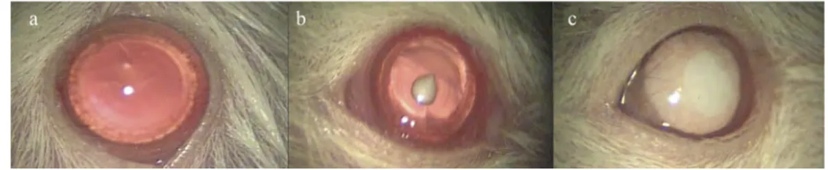
researcher (HTA), who was blinded to the treatment type, evaluated and noted the cataract levels. Cataract levels in lenses were graded as follows: 0- absence of cataract (com-pletely clear lens), 1- partially developed cataract, 2-mature cataract (Figure 1).
The extracted lens tissues were placed in liquid nitrogen and then kept in a freezer at−80°C degrees until required for analysis. The intracardiac blood samples were centrifuged and serum samples were separated and kept in a freezer at−80°C degrees until required for analysis. Advanced oxidative pro-tein product (AOPP) levels, total sulfhydryl (TSH) levels and nitric oxide metabolites (NOx) levels were analyzed in both lens and serum samples.
NOx assay
NOx (nitrite-nitrate) of lens tissues and serum were measured using the spectrophotometric method of Miranda et al.18Lens tissues were homogenized in phosphate buffer (pH: 7.4) and deproteinized with 96% ethanol at 1:2 (v/v). Homogenates were vortexed for 5 minutes and then mixed samples were centrifuged at 10,000 g at 4°C. Then, the collected superna-tants were analyzed to detect NOx levels.
For NOx analysis in tissues, 100μL of supernatant, 100 μL of vanadium (III) chloride and the following Griess reagents, 50μL of sulfanilamide and 50 μL of N-(1-naphtyl) ethylene-diamine dihydrochloride (NEED) were pipette into each microplate well and then incubated for 30 minutes at 37°C. The absorbance of NOx was read at 540 nm. NOx levels were calculated from the linear standard curves prepared by sodium nitrate (10–100 μM range). The results of NOx for lens tissue and serum were expressed as mM/g andΜmol/L, respectively.
TSH levels
Serum and lens tissues were homogenized in the phosphate buffer at pH 7.4 and then homogenates were centrifuged at 10,000 g for 10 minutes. Supernatants were mixed with an equal volume of 6% sodium dodecyl sulphate (SDS). Then, 200μL of 0.25 M Tris-HCl, pH 8.2, containing 20 mM EDTA,
25 μL of the samples with SDS or serum, and 10 μL of
Ellman’s reagent were pipette into microplate wells respec-tively, and incubated for 15 minutes at room temperature. After incubation, the plate was read at 412 nm using a micro-plate reader. The TSH levels of serum and lens tissue were calculated from a linear standard curve constructed using
Figure 1.Grading of the rat lenses. A-Clear lens (grade 0), B-Partially developed cataract (grade 1), C-Mature cataract (grade 2).
reduced glutathione. The results of TSH for lens tissue and serum were expressed asμM/mg and μmol/L, respectively.
AOPP assay
AOPP levels in serum were measured according to the spectro-photometric method of Witko-Sarsat et al.19 To measure the levels of AOPP of serum, 200μL of serum was reconstituted with phosphate-buffered saline (PBS). After that, 10μL of potassium iodide and 20μL of acetic acid were added. The samples were vortexed, and a spectrophotometric measurement was obtained with PBS as a blank at 340 nm absorbance. Dilutions of the chloramine T standard (0–100 μM) were used for calculation of sample concentration. The results of AOPP for lens tissue and serum were expressed asμM/mg and μmol/L, respectively.
Statistical analysis
The SPSS software for Windows version 22.0 (Chicago, IL) were used for statistical analysis. The results were shown as median (minimum-maximum). Shapiro-Wilk test was used to deter-mine whether the continuous variables were parametric or non-parametric. Kruskal-Wallis variance analysis was performed to determine whether there were significant differences of markers of the serum and lens samples among the four groups. Tamhane’s T2 post hoc test was also used for pairwise isons. Fisher’s exact test was performed for the pairwise compar-ison of cataract development between the groups. A p value less than 0.05 was accepted as statistically significant.
Results
Morphologic evaluation of cataract formation
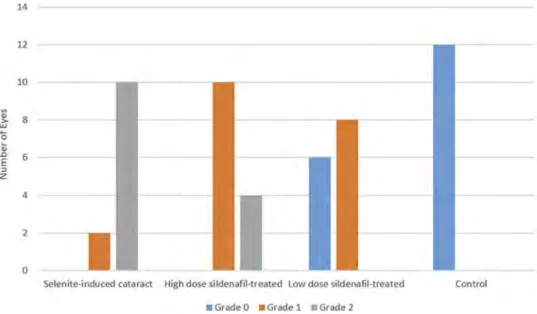
In the selenite-induced cataract group, cataracts developed in all lenses, with the majority of them being mature; this
indicates success in the creation of the selenite-induced catar-act model in the present study. In the low-dose sildenafil-treated group, cataracts developed partially in 8 eyes, and 6 eyes were completely clear. In the high-dose sildenafil-treated group, cataracts developed partially in 10 eyes and 4 eyes experienced mature cataracts. In the control group, all lenses were clear. The results of cataract development in the rats are shown inFigure 2.
Evaluation of indicators of protein oxidation
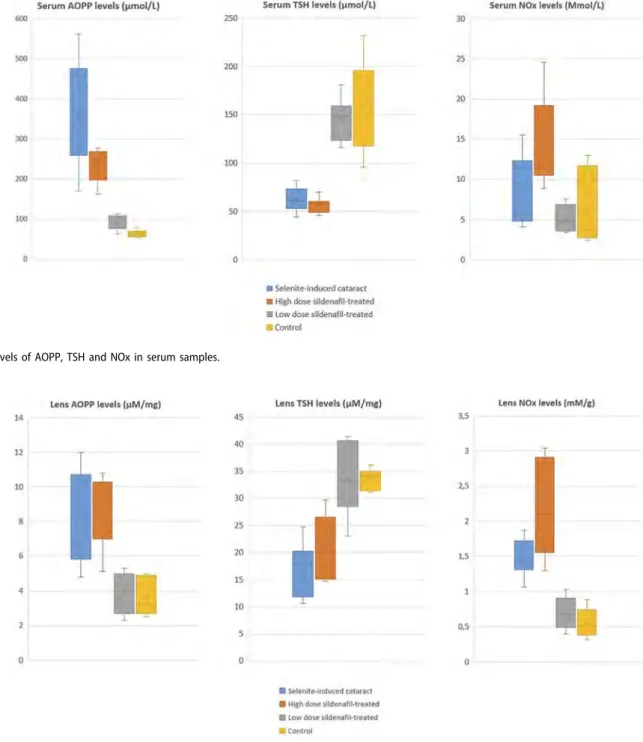
The collected data of AOPP, TSH and NOx levels in serum samples are shown in detail inFigure 3. Serum AOPP, TSH and NOx levels were significantly different between the four groups (p < .001, p < .001, and p < .001, respectively). The rats in group 3 had lower serum AOPP levels than those in groups 1 and 2 (p = .002 and p = .004, respectively). The rats in groups 1 and 2 had similar serum AOPP levels (p = .421). The rats in group 3 had higher serum TSH levels than those in groups 1 and 2 (p = .005 and p = .004, respectively). The rats in groups 1 and 2 had similar serum TSH levels (p = .672). Serum NOx levels in group 2 were significantly higher than those in groups 1 and 3, (p = .002 and p = .004, respectively). The rats in groups 1 and 3 had similar serum NOx levels (p = .364). Group 4, the control group, had the highest serum TSH levels and the lowest serum AOPP and serum NOx levels among the four groups.
The collected data of AOPP, TSH and NOx levels in lens tissues are shown in detail inFigure 4. Lens AOPP, TSH and NOx levels were significantly different between the four groups (p = .001, p = .001, and p < .001, respectively). The rats in group 2 had the higher lens AOPP levels than those in groups 3 and 4 (p = .003 and p = .002, respectively). The rats in groups 3 and 4 had similar lens AOPP levels (p = .978). The rats in group 3 had higher lens TSH levels than those in groups 1 and 2 (p = .003 and p = .050, respectively). The
Figure 2.Morphological evaluation of cataract formation of isolated lenses in each group.
Fisher Exact Test was used for pairwise comparison.(Group 1 versus Group 3,p < .001. Group 2 versus Group 3, p = .005. Group 2 versus Group 4, p < .001. Group 1 versus Group 2,p = .008. Group 3 versus Group 4, p = .002. Group 1 versus Group 4, p < .001).
14 12 10 :,; > w 8 0 ~
"
.0 6 E => z 4 2 0I
Selenite-induced cataract High dose slldenafil•treated Low dose slldenafil-treated • Grade O • Grade 1 • Grade 2
rats in groups 1 and 2 had similar lens TSH levels (p = .442). The rats in group 3 had lower lens NOx levels than those in groups 1 and 2 (p = .003 and p = .003, respectively). The rats in groups 3 and 4 showed similar lens NOx levels (p = .964).
Discussion
Although selenium is an important trace element, it is well-known that high concentrations of selenium might be toxic. The potential mechanism of selenium toxicity is not fully understood; however, it is often ascribed to its capability to induce oxidative stress. Developing nuclear cataracts follow-ing administration of sodium selenite is proposed to be
a consequence of lens glutathione (GSH) loss.20Loss of GSH decreases the redox buffering capacity and increase the oxida-tive stress sensitivity of the crystalline lens. Another proposed mechanism is that the loss of intracellular calcium (Ca2+) homeostasis could lead to the development of cataracts by increasing cytosolic Ca2+ levels and thus activating calpain.
Previous studies have shown that calpain-induced
β-crystalline proteolysis could play an important role in lens maturation and cataractogenesis.12
Several studies demonstrated that selenite-induced cataracto-genesis could be inhibited by antioxidant substances, for example, caffeic acid phenethyl ester, 2-ketoglutarate, and Ocimum sanctum extract.21–23These studies proposed that the
Figure 3.Levels of AOPP, TSH and NOx in serum samples.
Figure 4.Levels of AOPP, TSH and NOx in lens samples.
CURRENT EYE RESEARCH
9
1085Serum AOPP levels (µmol/L) Serum TSH levels (µmol/L) Serum NOx levels (Mmol/L)
600 250 30 500 25 200 400
.
1
w 150 300 15'
100 T lOO,
.
10•
I
•
50 100,..
_
0 0 0 • Selenlte-lnduc.ed c-araracr • High dose sllde:nafTHteat~d • low dose slldenafll-u-eated •Controllens AOPP levels (µM/mg) Lens TSH levels (µM/mg) lens NOx levels (mM/g)
14 45 3,5 40 12 35
II
10 2,5 30,
1
8 25 6 20 l.5'
,
.
15 10•
•
2 0,5 0 0 0 • S!ltnU~induc~d t.ll.W-IC't • Hllf\ do«' Slldi!:Nfil•trHtctd • LOW doir 111dt:n1f1l•tr~.atedcataractogenic effect was provided by protecting normal oxidant levels. A previous study also investigated the anti-cataractogenic effect of green tea (Camellia sinensis) and revealed that green tea had a significant anti-cataract potency because of its ability to support the antioxidant defense system.24Doganay et al. revealed that resveratrol had a protective effect against selenite induced cataract in rats. The authors proposed that an increase in GSH levels and a decrease in malondialdehyde (MDA) levels in rats that received resveratrol indicated the protective effect.25Yagci et al showed that melatonin, an endogenous antioxidant, could prevent the development of cataracts induced by selenite in rats. They demonstrated that MDA and other oxidative stress markers such as xanthine oxidase and protein carbonyls decreased in the melatonin-treated group.26Other antioxidant agents such as saf-fron, C-phycocyanin, a-lipoic acid and fisetin, ebselen, cysteamine, pirenoxine, acetyl L-carnitine, vitamin E, disulfonic stilbene and hydrogen-rich saline, N-acetylcysteine, caffeine, broccoli, hesper-etin and coenzyme Q have previously been reported to inhibit selenite-induced cataract development.27–41
Sildenafil, a phosphodiesterase-5 (PDE-5) enzyme inhibitor, has been used increasingly in clinical practice. Sildenafil is com-monly used to treat erectile dysfunction and pulmonary hyper-tension (PH), because it induces vasodilatation. Previous studies have shown that sildenafil also inhibits platelet aggregation, and has antioxidant and anti–inflammatory properties.42,43Recent human and animal studies have revealed that sildenafil could improve the clinical course of congestive heart failure and ovar-ian ischemia/reperfusion injury because of its antioxidant properties.44–49 However, the effect of sildenafil on cataract development has not yet been investigated.
Sildenafil shows its primary effect through increasing cellular cGMP by inhibiting PDE-5. Previous studies considered that cGMP had antioxidant effect in two main ways. The first mechanism has been explained that increased cGMP activates protein kinase G (PKG), which inhibits nitric oxide (NO) synthase through a negative feedback mechanism.50,51Ito et al.52 demonstrated that short term inhibition of NO synthase could prevent selenite-induced cataracts by reducing oxidative stress. The second mechanism has been explained as the activation of PKG leading to opening of mitochondrial potassium-ATP chan-nels and blocking Ca++ oscillation from the endoplasmic reticulum.53 Moreover, the opening of mitochondrial potas-sium-ATP channels results in inhibition of mitochondrial cal-cium import.53,54 Consequently, cytosolic free calcium is reduced and thus calpains, accused of causing cataract formation by increasing oxidative stress, are inactivated.53Furthermore, as the third possible mechanism, some researchers revealed that cGMP could contribute to antioxidant activity by increasing antioxidant gene expression.55,56
In 1996, a new oxidative stress biomarker was identified in the plasma of patients with chronic uremia known as AOPP. The levels of which correlate with highly oxidized protein concentrations, especially albumin.19 Recent studies evaluat-ing the anti-cataractogenic effects of varyevaluat-ing substances on the selenite-induced cataract model commonly used MDA as an oxidative stress marker.57–59 However, two different studies evaluating the accuracy of oxidative markers emphasized that AOPP was more accurate in reflecting oxidative stress than MDA.19,60 Considering that cataractogenesis mainly consists
of lens protein oxidation and precipitation, we thought that AOPP could better reflect the precipitation of oxidized lens proteins causing cataracts than MDA, and, therefore, we pre-ferred AOPP to demonstrate the level of protein oxidation in the current study.
Sulfhydryl (SH) groups are composed of a sulfur atom and a hydrogen atom linked to a carbon atom. TSH is the sum of both intracellular and extracellular SH groups, which are in a free form in glutathione or bound to plasma proteins, particularly albumin. TSH is the major part of the antioxidant defense system.61 SH are found in glutathione (GSH) in
plasma, and in cysteine (CysSH), cysteinylglycine
(CysGlySH) and homocysteine (HcySH), which are involved in the antioxidant system.62 Although several studies27,34,63 assessing the anti-cataractogenic effects of varying substances on selenite-induced cataract model commonly used glu-tathione levels to reflect antioxidant status, we considered TSH levels as being a more sensitive marker than glutathione because of the aforementioned reasons; therefore, we pre-ferred TSH levels to reflect antioxidant activity.
NOx is a well-known indicator of oxidative stress.64 Previous studies revealed that higher levels of NO and NOx were obtained in the event of oxidative stress and they also demonstrated that NO and NOx might be associated with cataract development.52,65 Furthermore, considering that sil-denafil increases cGMP by inhibiting PDE-5, and thus cGMP activates PKG, we thought that activated PKG could inhibit NO synthase through a negative feedback mechanism, similar to the views of other researchers.50,51Therefore, we preferred NOx levels to better reflect the effect of sildenafil on oxidative stress induced by selenite.
In the current study, the lower levels of oxidative markers, including AOPP and NOx, and the higher TSH levels indi-cated the higher antioxidant activity in rats treated with low dose sildenafil. All these laboratory findings were able to explain the lesser cataract development in rats treated with low-dose sildenafil. Otherwise, high-dose sildenafil showed no antioxidant activity, which was supported by the higher inci-dence of cataract development and higher levels of protein oxidation markers in rats that received high-dose sildenafil. The low (0.7 mg/kg) and high (1.4 mg/kg) doses of sildenafil in this study were determined by referencing previously used doses to prevent organ ischemia-reperfusion injury.14,49Some researchers reported that low-dose (0.7 mg/kg) and high-dose (1.4 mg/kg) sildenafil showed similar antioxidant activity,14 but others reported that low-dose sildenafil had greater anti-oxidant activity than sildenafil at high doses, similar to the present study.49
The present study demonstrates that low-dose sildenafil is more favorable to prevent cataract development than high-dose sildenafil, and underlines the possible mechan-isms of the anti-cataract effect of sildenafil. However, experimental selenite cataracts differ from actual human cataracts as mentined earlier. Therefore, the outcomes obtained in our experimental study cannot be directly extrapolated to the development of human cataracts. Furthermore, in our study, sildenafil was administered orally, but further studies are needed to clarify the optimal application route and dosage of sildenafil.
In conclusion, our preliminary study is encouraging, but further investigations that aim to determine whether a similar anticataractogenic potential can be revealed in humans and clarify the exact molecular mechanisms of the protective effects of sildenafil on cataract development should be designed.
Declaration of Interest
The authors report no conflicts of interest.
References
1. Pascolini D, Mariotti SP. Global estimates of visual impairment. Br J Ophthalmol. 2012;96(5):614–18. doi: 10.1136/bjophthalmol-2011-300539. 2010.
2. Zhang JS, Xu L, Wang YX, You QS, Wang JD, Jonas JB. Five-year incidence of age-related cataract and cataract surgery in the adult population of greater beijing: the beijing eye study. Ophthalmology.
2011;118(4):711–18. doi:10.1016/j.ophtha.2010.08.021.
3. Nirmalan PK, Krishnadas R, Ramakrishnan R, Thulasiraj RD, Katz J, Tielsch JM, Robin AL. Lens opacities in a rural population of southern india: the aravind comprehensive eye study. Invest Ophthalmol Vis Sci. 2003;44(11):4639–43. doi: 10.1167/iovs.03-0011.
4. Bergamini CM, Gambetti S, Dondi A, Cervellati C. Oxygen, reactive oxygen species and tissue damage. Curr Pharm Des.
2004;10(14):1611–26. doi:10.2174/1381612043384664.
5. Spector A. Oxidative stress-induced cataract: mechanism of action. Faseb J.1995;9(12):1173–82. doi:10.1096/fasebj.9.12.7672510. 6. Kyselova Z. Different experimental approaches in modelling
cat-aractogenesis: an overview of selenite-induced nuclear cataract in rats. Interdiscip Toxicol.2010;3(1):3–14. doi: 10.2478/v10102-010-0005-3.
7. Letavayova L, Vlckova V, Brozmanova J. Selenium: from cancer prevention to DNA damage. Toxicology. 2006;227(1–2):1–14. doi:10.1016/j.tox.2006.07.017.
8. Combs GF Jr., Midthune DN, Patterson KY, Canfield WK, Hill AD, Levander OA, Taylor PR, Moler JE, Patterson BH. Effects of selenomethionine supplementation on selenium status and thyroid hormone concentrations in healthy adults. Am J Clin Nutr.2009;89(6):1808–14. doi:10.3945/ajcn.2008.27356.
9. Rayman MP. The importance of selenium to human health. Lancet. 2000;356(9225):233–41. doi:10.1016/S0140-6736(00) 02490-9.
10. Ostadalova I, Babicky A, Obenberger J. Cataract induced by administration of a single dose of sodium selenite to suckling rats. Experientia.1978;34(2):222–23. doi:10.1007/BF01944690. 11. Shearer TR, David LL, Anderson RS, Azuma M. Review of selenite
cataract. Curr Eye Res. 1992;11(4):357–69. doi:10.3109/ 02713689209001789.
12. Shearer TR, Ma H, Fukiage C, Azuma M. Selenite nuclear catar-act: review of the model. Mol Vis.1997;3:8.
13. Corbin JD, Francis SH. Pharmacology of phosphodiesterase-5 inhibitors. Int J Clin Pract.2002;56:453–59.
14. Beheshtian A, Salmasi AH, Payabvash S, Kiumehr S, Ghazinezami B, Rahimpour S, Tavangar SM, Dehpour AR. Protective effects of sildenafil administration on testicular tor-sion/detorsion damage in rats. World J Urol. 2008;26 (2):197–202. doi:10.1007/s00345-008-0243-6.
15. Koupparis AJ, Jeremy JY, Muzaffar S, Persad R, Shukla N. Sildenafil inhibits the formation of superoxide and the expression of gp47 nad[p]h oxidase induced by the thromboxane a2 mimetic, u46619, in corpus cavernosal smooth muscle cells. BJU Int.
2005;96(3):423–27. doi:10.1111/j.1464-410X.2005.05643.x. 16. Kucuk A, Yucel M, Erkasap N, Tosun M, Koken T, Ozkurt M,
Erkasap S. The effects of pde5 inhibitory drugs on renal ischemia/
reperfusion injury in rats. Mol Biol Rep. 2012;39(10):9775–82. doi:10.1007/s11033-012-1843-1.
17. Yildiz H, Durmus AS, Simsek H, Yaman I. Effects of sildenafil citrate on torsion/detorsion-induced changes in red blood cell and plasma lipid peroxidation, antioxidants, and blood hematology of male rats. Eur J Obstet Gynecol Reprod Biol.2011;159(2):359–63. doi:10.1016/j.ejogrb.2011.07.023.
18. Miranda KM, Espey MG, Wink DA. A rapid, simple spectro-photometric method for simultaneous detection of nitrate and nitrite. Nitric Oxide.2001;5(1):62–71. doi:10.1006/niox.2000.0319. 19. Witko-Sarsat V, Friedlander M, Capeillere-Blandin C, Nguyen-Khoa T, Nguyen AT, Zingraff J, Jungers P, Descamps-Latscha B. Advanced oxidation protein products as a novel marker of oxidative stress in uremia. Kidney Int.1996;49(5):1304–13. doi:10.1038/ki.1996.186. 20. Fris M, Tessem MB, Saether O, Midelfart A. Biochemical changes
in selenite cataract model measured by high-resolution mas h nmr spectroscopy. Acta Ophthalmol Scand. 2006;84(5):684–92. doi:10.1111/j.1600-0420.2006.00716.x.
21. Varma SD, Hegde KR. Effect of alpha-ketoglutarate against sele-nite cataract formation. Exp Eye Res. 2004;79(6):913–18. doi:10.1016/j.exer.2004.06.012.
22. Gupta SK, Srivastava S, Trivedi D, Joshi S, Halder N. Ocimum sanctum modulates selenite-induced cataractogenic changes and prevents rat lens opacification. Curr Eye Res.2005;30(7):583–91. doi:10.1080/02713680590968132.
23. Doganay S, Turkoz Y, Evereklioglu C, Er H, Bozaran M, Ozerol E. Use of caffeic acid phenethyl ester to prevent sodium-selenite-induced cataract in rat eyes. J Cataract Refract Surg. 2002;28 (8):1457–62. doi:10.1016/S0886-3350(02)01242-7.
24. Gupta SK, Halder N, Srivastava S, Trivedi D, Joshi S, Varma SD. Green tea (Camellia sinensis) protects against selenite-induced oxidative stress in experimental cataractogenesis. Ophthalmic Res.2002;34(4):258–63. doi:10.1159/000063881.
25. Doganay S, Borazan M, Iraz M, Cigremis Y. The effect of resveratrol in experimental cataract model formed by sodium selenite. Curr Eye Res.2006;31(2):147–53. doi:10.1080/02713680500514685.
26. Yagci R, Aydin B, Erdurmus M, Karadag R, Gurel A, Durmus M, Yigitoglu R. Use of melatonin to prevent selenite-induced cataract formation in rat eyes. Curr Eye Res. 2006;31(10):845–50. doi:10.1080/02713680600899663.
27. Makri OE, Ferlemi AV, Lamari FN, Georgakopoulos CD. Saffron administration prevents selenite-induced cataractogenesis. Mol Vis.2013;19:1188–97.
28. Kan E, Kilickan E, Ayar A, Colak R. Effects of two antioxidants; alpha-lipoic acid and fisetin against diabetic cataract in mice. Int Ophthalmol. 2015;35(1):115–20. doi: 10.1007/s10792-014-0029-3.
29. Kumari RP, Sivakumar J, Thankappan B, Anbarasu K. C-phycocyanin modulates selenite-induced cataractogenesis in rats. Biol Trace Elem Res. 2013;151(1):59–67. doi:10.1007/ s12011-012-9526-2.
30. Lee SM, Jeong EM, Jeong J, Shin DM, Lee HJ, Kim HJ, Lim J, Lee JH, Cho SY, Kim MK, et al. Cysteamine prevents the devel-opment of lens opacity in a rat model of selenite-induced cataract. Invest Ophthalmol Vis Sci. 2012;53(3):1452–59. doi:10.1167/ iovs.11-8636.
31. Aydemir O, Guler M, Kaya MK, Deniz N, Ustundag B. Protective effects of ebselen on sodium-selenite-induced experimental catar-act in rats. J Catarcatar-act Refrcatar-act Surg. 2012;38(12):2160–66. doi:10.1016/j.jcrs.2012.07.022.
32. Elanchezhian R, Ramesh E, Sakthivel M, Isai M, Geraldine P, Rajamohan M, Jesudasan CN, Thomas PA. Acetyl-l-carnitine prevents selenite-induced cataractogenesis in an experimental ani-mal model. Curr Eye Res. 2007;32(11):961–71. doi:10.1080/ 02713680701673470.
33. Hu CC, Liao JH, Hsu KY, Lin IL, Tsai MH, Wu WH, Wei TT, Huang YS, Chiu SJ, Chen HY, et al. Role of pirenoxine in the effects of catalin on in vitro ultraviolet-induced lens protein turbidity and selenite-induced cataractogenesis in vivo. Mol Vis.
2011;17:1862–70.
34. Mathew JP, Thomas VC, Thomas I. Selenite cataract and its attenuation by vitamin e in wistar rats. Indian J Ophthalmol.
2003;51:161–70.
35. Yilmaz G, Demirel-Yilmaz E, Turan B. Disulfonic stilbene pre-vents selenite-induced cataract in rat pup lens. Biol Trace Elem Res.2000;75(1–3):129–38. doi:10.1385/BTER:75:1-3.
36. Yang CX, Yan H, Ding TB. Hydrogen saline prevents selenite-induced cataract in rats. Mol Vis.2013;19:1684–93. 37. Wang S, Zhang J, Jiang T, Zheng L, Wang Z, Zhang J, Yu P. Protective
effect of coenzyme q(10) against oxidative damage in human lens epithelial cells by novel ocular drug carriers. Int J Pharm.2011;403 (1–2):219–29. doi:10.1016/j.ijpharm.2010.10.020.
38. Nakazawa Y, Oka M, Bando M, Takehana M. Hesperetin prevents selenite-induced cataract in rats. Mol Vis.2015;21:804–10. 39. Maddirala Y, Tobwala S, Karacal H, Ercal N. Prevention and
reversal of selenite-induced cataracts by n-acetylcysteine amide in wistar rats. BMC Ophthalmol. 2017;17(1):54. doi:10.1186/ s12886-017-0443-1.
40. Varma SD, Hegde KR, Kovtun S. Inhibition of selenite-induced cataract by caffeine. Acta Ophthalmol. 2010;88(7):e245–249. doi:10.1111/aos.2010.88.issue-7.
41. Vibin M, Siva Priya SG, N. Rooban B, Sasikala V, Sahasranamam V, Abraham A. Broccoli regulates protein altera-tions and cataractogenesis in selenite models. Curr Eye Res.
2010;35(2):99–107. doi:10.3109/02713680903428991.
42. Balarini CM, Leal MA, Gomes IB, Pereira TM, Gava AL, Meyrelles SS, Vasquez EC. Sildenafil restores endothelial function in the apolipoprotein e knockout mouse. J Transl Med.2013;11:3. doi:10.1186/1479-5876-11-3.
43. Ghofrani HA, Pepke-Zaba J, Barbera JA, Channick R, Keogh AM, Gomez-Sanchez MA, Kneussl M, Grimminger F. Nitric oxide pathway and phosphodiesterase inhibitors in pulmonary arterial hypertension. J Am Coll Cardiol. 2004;43(12Suppl S):68S–72S. doi:10.1016/j.jacc.2004.02.031.
44. Rodrigues BP, Campagnaro BP, Balarini CM, Pereira TM, Meyrelles SS, Vasquez EC. Sildenafil ameliorates biomarkers of genotoxicity in an experimental model of spontaneous atherosclerosis. Lipids Health Dis. 2013;12:128. doi:10.1186/ 1476-511X-12-128.
45. Leal MA, Balarini CM, Dias AT, Porto ML, Gava AL, Pereira TM, Meyrelles SS, Vasquez EC. Mechanisms of enhanced vasoconstric-tion in the mouse model of atherosclerosis: the beneficial effects of sildenafil. Curr Pharm Biotechnol. 2015;16(6):517–30. doi:10.2174/138920101606150407113458.
46. Dias AT, Cintra AS, Frossard JC, Palomino Z, Casarini DE, Gomes IB, Balarini CM, Gava AL, Campagnaro BP, Pereira TM, et al. Inhibition of phosphodiesterase 5 restores endothelial func-tion in renovascular hypertension. J Transl Med. 2014;12:250. doi:10.1186/s12967-014-0250-x.
47. Dias AT, Rodrigues BP, Porto ML, Gava AL, Balarini CM, Freitas FP, Palomino Z, Casarini DE, Campagnaro BP, Pereira TM, et al. Sildenafil ameliorates oxidative stress and DNA damage in the stenotic kidneys in mice with renovascular hypertension. J Transl Med.2014;12:35. doi:10.1186/1479-5876-12-35.
48. Fahning BM, Dias AT, Oliveira JP, Gava AL, Porto ML, Gomes IB, Nogueira BV, Campagnaro BP, Pereira TM, Vasquez EC, et al. Sildenafil improves vascular endothelial struc-ture and function in renovascular hypertension. Curr Pharm Biotechnol. 2015;16(9):823–31. doi:10.2174/1389201016 666150610161330.
49. Celik M, Aksoy AN, Aksoy H, Aksoy Y, Halici Z. Sildenafil reduces ischemia-reperfusion injury in rat ovary: biochemical and histopathological evaluation. Gynecol Obstet Invest.2014;78 (3):162–67. doi:10.1159/000363747.
50. John TA, Ibe BO, Raj JU. Regulation of endothelial nitric oxide synthase: involvement of protein kinase g 1 beta, serine 116 phosphorylation and lipid structures. Clin Exp Pharmacol Physiol.2008;35(2):148–58. doi:10.1111/j.1440-1681.2007.04801.x. 51. Adebola TJ, Usha R. Inhibitors caveolin-1 and protein kinase g show differential subcellular colocalization with nitric oxide synthase. Afr Health Sci.2011;11:526–34.
52. Ito Y, Nabekura T, Takeda M, Nakao M, Terao M, Hori R, Tomohiro M. Nitric oxide participates in cataract development in selenite-treated rats. Curr Eye Res. 2001;22(3):215–20. doi:10.1076/ceyr.22.3.215.5516.
53. Francis SH, Busch JL, Corbin JD. Cgmp-dependent protein kinases and cgmp phosphodiesterases in nitric oxide and cgmp action. Pharmacol Rev.2010;62(3):525–63. doi:10.1124/pr.110.002907. 54. Salloum FN, Takenoshita Y, Ockaili RA, Daoud VP, Chou E,
Yoshida K, Kukreja RC. Sildenafil and vardenafil but not nitro-glycerin limit myocardial infarction through opening of mito-chondrial k(atp) channels when administered at reperfusion following ischemia in rabbits. J Mol Cell Cardiol. 2007;42 (2):453–58. doi:10.1016/j.yjmcc.2006.10.015.
55. Stephens RS, Rentsendorj O, Servinsky LE, Moldobaeva A, Damico R, Pearse DB. Cgmp increases antioxidant function and attenuates oxidant cell death in mouse lung microvascular endothelial cells by a protein kinase g-dependent mechanism. Am J Physiol Lung Cell Mol Physiol. 2010;299(3):L323–L333. doi:10.1152/ajplung.00442.2009.
56. Wang R, Islam BN, Bridges A, Sharman SK, Hu M, Hou Y, Somanath PR, Venable L, Singh N, Kim S. Cgmp signaling increases antioxidant gene expression by activating forkhead box o3a in the colon epithelium. Am J Pathol. 2017;187(2):377–89.
doi:10.1016/j.ajpath.2016.10.016.
57. Fang W, Ye Q, Yao Y, Xiu Y, Gu F, Zhu Y. Protective effects of trimetazidine in retarding selenite-induced lens opacification. Curr Eye Res.2019;44(12):1325–1336.
58. Choi JI, Kim J, Choung SY. Polyphenol-enriched fraction of vaccinium uliginosum l. Protects selenite-induced cataract forma-tion in the lens of sprague-dawley rat pups. Mol Vis.
2019;25:118–28.
59. Turgut B, Ergen I, Ilhan N. The protective effect of sesamol in the selenite-induced experimental cataract model. Turk J Ophthalmol.
2017;47(6):309–14. doi:10.4274/tjo.42385.
60. Yildirim Z, Yildirim F, Ucgun NI, Kilic N. The evaluation of the oxidative stress parameters in nondiabetic and diabetic senile cataract patients. Biol Trace Elem Res. 2009;128(2):135–43. doi:10.1007/s12011-008-8258-9.
61. Mungli P, Shetty MS, Tilak P, Anwar N. Total thiols: biomedical importance and their alteration in various disorders. Online J Health Allied Sci.2009;8(2):2.
62. Rossi R, Giustarini D, Milzani A, Dalle-Donne I. Cysteinylation and homocysteinylation of plasma protein thiols during ageing of healthy human beings. J Cell Mol Med. 2009;13(9B):3131–40. doi:10.1111/j.1582-4934.2008.00417.x.
63. Ishimori N, Oguchi J, Nakazawa Y, Kobata K, Funakoshi-Tago M, Tamura H. Roasting enhances the anti-cataract effect of coffee beans: ameliorating selenite-induced cataracts in rats. Curr Eye Res.2017;42(6):864–70. doi:10.1080/02713683.2016.1262877. 64. Pitocco D, Zaccardi F, Di Stasio E, Romitelli F, Santini SA,
Zuppi C, Ghirlanda G. Oxidative stress, nitric oxide, and diabetes. Rev Diabet Stud. 2010;7(1):15–25. doi:10.1900/ RDS.2010.7.15.
65. El-Gharabawy RM, Ahmed AS, Al-Najjar AH. Cataract induction by administration of nitroglycerin in cardiac patients through imbalance in redox status. Ther Clin Risk Manage.