PEPTIDE BASED LIGAND DISCOVERY TO PREVENT
PROTEIN AGGREGATION IN NEURODEGENERATIVE
DISEASE CONDITIONS
A THESIS SUBMITTED TO
THE GRADUATE SCHOOL OF ENGINEERING AND SCIENCE
OF BILKENT UNIVERSITY
IN PARTIAL FULFILLMENT OF THE REQUIREMENTS FOR
THE DEGREE OF
MASTER OF SCIENCE
IN
MATERIALS SCIENCE AND NANOTECHNOLOGY
By
ÖZGE BEĞLİ
PEPTIDE BASED LIGAND DISCOVERY TO PREVENT PROTEIN AGGREGATION IN NEURODEGENERATIVE DISEASE CONDITIONS
By Özge Beğli September 2019
We certify that we have read this dissertation and that in our opinion it is fully adequate, in scope and in quality, as a thesis for the degree of Master of Science.
_______________________________ Urartu Özgür Şafak Şeker (Advisor)
_______________________ Michelle Marie Adams _______________________
Çağdaş Devrim Son
Approved for the Graduate School of Engineering and Science:
_________________________________ Ezhan Karaşan
i
ABSTRACT
PEPTIDE BASED LIGAND DISCOVERY TO PREVENT
PROTEIN AGGREGATION IN NEURODEGENERATIVE
DISEASE CONDITIONS
Özge Beğli
M.S. in Materials Science and Nanotechnology
Advisor: Urartu Özgür Şafak Şeker
September, 2019
Neurodegenerative diseases such as Alzheimer’s disease, Parkinson’s disease and Huntington’s disease are cognitively and physically debilitating and progressive diseases due to the gradual and irreversible loss of discrete neuronal populations in the brain. In addition to millions of people worldwide suffering from them, the prevalence of the neurodegenerative diseases dramatically increases with the increasing lifespan of the population. Most of the current therapeutic strategies either target toxic aggregates in neurons or support the healthy cells in diseased region. However, these interventions provide only symptomatic relief and deceleration of disease progression. Besides, aggregation involves a locking phase in which irreversible transition of soluble monomeric and oligomeric molecules into insoluble fibrous structures occurs. During aggregation, fragmentation of mature fibrils leads to the formation of new oligomeric structures possessing seeding
ii
activity. The seeds behaving as a nucleation unit trigger other structures to join the accumulated proteins.
Synthetic biology is an emerging field that suggests therapeutic solutions for several diseases. Development of synthetic proteins such as artificial transcription factors and improved antibodies, artificial cell transplants with controlled secretion, designed inhibitory RNA molecules and antisense oligonucleotides, gene circuits and logic gates, synthetic viruses as an advanced delivery system and genome editing technologies using programmable nucleases are revolutionary approaches for the diagnosis and treatment of diseases. With the utilization of a variety of advanced tools, synthetic biology is extremely promising to treat neurodegenerative disorders too. In this study, biotechnological approaches and tools such as gene cloning, yeast surface display and phage display library have been used to target neurodegenerative proteins before aggregation takes place. Neurodegenerative proteins were cloned into a plasmid DNA within bacteria and displayed on the surface of Saccharomyces cerevisiae cells. A phage display library has been screened against those neurodegenerative proteins and binding peptides of these proteins have been selected following recursive rounds of binding and washing steps. Peptides that bind to neurodegenerative proteins with high affinity possess the potential to block them and prevent the initiation of aggregation. Beside to the promising results of neuroprotective and neurorestorative interventions, this strategy can provide prevention of aggregation which is the underlying cause of neurodegeneration.
Keywords: Neurodegenerative diseases, synthetic biology, amyloid, yeast surface display, phage library display, protein-ligand interactions
iii
ÖZET
NÖRODEJENERATİF HASTALIK KOŞULLARINDA
AGREGASYONU ÖNLEMEK İÇİN PEPTİT TEMELLİ LİGAND
KEŞFİ
Özge BEĞLİ
Malzeme Bilimi ve Nanoteknoloji, Yüksek Lisans
Tez Danışmanı: Urartu Özgür Şafak Şeker
Eylül, 2019
Alzheimer Hastalığı, Parkinson Hastalığı, Huntington Hastalığı gibi nörodejeneratif hastalıklar, beyindeki farklı nöron popülasyonlarının kademeli ve tersine çevrilemez şekildeki kaybına bağlı olarak gelişen, bilişsel ve bedensel olarak zayıflatıcı ve ilerleyici hastalıklardır. Dünya üzerinde bu hastalıklara sahip olan milyonlarca insanın yanı sıra, ortalama yaşam süresinin de artmasıyla bu hastalıkların yaygınlıkları artmaktadır. Var olan terapötik stratejilerin çoğu ya toksik kümelenmeleri hedeflemektedir ya da beynin hastalıklı bölgesindeki sağlıklı hücreleri desteklemeyi amaçlamaktadır. Fakat bu girişimler sadece semptomların hafifletilmesini ve hastalığın ilerleyişinin yavaşlatılmasını sağlamaktadır. Buna ek olarak, protein agregasyonu, çözülebilir monomerik ve oligomerik yapılardan çözülemez fibröz yapılara tersine çevrilemez bir geçişin görüldüğü kenetlenme fazını (locking phase) içerir. Agregasyon sırasında, olgunlaşmış fibrillerin fragmentasyonu yeni oligomerik yapıların oluşumuna sebep olur. Bu oluşumlar birer çekirdeklenme
iv
birimi gibi davranır ve diğer yapıları da tetikleyerek kümelenmiş proteinlere katılmalarına yol açar.
Sentetik biyoloji birçok hastalığa tedavi edici çözümler sunan ve hızla gelişmekte olan bir alandır. Artifisiyel transkripsiyon faktörleri ve geliştirilmiş antikorlar gibi sentetik proteinler,, artifisiyel hücre transplantasyonu ile kontrollü molekül salınımı, tasarlanmış inhibitör RNA molekülleri ve antisens oligonükleotidler ile gen baskılama, gen devreleri ve mantık kapıları, gelişmiş taşıma sistemleri olarak kullanılan sentetik virüsler ve programlanabilir nükleazlar kullanılarak genom düzenleme teknolojileri, hastalıkların teşhis ve tedavisinde devrimsel nitelikte yaklaşımlar sunmaktadır. Çeşitli gelişmiş araçları kullanması yönüyle, sentetik biyoloji, nörodejeneratif hastalıkların da tedavisi için fazlasıyla umut vadedmektedir. Bu çalışmada, nörodejeneratif proteinleri agragasyon oluşmadan önce hedeflemek için gen klonlama, mayada yüzey gösterimi ve faj gösterim kütüphanesi gibi biyoteknolojik yaklaşım ve araçlar kullanılmıştır. Nörodejeneratif proteinler bakteri hücrelerinde plasmid DNAsına klonlanmış ve Saccharomyces cerevisiae hücrelerinin yüzeyinde gösterilmiştir. Birbirini izleyen bağlama ve yıkama aşamalarıyla, faj gösterim kütüphanesi içerisinden bu proteinlere karşı peptid seçimi yapılmıştır. Nörodejeneratif proteinlere yüksek afiniteyle bağlanan peptidler, bu proteinleri bloke edecek ve agregasyonu önleyecek nitelik taşımaktadır. Nöroprotektif ve nörorestoratif müdahalelerin umut vadeden sonuçlarına ek olarak, bu çalışmadaki strateji nörodejenerasyonun altında yatan sebep olan agregasyonu önleme potansiyeline sahiptir.
Anahtar kelimeler: Nörodejeneratif hastalıklar, sentetik biyoloji, amiloid, maya yüzey gösterimi, faj gösterim kütüphanesi, protein ligand etkileşimleri
v
Acknowledgement
Being a part of Synthetic Biosystems Laboratory under the supervision of Asst.Prof.Urartu Özgür Şafak Şeker is one of the greatest chances of my life. I want to express my deepest gratitude to my advisor for his guidance and mentorship throughout my master’s degree. I will never forget his understanding and patience when I was dealing with lots of failures. I am so lucky to have such a role model doing his best to provide opportunities to his students and to encourage young scientists.
I am thankful to my jury members, Michelle Marie Adams and Çağdaş Devrim Son for their helpful and understanding attitudes and for their invaluable feedbacks.
I especially want to thank to my project mate, Cemile Elif Özçelik for her important contributions to improve my thesis. I am thankful to Recep Erdem Ahan for being a great iGEM advisor and all his helps in the experiments.
I am grateful to Synthetic Biosystems Laboratory for bringing a lot of valuable people in my life. During my undergraduate years, some senior students of our lab were my first guides. I am grateful to Ebru Şahin Kehribar, Behide Saltepe and Elif Duman for their informative and helpful attitudes and nice friendships. I am very happy to know Esra Yuca, Ebuzer Kalyoncu and Onur Apaydın who have always been helpful and kind to me. I am thankful to Tolga Tarkan Ölmez for his advices on yeast experiments and nice talks about life and science. It is great to know such a person like Eray Ulaş Bozkurt with a fantastic sense of humor and music taste. I want to thank for all good times I have shared with İlkay Çisil Köksaldı, Merve Erden, Gökçe Özkul, and Zafer Koşar. I want to thank Ahmet Hınçer for his friendship, contributions to this study and the motivation he has given. Additionally, being an iGEM advisor was an unforgettable experience thanks to Göktuğ Attar, Deniz Çayırtepe, İlayda Şenyüz, Ahmet Hınçer and Mehmet Ali Hoşkan.
It is a great feeling to have lots of special friends to mention here. I feel very lucky to have unforgettable memories with Nedim Hacıosmanoğlu during our iGEM advanture
vi
in Boston. Saygın Bilican will always be my biggest protective factor. I will never feel alone as long as I have Sinem Uluocak in my life. I owe most of the great times I have spent during my master’s to Sinem and Erdem Dinçer. Efe Işılak is the most colorful personality in my life. I will never forget our unique friendship initiated with a single gossip. I would like to thank Didem Dede for our unforgettable memories as house mates and for her matchless music lists. I will always remember our rich breakfast sessions with Engin Can Sürmeli, Koray Yavuz, Didem Dede and Efe Işılak. I want to express my admiration to musician and traveler friend, Erkin Hadimoğlu. I have listened to his compositions more than he can imagine.
I want to declare my deep love to Kukumber team. I have already missed the puzzle sessions that Büşra Merve Kırpat has introduced to us. Our coffee times, finding her grammatical mistakes and our one-to-one therapy sessions were unforgettable. Despite of the mystery behind her poker face, Merve Yavuz has always been a reliable friend. I will remember her as a talented musician with a smiling face, sarcastic tone and the grocery store that she hides in her office cabinet. I can listen to Julian’s speaking in Turkish for hours. I had never thought that I could establish a strong friendship within such a short time until I met with Julian Ostaku. I hope all of his positive foresights will come true. Sıla Köse is my biggest support in the lab and my fellow traveler during our great trips to London and İstanbul. Her truthful criticisms, beautiful smile, scientific achievements, love for animals and artistic personality are just some of the things that I admire about her.
I have a lot of special people in my life that are proud of me. Being successful is much more meaningful to me when shared with those people. I am thankful to Rüstem Ayhan, Havva Ayhan, Salim Kahraman, Hüseyin Çelik, Sabiha Ayhan, Emel Atamaner, Süleyman Kınalı, Aysel Taşer and Hayriye Demir Açıkgöz for their endless support. I am grateful to my psychologist friends Aylin Şahin and İlknur Avcı Yurtsever who make Dışkapı a prestigious and enjoying place. Additionally, I feel very lucky to meet
vii
Önder Albayram who is a great scientist and has enlightened me in the fields of molecular psychiatry and neuroscience.
We have a much more special connection beyond friendship with Filiz Durgun. We had shared a room for five years which were the happiest times of my life. I am sure that we will always make each other motivated by listening to Çıtır Kızlar and laugh a lot whenever we remember the ‘egg family’. I am also thankful to Durgun family for their supports through phone calls and for their hospitality.
I am grateful to my family for their eternal support. Seyhan Beğli is my guardian angel, Habib Beğli is my irreplaceable guide and my rabbit, Destina is the meaning of my life. Also, I want to thank the best psychologist ever. In addition to being a great sister, Seda Beğli is the person that cares about me most. Without her support and motivation, I could not finish this thesis.
I would like to thank TUBİTAK for financially supporting this study with project number 216S127.
viii
1 Table of Contents
INTRODUCTION ... 1
1.1 Neurodegenerative Diseases ... 1
1.2 Synthetic Biology Based Solutions for Neurodegenerative Diseases ... 3
1.3 Accumulation of Proteins in Neurodegenerative Diseases ... 4
1.4 Diagnostic Studies for Neurodegenerative Disorders ... 7
1.5 Therapeutic Interventions ... 9 1.5.1 Gene Therapy ... 9 1.5.2 Cell-based therapies ... 10 1.6 Alzheimer’s Disease ... 11 1.7 Parkinson’s Disease ... 13 1.8 Huntington’s Disease ... 14
1.9 Biotechnology Tools for Protein Studies ... 16
1.9.1 Yeast Surface Display ... 16
1.9.2 Phage Display Library ... 19
1.10 Aim of the study ... 20
2 EXPERIMENTAL ... 22
2.1 Yeast maintenance and growth conditions ... 22
2.2 Bacterial cell maintenance and growth conditions ... 24
2.3 Preparation of Mediums ... 24
2.3.1 LB (Lysogeny Broth) and LB-agar ... 24
2.3.2 YPD (Yeast Extract, Peptone, Dextrose) and YPD-agar ... 24
2.3.3 Synthetic Drop-out Medium ... 25
2.3.4 Growth medium ... 26
2.3.5 Induction medium ... 26
2.4 Preparation of Buffers and Solutions ... 26
2.5 METHODS ... 32
2.5.1 Immunocytochemistry (ICC) ... 32
2.5.2 Protein Isolation from yeast cells ... 33
2.5.3 Genomic DNA isolation from yeast cells ... 33
2.5.4 Preparation of electrocompetent yeast cells ... 34
ix
2.5.6 Chemical transformation of DNA into yeast cells ... 35
2.5.7 Polymerase Chain Reaction (PCR) ... 36
2.5.8 Plasmid isolation from bacteria ... 37
2.5.9 DNA extraction from agarose gel ... 38
2.5.10 Plasmid Isolation from Yeast Cells ... 38
2.5.11 Western blot ... 39
2.5.12 Plasmid construction and cloning of constructs ... 40
2.5.13 Surface Display of Neurodegenerative Proteins ... 41
2.5.14 Preparation of Chemically Competent Bacterial Cells ... 42
2.5.15 Transformation of DNA into Chemically Competent E.coli Cells ... 42
3 RESULT AND DISCUSSION ... 44
3.1 Cloning of Constructs ... 44
3.1.1 Cloning of Htt-25Q ... 44
3.1.2 Cloning of Htt-46Q ... 46
3.1.3 Cloning of Htt-103Q ... 47
3.1.4 Cloning of amyloid40 and amyloid 42 ... 47
3.1.5 Cloning of repeated amyloid 40 and amyloid 42 ... 50
3.1.6 Cloning of -synuclein... 51
3.2 Transformation into Yeast Cells and Verifications regarding plasmid presence and protein production ... 52
3.3 Immunocytochemistry (ICC) Experiments ... 58
3.3.1 ICC analysis of Htt-25Q ... 60
3.3.2 ICC analysis of Htt-46Q ... 63
3.3.3 ICC analysis of Htt-103Q ... 65
3.3.4 ICC analysis of amyloid 40 ... 66
3.3.5 ICC analysis of amyloid 42 ... 67
3.3.6 ICC analysis of amyloid 40x2 ... 68
3.3.7 ICC analysis of amyloid 42x2 ... 70
3.3.8 ICC analysis of -synuclein ... 71
3.4 Fluorescent Profiles ... 72
3.4.1 Fluorescent profile of Htt-25Q ... 72
3.4.2 Fluorescent profile of Htt-46Q ... 73
3.4.3 Fluorescent profile of Htt-103Q ... 74
x
3.4.5 Fluorescent profile of amyloid 42 ... 76
3.4.6 Fluorescent profile of amyloid 40x2 ... 77
3.4.7 Fluorescent profile of -synuclein ... 78
3.5 Genome Integration Cassette ... 84
4 CONCLUSION AND FUTURE PERSPECTIVES ... 89
5 BIBLIOGRAPHY ... 91
APPENDIX A ... 98
DNA sequences of the proteins and parts that have been used in this study ... 98
APPENDIX B ... 104
List of the primers that have been used in this study ... 104
APPENDIX C ... 107
Maps of the plasmids that have been used in this study ... 107
APPENDIX D ... 111
xi
List of Figures
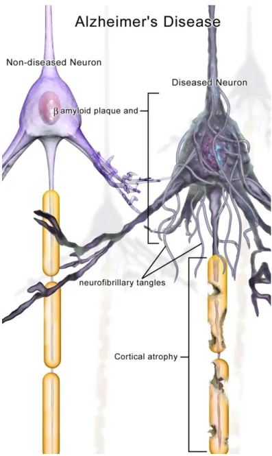
Figure 1.1: An illustration depicting the neuronal pathology in Alzheimer’s Disease (image credit Bruce Blausen;
commons.wikimedia.org/wiki/File:Blausen_0017_AlzheimersDisease.png). ... 2 Figure 1.2:An overview of synthetic biology based solutions for the treatment of neurodegenerative diseases. Reprinted with permission from reference [11]. ... 4 1.3: An illustration depicting the formation of insoluble fibrils from soluble
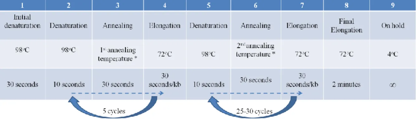
oligomeric structures because of misfolding... 5 Figure 1.4: An illustration summarizing the process of formation and propagation of aggregation in neurodegenerative diseases. Reprinted with the permission from publisher of the study [13]. ... 6 Figure 1.5: A drawing that depicts the proteins involved in a-agglutination system in Saccharomyces cerevisiae cells and the fused peptides and tags to be displayed on the surface. Figure is adapted from the reference [107]. ... 18 Figure 1.6: An illustration of Saccharomyces cerevisiae strain, EBY100. Aga1p is expressed on its genomic DNA and Aga2p fused with protein of interest (POI) is expressed on the plasmid DNA to establish surface display system with the POI. ... 19 Figure 1.7: An illustration of Saccharomyces cerevisiae strain, EBY100. Aga1p is expressed on its genomic DNA and Aga2p fused with protein of interest (POI) is expressed on the plasmid DNA to establish surface display system with the POI. ... 21 Figure 2.1: Reaction conditions of Polimerase Chain Reaction with Q5 polymerase 36 Figure 2.2: Reaction conditions of Polimerase Chain Reaction with Q5 polymerase in case primers with long overhang regions are used... 37 Figure 2.3:Reaction conditions of Polimerase Chain Reaction with pfu polymerase for verification of clonings by using the colony to serve as the template. ... 37 Figure 2.4: The flow chart indicating the construction of double-stranded amyloid sequences... 41 Figure 3.1: Agarose gel image of the first PCR to amplify Htt-25Q sequence by using forward primer 1 (to add HA tag and PstI restriction site) and reverse primer (to add
xii
XhoI restriction site) Lane 1: 2-log ladder Lane2-6: 25Q (Expected band length ~300 bp) Lane 7: negative control ... 45 Figure 3.2: Agarose gel image of the second PCR to amplify Htt-25Q by using forward primer 2 (to add SpeI restriction site) and reverse primer Lane 1-5: 25Q (Expected band length ~300 bp) Lane 6: negative control Lane7: 2-log ladder ... 45 Figure 3.3: Agarose gel image of the restriction enzyme digestion of pETcon
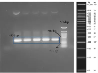
backbone and Htt-25Q sequence. Lane 1: NEB 2-log ladder, lane 2: digested pETcon, lane 3: empty, lane 4: digested Htt-25Q. Expected band lengths: 6 kb and 350 bp. ... 46 Figure 3.4: Agarose gel image showing the result of PCR to amplify Htt-46Q. Lane 1: negative control, Lane2-6: 46Q (Expected band length ~350 bp) Lane 7: 50-bp ladder ... 46 Figure 3.5: Products of double restriction enzyme digestion of Htt-46Q by using XhoI and SpeI. Lane 1: 50-bp ladder, Lane2-3: digested 46Q (Expected band length ~350 bp) ... 47 Figure 3.6: Agarose gel image showing the result of PCR to amplify Htt-103Q. Lane 1: NEB 2-log ladder, Lane2-6: 103Q (Expected band length ~500 bp) ... 47 Figure 3.7: Agarose gel image showing the result of the PCR to amplify amyloid 40 and amyloid 42 after annealing complementary oligo sequences form the double stranded DNA piece. Lane 1: negative control, lane 2-4: amyloid β40, lane 5: 50 bp ladder, lane 6: negative control, lane 7-9: amyloid 42. Expected band length: ~140 bp ... 48 Figure 3.8: Agarose gel image showing the result of gradient PCR to find the
optimum annealing temperature to eliminate the formation of amplicon in the negative controls for amyloid β42. Nonspecific bands disappeared at higher temperatures like 71oC which was used as the annealing temperature in the next experiments. ... 49 Figure 3.9: Agarose gel image showing the result of PCR to amplify amyloid 40 and amyloid 42 sequences and add homology regions corresponding to pETcon
backbone for Gibson assembly. Expected band lengths: ~200 bp. ... 49 Figure 3.10: Schematic representation of how two amyloid 40 or amyloid 42
seqeunces joined together. One of the primers provided homology with pETcon backbone and the other primer provided a common region containing GS linker and MluI cut site. ... 50
xiii
Figure 3.11: Agarose gel image showing the result of PCR to amplify amyloid 40 sequences with proper homology regions for gibson assembly to join two amyloid 40 sequences successively. Lane 1-6: amyloid 40 first fragment, lane 7: negative
control, lane 8: NEB 2-log ladder, lane 9-14: amyloid 40 second fragment, lane 15: negative control. Expected band length: ~200 bp. ... 50 Figure 3.12: Agarose gel image showing the result of PCR to amplify amyloid 42 sequences with proper homology regions for gibson assembly to join two amyloid 42 sequences successively. Lane 1: control, lane 2: amyloid 42 first fragment, lane 3: 50 bp ladder, lane 4: control, lane 5: amyloid 42 second fragment. Expected band
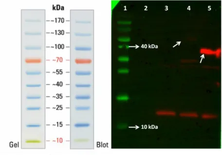
length: ~200 bp. ... 51 Figure 3.13: Agarose gel image showing the result of double restriction enzyme digestion reaction to verify the presence of -synuclein sequence within pETcon backbone using one enzyme digesting -synuclein sequence in the middle. Lane 1: NEB 2-log ladder, lane 2-3: colony #1, lane 4-5: colony #2, lane 6-7: colony #3, lane 8-9: colony #4, lane 10-11: colony #5. ‘-’ stands for not digested, ‘+’ stands for digested. ... 52 Figure 3.14: Agarose gel image showing the result of colony PCR on yeast colonies transformed with the indicated constructs above. Since two primers flanking Aga2p and gene of interest were used, amplicon lengths changed accordingly... 53 Figure 3.15: Agarose gel image showing the results of colony PCR experiments using colonies transformed with plasmids containing amyloid 40 and amyloid 40 conjugated with sfGFP followed by a series of different treatments. ... 54 Figure 3.16: Pellets of Saccharomyces cerevisiae EBY100 cells transformed with amyloid 40 conjugated with sfGFP (left) and amyloid 40 (right) after induction with galactose. ... 55 Figure 3.17: Fluorescent microscope images of Htt-46Q, Htt103-Q and amyloid 40 all conjugated with sfGFP. The ones on upperlane are bright field images. ... 56 Figure 3.18: The result of the experiment to see whether proteins of interest were interacting within the cells or on the surface and leading to cell aggregation. ... 57 Figure 3.19: Result of western blot analysis of proteins with c-myc tag produced by listed cells. ... 58 Figure 3.20: Result of western blot analysis of proteins with c-myc tag produced by cells transformed with pETcon with -synuclein, repeated amyloid 40, amyloid 42, and amyloid 40 respectively. ... 58
xiv
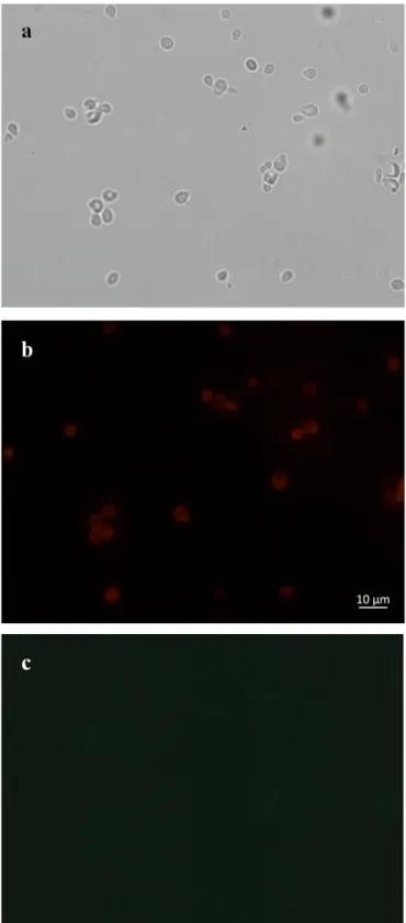
Figure 3.21: Bright field (a) and fluorescence microscopy (b,c) images of EBY100 cells transformed with pETcon plasmid expressing Htt-25Q upon galactose
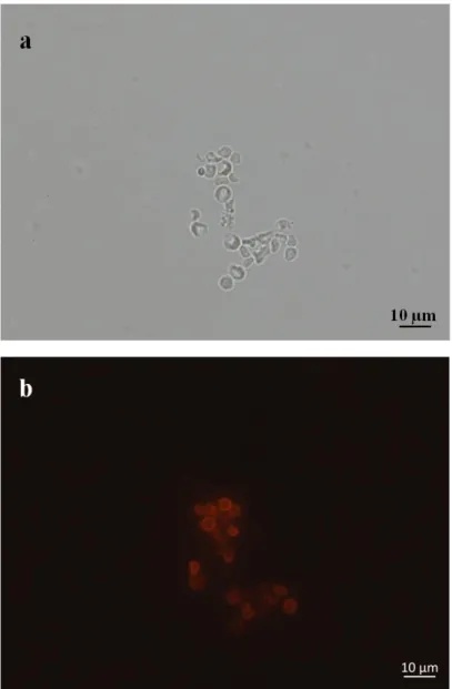
induction. ... 60 Figure 3.22: Bright field (a) and fluorescence microscopy with green filter (b) and red filter (c) images of control cells which were EBY100 cells transformed with pETcon plasmid expressing Htt-25Q upon galactose induction but not treated with primary antibody, anti c-myc antibody ... 61 Figure 3.23: Bright field (a) and fluorescence microscopy with red filter (b) images of control cells which were control EBY100 without pETcon plasmid upon galactose induction. ... 62 Figure 3.24: Bright field (a) and fluorescence microscopy (b) images of EBY100 cells transformed with pETcon plasmid expressing Htt-46Q upon galactose
induction. ... 63 Figure 3.25: Bright field (a) and fluorescence microscopy with green filter (b) and red filter (c) images of control cells which were EBY100 cells transformed with pETcon plasmid expressing Htt-46Q upon galactose induction but not treated with primary antibody, anti c-myc antibody. ... 64 Figure 3.26: Bright field (a) and fluorescence microscopy (b) images of EBY100 cells transformed with pETcon plasmid expressing Htt-103Q upon galactose
induction. ... 65 Figure 3.27: Bright field (a) and fluorescence microscopy (b) images of EBY100 cells transformed with pETcon plasmid expressing amyloid 40 upon galactose induction. ... 66 Figure 3.28: Bright field (a) and fluorescence microscopy (b) images of EBY100 cells transformed with pETcon plasmid expressing amyloid 42 upon galactose induction. ... 67 Figure 3.29: Bright field (a) and fluorescence microscopy (b) images of EBY100 cells transformed with pETcon plasmid expressing amyloid 40x2 upon galactose induction. ... 68 Figure 3.30: The image showing the difference coming from the EBY100 cells transformed with pETcon plasmid expressing amyloid 40x2 upon galactose induction depending on the low (a) or high (b) exposure. ... 69
xv
Figure 3.31: Bright field (a) and fluorescence microscopy (b) images of EBY100 cells transformed with pETcon plasmid expressing amyloid 42x2 upon galactose induction. ... 70 Figure 3.32: Bright field (a) and fluorescence microscopy (b) images of EBY100 cells transformed with pETcon plasmid expressing -synuclein upon galactose induction. ... 71 3.33: Linear map demonstrating the parts for the expression of Htt-25Q protein within Saccharomyces cerevisiae cells ... 72 Figure 3.34: Fluorescent profile of EBY100 cells transformed with pETcon plasmid expressing Htt-25Q upon galactose induction. Using the data points on the line indicated (a), graph of the fluorescent profile of data points (b) was drawn. ... 72 3.35: Linear map demonstrating the parts for the expression of Htt-46Q protein within Saccharomyces cerevisiae cells ... 73 Figure 3.36: Fluorescent profile of EBY100 cells transformed with pETcon plasmid expressing Htt-46Q upon galactose induction. Using the data points on the line indicated (a), graph of the fluorescent profile of data points (b) was drawn. ... 73 3.37: Linear map demonstrating the parts for the expression of Htt-46Q protein within Saccharomyces cerevisiae cells ... 74 Figure 3.38: Fluorescent profile of EBY100 cells transformed with pETcon plasmid expressing Htt-103Q upon galactose induction. Using the data points on the line indicated (a), graph of the fluorescent profile of data points (b) was drawn ... 74 3.39: Linear map demonstrating the parts for the expression of a40 protein within Saccharomyces cerevisiae cells ... 75 Figure 3.40: Fluorescent profile of EBY100 cells transformed with pETcon plasmid expressing amyloid 40 upon galactose induction. Using the data points on the line indicated (a), graph of the fluorescent profile of data points (b) was drawn. ... 75 3.41: Linear map demonstrating the parts for the expression of a42 protein within Saccharomyces cerevisiae cells ... 76 Figure 3.42: Fluorescent profile of EBY100 cells transformed with pETcon plasmid expressing amyloid42 upon galactose induction. Using the data points on the line indicated (a), graph of the fluorescent profile of data points (b) was drawn. ... 76 3.43: Linear map demonstrating the parts for the expression of a40x2 protein within Saccharomyces cerevisiae cells ... 77
xvi
Figure 3.44: Fluorescent profile of EBY100 cells transformed with pETcon plasmid expressing repeated amyloid 40 upon galactose induction. Using the data points on the line indicated (a), graph of the fluorescent profile of data points (b) was drawn. 77 3.45: Linear map demonstrating the parts for the expression of -synuclein protein within Saccharomyces cerevisiae cells ... 78 Figure 3.46: Fluorescent profile of EBY100 cells transformed with pETcon plasmid expressing -synuclein upon galactose induction. Using the data points on the line indicated (a), graph of the fluorescent profile of data points (b) was drawn. ... 78 Figure 3.47: Fluorescent profile of EBY100 cells transformed with pETcon plasmid expressing -synuclein upon galactose induction. Using the data points on the line indicated (a), graph of the fluorescent profile of data points (b) was drawn. ... 79 Figure 3.48: The sequences and 3D models of peptides against Htt-25Q, Htt-46Q and amyloid 40. ... 80 Figure 3.49: Linear map demonstrating the parts for the expression of the selected peptide as a fusion with sfGFP protein on pET22b expression vector and within BL21 cells. ... 80 Figure 3.50: Fluorescent signals of the BL21 cell pellets expressing sfGFP fused with the selected peptides against Htt-25Q, Htt-46Q and a40 on pET22b vector. ... 81 Figure 3.51: Agarose gel showing the result of colony PCR to screen the presence of sfGFP within the transformants. ... 82 Figure 3.52: A workflow showing the steps of binding assay. ... 82 Figure 3.53: The fluorescent microscope images of the slides containing EBY100 cells expressing amyloid 40 (sample) and empty EBY100 cells (negative control) both treated with purified fusion protein composed of sfGFP and the peptide against amyloid 40. ... 83 Figure 3.54: Agarose gel image of the result of PCR to obtain a backbone without any yeast marker to clone gene integration cassette. Expected band length: 5.2 kb.. 85 Figure 3.55: Agarose gel image showing the results of PCR amplifying pTDH3 promoter (lane 2-4) and ADH1 terminator (lane 5-7). Expected band length for pTDH3 is 750 bp, for ADH1 is 250 bp. ... 86 Figure 3.56:Agarose gel image showing the result of restriction enzyme digestion reaction with Acc65I and XhoI to clone pTDH3 and LEU2 marker after the
xvii
Figure 3.57:Agarose gel image showing the result of polymerase chain reaction that amplify LEU2 marker from pML107 plasmid to be cloned into genome integration cassette. Expected band length: 1.6 kb... 87 Figure 3.58: Schematic representation of genome integration cassette containing mOrange as the selection marker with the other parts and restriction sites. ... 88 Figure 3.59: Schematic representation of genome integration cassette containing LEU2 as the selection marker with the other parts and restriction sites. ... 88 Figure C.1: Plasmid map of pETcon vector in which Aga2 gene is fused with Htt-25Q ... 107 Figure C.2: Plasmid map of pETcon vector in which Aga2 gene is fused with Htt-46Q ... 107 Figure C.3: Plasmid map of pETcon vector in which Aga2 gene is fused with Htt-103Q ... 108 Figure C.4: Plasmid map of pETcon vector in which Aga2 gene is fused with
amyloid 40 ... 108 Figure C.5: Plasmid map of pETcon vector in which Aga2 gene is fused with
amyloid 42 ... 109 Figure C.6: Plasmid map of pETcon vector in which Aga2 gene is fused with
repeated amyloid 40 ... 109 Figure C.7: Plasmid map of pETcon vector in which Aga2 gene is fused with
repeated amyloid 42 ... 110 Figure C.8: Plasmid map of pETcon vector in which Aga2 gene is fused with -synuclein ... 110 Figure D.1:The result indicating the sequence analysis of Htt-25Q being cloned into pETcon backbone. ‘G...’ Stands for GS linker. ... 111 Figure D.2: The result indicating the sequence analysis of Htt-46Q being cloned into pETcon backbone. ‘GS...’ Stands for GS linker. ... 112 Figure D.3: The result indicating the sequence analysis of Htt-103Q being cloned into pETcon backbone. ‘G...’ Stands for GS linker. ... 113 Figure D.4: The result indicating the sequence analysis of amyloid 40 being cloned into pETcon backbone. ‘G...’ Stands for GS linker. ... 114 Figure D.5: The result indicating the sequence analysis of amyloid β42 being cloned into pETcon backbone... 115
xviii
Figure D.6: The result indicating the sequence analysis of repeated amyloid β40 being cloned into pETcon backbone. ‘G...’ stands for GS linker. ... 116 Figure D.7: The result indicating the sequence analysis of repeated amyloid 42 being cloned into pETcon backbone. ‘G...’ stands for GS linker. ... 117 Figure D.8: The result indicating the sequence analysis of repeated -synuclein being cloned into pETcon backbone. ‘G...’ stands for GS linker. ... 118 Figure D.9: The result indicating the sequence analysis of integration cassette being cloned into pETcon backbone with a forward primer. ... 119 Figure D.10: The result indicating the sequence analysis of integration cassette being cloned into pETcon backbone with a reverse primer. ... 120 Figure D.11: Statistics of sequence analysis of Htt-25Q. A region which is 718 bp in length including Htt-25Q sequence was chosen for statistical analysis. ... 121 Figure D.12: Statistics of sequence analysis of Htt-46Q. A region which is 677 bp in length including Htt-46Q sequence was chosen for statistical analysis. ... 121 Figure D.13: Statistics of sequence analysis of Htt-103Q. A region which is 811 bp in length including Htt-103Q sequence was chosen for statistical analysis. ... 122 Figure D.14: Statistics of sequence analysis of amyloid β40. A region which is 865 bp in length including amyloid β40 sequence was chosen for statistical analysis. .. 122 Figure D.15: Statistics of sequence analysis of amyloid β42. A region which is 870 bp in length including amyloid β42 sequence was chosen for statistical analysis. .. 123 Figure D.16: Statistics of sequence analysis of repeated amyloid β40. A region which is 938 bp in length including repeated amyloid β40 sequence was chosen for
statistical analysis. ... 123 Figure D.17: Statistics of sequence analysis of repeated amyloid β42. A region which is 956 bp in length including repeated amyloid β42 sequence was chosen for
statistical analysis. ... 124 Figure D.18: Statistics of sequence analysis of repeated -synuclein. A region which is 860 bp in length including repeated -synuclein sequence was chosen for statistical analysis. ... 124 Figure D.19: Statistics of sequence analysis of integration cassette by using a forward primer. A region which is 988 bp in length including promoter and marker gene sequence was chosen for statistical analysis. ... 124
xix
Figure D.20: Statistics of sequence analysis of integration cassette by using a reverse primer. A region which is 1000 bp in length including promoter and marker gene sequence was chosen for statistical analysis. ... 125 Figure D.21: The result indicating the sequence analysis of the peptide against Htt-25Q fused with sfGFP.. ... 124 Figure D.21: The result indicating the sequence analysis of the peptide against Htt-46Q fused with sfGFP.. ... 125 Figure D.22: The result indicating the sequence analysis of the peptide against a40 fused with sfGFP.. ... 125
xx
List of Tables
Table 2.1: Components required for growing yeast cells………...23 Table 3.1: A table indicating the presence or absence of the restriction sites within yeast parts. ‘-’ stands for the absence, and the numbers indicating the number of the restriction sites within corresponding part. ‘*’ shows that the restriction site is absent in the sequence the part, but it is present within the 100 bp flanking regions of that part………..84 Table A.1: The DNA sequences of the neurodegenerative proteins and the parts of integration cassette that have been used in this study in 5’ to 3’ orientation………..98 Table B.1: The sequences of primers that have been used in cloning experiments of neurodegenerative proteins and genome integration cassette………...104
1
INTRODUCTION
1.1 Neurodegenerative Diseases
Neurodegenerative diseases are progressive diseases associated with a gradual and irreversible loss in discrete neuronal populations of central nervous system [1]. Once neuronal loss reaches a severe stage that affects the cognitive functioning, patients develop dementia which is characterized with behavioral impairments and the problems in cognitive capabilities such as language, memory, reasoning and decision making [2, 3]. To enlighten the underlying causes of neurodegenerative diseases, numerous studies have suggested possible explanations disruption in different mechanisms or abnormalities in cell functioning such as oxidative damage due to free radicals [4], neuroinflammation [5], mitochondrial dysfunction [6], endoplasmic reticulum (ER) stress [7] and so on.
2
Figure 0.1.1: An illustration depicting the neuronal pathology in Alzheimer’s Disease (image credit Bruce Blausen; commons.wikimedia.org/wiki/File:Blausen_0017_AlzheimersDisease.png).
Alzheimer’s Disease (AD), Parkinson’s Disease (PD), Huntington’s Disease (HD), Amyotrophic Lateral Sclerosis (ALS), Metachromic Leukodystrophy (MLD), and Spinal Muscular Atrophy (SMA) are some examples of neurodegenerative diseases [8]. Despite of the fact that there are differences between the sites where the diseases initiate, the types of neurons that are affected, behavioral, motor and cognitive symptoms and the progress, there is a common factor leading to neurodegeneration in these diseases. Protein aggregation and the formation of inclusion bodies are
3
shared characteristics of neurodegenerative diseases. The behavior of misfolded proteins as a nucleation unit triggering the formation of polymeric structures accelerates the aggregation [9, 10].
1.2 Synthetic Biology Based Solutions for Neurodegenerative
Diseases
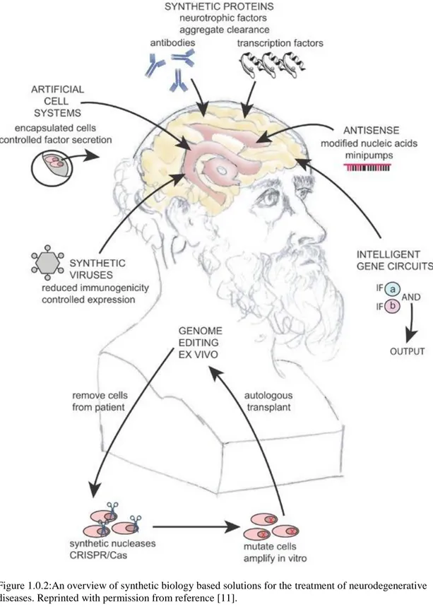
Synthetic biology has the potential to create solutions to existing limitations in detection and treatment. Most of the current approaches focus on the alleviation of the symptoms and clearance of the toxic accumulations. However, synthetic biology tools can target the underlying factors such as disease causing genes, and monomeric and oligomeric structure involved in aggregation. Genome editing technologies are promising especially in diseases caused by a single gene like Huntington’s disease. Logic gates can be utilized to recognize the agents causing the disease and to respond them by producing signals or genes to eliminate disease related formations. RNA inhibition systems and use of antisense oligonucleotides can silence the genes contributing to the diseases. Synthetic proteins such as artificial transcription factors and improved antibodies can decelerate the aggregate formation. Artificial cells can be programmed to release growth factors to support healthy neurons in the diseased brain regions. Stem cells can be engineered and transplanted to the region of interest as a neuroprotective and neurorestorative strategy. Synthetic viruses enable a safe and controlled delivery method. Gene circuits constructed with special parts can provide a controlled and temporal or constant expression of specific genes. Re-establishment of a healthy gut microbiome leads to neuroprotection by sending signals via gut-brain axis. Considering all advanced technologies, a comprehensive therapeutic intervention thanks to synthetic biology can target the real source of the neurodegeneration [11].
4
Figure 1.0.2:An overview of synthetic biology based solutions for the treatment of neurodegenerative diseases. Reprinted with permission from reference [11].
1.3 Accumulation of Proteins in Neurodegenerative Diseases
Amyloid beta (a) and tau in AD, alpha-synuclein (-syn) in PD and mutant huntingtin (mHtt) in HD are the proteins causing the formation of aggregates in
5
different neuronal populations of the brain [12]. Despite the contribution of different genes and proteins in different neurodegenerative diseases, misfolding, conformational changes in protein structure, disruptions in clearance mechanisms, accumulation of toxic aggregates considerably resemble to each other. Discovery of self-propagating behavior and seeding activity of misfolded proteins has been a milestone for the developments of diagnostic and therapeutic approaches [12, 13]. Accumulated proteins, known as amyloids, leading to neurodegeneration mostly contain structures called cross- which are composed of intermolecular -sheets [14]. From another perspective, protein misfolding involves a process of the rearrangement of the protein into an array of -strands that are stabilized through hydrophobic interactions and hydrogen bonds. -strands provide places for triggering other molecules to join the misfolded structure [13].
1.0.3: An illustration depicting the formation of insoluble fibrils from soluble oligomeric structures because of misfolding.
Dock-Lock mechanism explains the formation of insoluble aggregates from soluble monomeric and oligomeric structures in two main steps. The first step called ‘dock’ when the soluble monomeric structures dock to an amyloid template is a reversible
6
phase. However, in the ‘lock’ phase, conformational changes in the monomeric structures takes place and irreversibly associated fibrous structures are formed [14, 15].
Seeding-nucleation model proposed by Lansbury and colleagues explains aggregation in two stages. In the nucleation stage, a stable seed as a nucleation unit is formed. In the elongation stage, the seeds formed in nucleation phase grow rapidly since they trigger soluble monomeric structures to be incorporated into polymeric structures [16, 17]. Fragmentation of long polymers gives rise to the more seed structure leading to the acceleration of aggregation.
Figure 1.0.4: An illustration summarizing the process of formation and propagation of aggregation in neurodegenerative diseases. Reprinted with the permission from publisher of the study [13].
Another hypothesis claims that misfolded proteins behaving as infectious agents can be transmitted like prions. Several pathways have been suggested to mediate cell-to-cell transmission of improperly folded seeds such as exosome mediated transport, transmission via tunneling nanotubes, transfer through protein-protein interactions, mechanisms of endocytosis and exocytosis and so on [18-21]. Although it seems
7
logical to target the cell-to-cell transmission of the seeds, it is complicated to manipulate general mechanisms that can be involved in other cellular processes [13].
1.4 Diagnostic Studies for Neurodegenerative Disorders
In order to make effective therapeutic interventions, early diagnosis of neurodegenerative diseases is essential. However, neuropathy as a one of the most reliable diagnostic can be applied only after patients’ death [11, 22]. A major difficulty to detect neurodegenerative diseases is that protein biomarkers of neurodegenerative diseases are present in the body fluids like blood and cerebrospinal fluid in small amounts [23]. Limited understanding of neural circuits, complexity of the brain, presence of the blood brain barrier and difficulties of bypassing it for targeted delivery indicate that there is a need for a comprehensive theranostic approaches to diagnose and cure these diseases [11].
To track polyglutamine aggregation, traditional methods utilizes microscopy or detergent insolubility which do not clarify the mechanisms leading to protein misfolding. As a combination of fluorescence energy transfer (FRET) system and cell imaging, Pollitt et al. constructed a reporter system to track polyglutamine aggregation in HEK293 cell line and showed that aggregation-prone proteins with polyglutamine repeats gave FRET positive signal when they were fused with fluorescent proteins. In the study, 2800 biomolecules have been screened and the ones with a inhibitory activity that reversed the process of polyglutamine aggregation were characterized using Drosophila model for polyglutamine disease [24]. In a similar study in 2017, a biosensor cell line has been developed using fusions of tau domain with P301S mutation with two different fluorescent protein, cyan fluorescent protein (CFP) and yellow fluorescent protein (YFP). Application of tau seeds exogenously trigger the initiation of aggregation that results in close proximity of
8
fluorescent proteins and positive signal [25]. Holmes et al. designed a FRET-based sensor and assessed taupathy and tau seeding activity in the brains of transgenic P301S mice. They also carried out assays on cross-seeding activity between tau and a-synuclein [26].
yTRAP (yeast transcriptional reporting of aggregating proteins) is another novel system that has been constructed in yeast cells with a theranostic approach. The sensor system has been established in order to give an output depending on the solubility state of the protein of interest. Protein of interest that is prone to aggregation is fused to a synthetic promoter which can activate its cognate promoter only if the cell is not in aggregation state [27].
Since the cells undergoing neurodegeneration are located at different regions in the brain depending on the type of the disease, spatial and temporal control of gene expression is essential for gene therapies. Convection-enhanced delivery was first described in the study of Bobo et al [28]. This method facilitates direct delivery of the therapeutics to the brain by creating a pressure gradient at the tip of a catheter with a small diameter. The method has been developed with the integration of the interventional magnetic resonance imaging (iMRI) technology [29]. There are studies using iMRI-CED technology that focus on neurorestorative therapies for the delivery of neurotrophic factors to support brain health and gene replacement therapies to deliver genes responsible for GABA production and gene encoding the rate limiting enzyme in dopamine synthesis converting levodopa to dopamine to provide symptomatic relief to the patients with Parkinson’s disease [8, 30].
9
1.5 Therapeutic Interventions
1.5.1 Gene Therapy
Gene therapy is an approach with the purpose of prevention and treatment of the neurodegenerative diseases by means of neuroprotection, neurorestoration and disappearance of neuropathology. The main purpose of gene therapy is to either compensate a defective gene by replacing with a transgene or correcting it or supporting the healthy cells in the diseased region [8, 31]. There are viral and nonviral vectors being used as gene therapy agents [32]. Non-viral vectors including nucleic acids and especially naked plasmid DNA are mostly delivered as complexed with some delivery vehicles such as cationic polymers and lipids [33]. Due to rapid clearance and transient gene expression profile, non-viral vectors are not convenient agents to confer sufficient dosage effect for chronic neurodegeneration [31, 34]. Viral vectors have the ability to invade the cells and deliver the DNA content they are loaded with to the host cell. Their genome is altered to prevent their replication for safety issues [35]. Commonly used viral vectors are lentiviral vectors and adenoviral vectors. Lentiviruses which have larger DNA loading capacity are designed to be integrated into the genome apart from the adeno-associated viruses (AAV) [31, 36]. This unique feature provides stable expression pattern in long-term, but also brings the risk of insertional mutagenesis [37]. AAVs have been commonly used in clinical trials to deliver genes to the brain. There are several serotypes of AAVs; however, some of the serotypes have been so widespread that numerous people have developed immunity against them [38, 39]. Among those serotypes, AAV2 is one of the most preferred ones due to its clinically safe profile whereas AAV9 is more capable of crossing the blood-brain barrier as compared to the other
10
serotypes. It has been indicated that intravenous administration of AAV9 provides the opportunity for transgene delivery to adult motor neurons [40].
1.5.2 Cell-based therapies
Transplantation of engineered cells that secrete neurotrophic factors promoting growth and maintenance of neurons or enzymes that are deficient in neurodegenerative disease conditions is a neuroprotective and neurorestorative strategy [11, 41, 42]. Despite promising results for treating and preventing neurodegenerative diseases, there are still problems such as immunorejection of grafted cells, lack of stromal support and required growth factors and insufficient integration of the grafted cells. There are two technical concerns about cell replacement therapies for neurodegenerative diseases: which cell type should be convenient and which transplantation method leads to effective functioning of the grafted cells [43]. Human retinal pigment epithelial cell line engineered to secrete NGF has been indicated as an encapsulated therapeutic agent since it displays contact-independent growth characteristics and low nutritional requirements[44]. With the self-renewal capacities and ability to differentiate into several cell types, stem-cells are ideal sources for cell replacement therapies.
In addition to embryonic stem cells which are obtained during embryonic stages and induced pluripotent cells derived from adult somatic cells, glial cells and neurons originated from stem cells such as neural stem cells (NSC) and mesenchymal stem cells can be used in transplantation therapies. NSCs are advantageous regarding their rapid growth characteristics and stable gene expression pattern of therapeutic genes [45-47].
Vascular endothelial growth factor (VGEF) which is a growth factor with neuroprotective effects especially in case of brain injury has been promising in the
11
treatment of ALS. When VGEF is coupled with stem cell transplantation technology, improvements in therapeutic effects have been seen. Transplantation of NSCs that overexpress VEGF resulted in functional improvements, increased survival and delay in the disease onset [48].
As a therapeutic intervention to metachromatic leukodystrophy which is caused by a deficiency of an enzyme called arylsulfatase A, programmed haematopoietic cells that expressed the enzyme were transplanted and migrated to the brain. Complemented enzyme activity resulted in the clearance of the toxic clusters composed of the substrate of the enzyme [49].
1.6 Alzheimer’s Disease
World Health Organization (WHO) has reported that there are approximately 50 million cases of dementia increasing with the addition of 10 million cases each year. 60-70% of cases with dementia arise from Alzheimer’s disease. During the disease progression, degeneration of cholinergic neurons and accumulation of amyloid beta plaques have been observed. Familial Alzheimer’s disease is rare form being inherited in a Mendelian fashion and caused by mutations in either amyloid precursor protein or the enzymes that are responsible for its cleavage whereas both genetic and environmental factors contribute to the sporadic form of the disease [50, 51].
As a neuroprotective intervention, nerve growth factor (NGF) which supports cholinergic neurons in the basal forebrain that undergo degeneration due to the disease was delivered using AAV2 vector [52]. Safe profile of the vector and NGF expression in an active form have been indicated; however, the clinical trial failed to meet the expectations [53].
Neuroinflammation is one of the underlying factors in Alzheimer’s disease. Interleukin-2 as an inflammation controller was delivered within AAV vector to
12
mouse model for AD. Improvements in memory due to the alleviations in hippocampus have been observed [54, 55]. Another strategy to prevent aggressive progression of AD is to reduce the levels of tau and amyloid beta which have a tendency to form clusters within neurons. To do so, antisense oligonucleotides (ASO) was used to prevent tau aggregation, but this strategy needs to be assessed in animal models to make conclusions about their role in disease progression [56]. AAV mediated miRNA delivery to knockdown acyl-CoA:cholesterol acyltransferase 1 has succeeded in reducing amyloid beta levels [57, 58]. To reverse the effects of reduction in neprilysin enzyme that has a role in amyloid beta catabolism, AAV9 mediated delivery of the enzyme into hippocampus resulted in reduced amyloid beta levels [59]. Indeed, improved memory and learning skills reported by Iwata et al. [60].
Delivery of growth factors is another promising strategy to prevent neuropathology and to provide neuroprotection. The level of insulin growth factor (IGF) in the hippocampus of AD patients has been indicated. AAV8 mediated delivery of IGF caused a significant decrease in the levels of amyloid beta in a mouse model overexpressing amyloid precursor protein [61]. Lentiviral delivery of glial cell-line derived neurotrophic factor (GDNF) that supports brain cells resulted in the preservation cognitive skills such as memory and learning [62].
There are cell transplantation studies for AD that have focused on either the toxicity caused by amyloid beta or neuroprotective and neurorestorative precautions using growth factors to alleviate the symptoms. For instance, glucagon-like peptide-1 secreted by mesenchymal stem cells reduced amyloid beta deposition and toxicity [63-66]. While the secretion of vascular endothelial growth factor (VGEF) by encapsulated cells resulted in improvements in cognition and reduction in amyloid
13
beta deposition and hyperphosphorylated tau [67], secretion of ciliary neurotrophic factor resulted in improved cognition and stabilization of synaptic proteins [68].
1.7 Parkinson’s Disease
According to European Brain Council (EBC), approximately 6.3 million people worldwide have Parkinson’s Disease (PD) which is the second most common neurodegenerative disease after AD. Parkinson’s Disease (PD) is characterized by symptoms such as impairments in motor function, resting tremor, slow movements and stiffness. Loss of dopaminergic neurons in the brain region called substantia nigra that modulates movement and the reduction in dopamine levels in the striatum are underlying factors of neuropathology. Lewy bodies which are cytoplasmic inclusion bodies are composed of abnormal fibrillar assemblies [69].
In 1998, Spillantini et al. have indicated the composition of lewy bodies are ubiquitin and mostly a-synuclein through several immunostaining experiments[70].
Current therapies include deep brain stimulation (DBS) and supplying dopamine via drugs such as levodopa which is a dopamine precursor. Although these therapies are effective interventions considering symptomatic relief to some extent, they are insufficient to target the underlying pathology of the disease. Levodopa that is used for dopamine replacement gets less effective as PD progresses and DBS can be emotionally and cognitively deteriorating [71].
At early stages of the disease, dopamine replacement therapy is efficient; however, even increased dosages do not result in same therapeutic outcome as PD progresses. One strategy to overcome this insufficiency is to deliver the rate-limiting enzyme of the dopamine synthesis as a pro-drug approach [8]. The enzyme is called aromatic L-amino acid decarboxylase (AACD) which catalyzes the conversion of levodopa to dopamine during dopamine synthesis. AAV2 mediated delivery of AACD resulted in
14
statistically significant improvements in behavioral symptoms, lowered the need for levodopa and its side effects [72-76].
There are also neurorestorative strategies focusing on the delivery of GDNF to support neurons. Viral mediated delivery has been combined with iMRI-guided CED technology and utilized in phase 1 trial [77].
There are also studies focusing on cell transplantation strategies for neuroprotective and neurorestorative purposes. Encapsulated cells supplying dopamine precursor and dopamine were transplanted into striatum to alleviate the syptoms of PD. Although this intervention had been effective for several years, it eventually became ineffective since degeneration of dopaminergic neurons continued during this period [78-81]. It has been indicated that dopamine supply is temporarily effective, thus protective precautions before the neurodegeneration takes place can be more successful [82]. Supporting this claim, loss of dopaminergic neurons could be prevented by implating encapsulated cells that released certain dose of GDNF daily into the substantia nigra [83].
1.8 Huntington’s Disease
Huntington’s Disease (HD) shares similarities with AD and PD regarding the improper processing of proteins and neurons that are vulnerable to these proteins forming clusters. However, in contrast to complex and unknown underlying mechanisms of AD and PD, HD is an autosomal dominant disorder with a single cause which is a mutation leading to the expansion of trinucleotide CAG repeat coding glutamine [84, 85]. In healthy individuals’ genome, the number of CAG repeats on the exon 1 of chromosome 4 varies between 6 and 34. Repeat size longer than 36 repeats leads to a problematic, unstable and improperly expanded protein [86, 87].
15
HD is one of the nine polyglutamine diseases that result in neurodegeneration [85, 88]. As a disease inherited with an autosomal dominant pattern, there is 50% risk of transmitting the disease to the offspring. HD occurs less frequently compared to AD and PD with a ratio of 1:10000 people. Symptoms include cognitive impairments, incoordinate involuntary muscle contractions, behavioral changes and chorea which can be defined as unpredicted body movements [89].
AAV mediated generation of a novel mouse model for HD not only enlightened the mechanism of neuropathology but also enabled the exploration of novel therapeutic interventions. AAV-DJ model indicated changes in behavior, increased in the expression of inflammatory cytokines, and occurrence of neural apoptosis.
AAV9 mediated miRNA delivery targeting exon 48 of human mutant huntingtin eliminated 50-60% of mutant HTT mRNA and mutant huntingtin protein six months after the injection in striatum of sheep model [90]. AAV mediated SIRT3 gene displayed neuroprotective effects by preventing mitochondrial oxidative stress and providing neuronal support [91]. Evers et al, also reported reduced levels of mutant huntingtin protein and mRNA in minipigs as a response to delivery of AAV5 mediated delivery of Huntington-lowering gene [92].
Since disruption of a single gene causes HD, the disease is in the focus of therapeutic applications using gene editing technologies such as CRISPR/Cas9. There are several strategies to silence mHtt expression. One approach is to target promoter region or the beginning of the gene; however, preclinical studies have suggested a global silencing of the mutant and wild-type alleles since it has been shown that complete deletion of HTT gene is not deleterious [93-95]. Exon 1 coding the protein with polyglutamine repeats can be removed completely by using two guide RNAs to
16
targeting the both sides of the exon [96, 97]. Using next generation sequencing technology, allele-specific strategies can also be developed [98].
Taking neuroprotective precautions is more probable in case of HD since genetic screening can determine the individuals carrying the risk of developing the disease [82]. Implantation of cells producing NGF and CNTF not only preserved neuronal populations of striatum such as GABAergic and cholinergic neurons, but also improved motor and cognitive capabilities [99-101]. CNTF secreting cells encapsulated within polymer structures were transplanted into striatum of monkey models and reduced the striatal damage while protecting and increasing the GABAergic and cholinergic neurons [102, 103].
1.9 Biotechnology Tools for Protein Studies
1.9.1 Yeast Surface Display
Displaying recombinant proteins on the cell surface of Saccharomyces cerevisiae is an advanced technology to study and engineer proteins, to optimize affinity of antibodies, to improve catalytic activity of enzymes, to discover binding epitopes, to assess interactions of proteins with small molecules or other proteins and so on [104-107]. Among several proteins anchored to the yeast cell surface such as Flo1p, Cwp1p, Ag1p, Aga2p is the most preferred protein for the fusion of protein of interest [108].
Utilization of yeast display technology provides some advantages over the other display methods. In addition to the ease of handling of a unicellular organism, machineries for eukaryotic expression, protein translation and processing make it possible to study a range of recombinant proteins with proper folding and functions. The protein translation and secretion mechanisms of yeast cells resembling
17
mammalian cells have made this single-cell microbe a convenient platform to perform library applications and to study eukaryotic proteins too [107]. Yeast two-hybrid system has enabled the studies on protein-protein interactions [109]. Thanks to innate homologous recombination mechanism, genetic manipulations can be done on yeast genome via transformation of a PCR product with upstream and downstream homologies of the target site as short as 40-50 nucleotides for purposes like gene deletion, gene insertion or tag addition [110, 111]. To be classified as ‘generally regarded as safe’, Saccharomyces cerevisiae cells are ideal organisms for applications of food and pharmaceutical industry [112]. However, different glycosylation pattern as compared to mammalian cells [113, 114] and the limited library size as compared to platforms using phage and bacteria [115] are some drawbacks of yeast display system.
Aga1p and Aga2p are proteins that are anchored to the cell surface of yeast cells with mating type a. Those proteins are subunits of a-agglutinin which modulates cell adhesion during yeast cell mating [116, 117]. While Aga1p is anchored to the surface through beta-glucan covalent bonds, Aga1p and Aga2p form two disulfide bonds before being secreted into the extracellular space and placed on the cell surface [107]. This placement results in the display of the protein of interest that is conjugated to Aga2p on the yeast surface.
18
Figure 1.0.5: A drawing that depicts the proteins involved in a-agglutination system in Saccharomyces
cerevisiae cells and the fused peptides and tags to be displayed on the surface. Figure is adapted from
the reference [107].
Aga1 gene that encodes Aga1p is integrated into the genome of the strain of Saccharomyces cerevisiae designed for surface display applications. A circular plasmid with an auxotrophic marker for the selection contains Aga2 gene encoding the Aga2p is fused with the protein of interest. Both Aga1 and Aga2 are regulated by a galactose inducible promoter. Therefore, surface display of the proteins is not constitutive. The conditional expression prevents the potential formation of cytotoxic protein products [107, 114].
19
Figure 1.0.6: An illustration of Saccharomyces cerevisiae strain, EBY100. Aga1p is expressed on its genomic DNA and Aga2p fused with protein of interest (POI) is expressed on the plasmid DNA to establish surface display system with the POI.
1.9.2 Phage Display Library
Phages, also known as bacteriophages, are the viruses that infect the bacterial cells. These biological entities have numerous unique features such as resistance to harsh environments, ease of handling, inexpensive production in large quantities and immunostimulatory effects [118, 119].
Phage display is an advanced biotechnology tool that utilizes phage particles to display foreign peptides on their surface by fusing the sequence encoding the peptide into the genes encoding viral coat protein [120]. Phage libraries which are constructed by integrating randomized oligonucleotides into the sequence of viral coat proteins have the potential to display a variety of peptides. The technique has been used in several applications such study of protein-protein interactions, drug development and discovery of target molecules like antigens, antibodies and receptors [119].
20
Phages have a safe profile since viruses have had interactions with mammalians throughout an evolutionary period. Additionally, the genetic diversity of the phages and the large library size are the advantageous sides of this tool over the other display platforms [119, 121, 122].
Screening strategy for phage display libraries is based on biopanning which is a process of recursive binding and washing steps to find the peptides which bind to the target with high affinity. In biopanning, phages with a pool of peptides are incubated with the protein of interest. Then, harsh washing steps are applied to eliminate nonspecific and weak binders. Several methods such as changing pH, use of a proteolytic enzyme addition of a competitive ligand or a denaturing agent can be used for retrieval of the phages. They are not damaged upon these applications since they are stable under tough conditions [123]. Following the retrieval step, bacterial cells are infected with phages; however, the library might be still heterogeneous. Therefore, a few affinity selection steps improve the peptide selection process. DNA sequence encoding the selected peptides can be obtained via DNA sequencing [119].
1.10 Aim of the study
Most of the current studies on the treatment of neurodegenerative diseases focus on the alleviation of the symptoms and clearance of the toxic accumulations after the aggregates has been formed. The purpose of this study is to develop peptides that can block the monomeric structures before the aggregation takes place. The proteins that have been used in this study are amyloid , -synuclein and mutant Huntingtin proteins causing aggregate formation in Alzheimer’s Disease, Parkinson’s Disease and Huntington’s Disease, respectively. To develop blocking peptides as drug candidates, methods of biotechnology such as gene cloning, yeast surface display and phage library display have been used. Disease causing proteins have been displayed
21
on the surface of Saccharomyces cerevisiae cells. The peptides interacting with disease causing proteins have been selected among a library displayed on the phages. The potential of those candidate peptides will be assessed through ex vivo binding assays and using neural cell lines and animal models. Peptides that have been selected are also promising to be integrated into biosensors as recognition units.
Figure 1.0.7: An illustration of Saccharomyces cerevisiae strain, EBY100. Aga1p is expressed on its genomic DNA and Aga2p fused with protein of interest (POI) is expressed on the plasmid DNA to establish surface display system with the POI.
22
CHAPTER 2
2
EXPERIMENTAL
2.1 Yeast maintenance and growth conditions
Yeast cells can be grown both in liquid medium and on agar plates. A minimal medium including a nitrogen source, a carbon source like glucose, and trace metals is enough to grow the wild-type strain. Yeast cells have the metabolic pathways to produce all amino acids they need; however, extra suppliance of proteins, amino acids, yeast extract, vitamins or nucleotide precursors accelerates the growth.
The wild type yeast strains that have the capacity of meeting nutritional requirements are named as prototroph. The mutant strains with mutations in the genes that belong to the pathways being responsible for the synthesis of the nutritions with survival importance are called auxotroph. The genes coding the enzymes of these pathways that are involved in the production of metabolically crucial monomers are called auxotrophic markers. Auxotrophic genes do not only meet nutritional requirements but also have a role as biological parts within cassettes utilized in genomic manipulations or plasmids as a marker genes. The presence of auxotrophic mutant strains enables doing complementation assays by meeting the auxotrophy deficiency via a DNA fragment to be inserted into the genome or a plasmid including the missing auxotrophy marker. LYS2, URA3, MET15, TRP1, LEU2, HIS3 are the examples of commonly used auxotrophy markers.