Introduction
Moulds are of great importance not only with respect to health and industry but also in terms of economics due to their metabolic properties. In industrial microbiology, the ability to produce organic acids such as citric acid and itaconic acid, some enzymes, pigments and antibiotics by moulds has been exploited (Topal, 1984). In addition, these organisms
are important contaminants of food and agricultural products due to their presence in the soil and air. As well as their negative effects on food, moulds are capable of producing mycotoxins. One to their importance, the microfloristic study of moulds is well in progress around the world. The most common strains reported belong to Penicillium Link:Fr and Aspergillus Fr.:Fr. genera (Asan, 2000).
Colonial and Morphological Characteristics of Some
Aspergillus Fr.:Fr. Species Isolated from Vineyards in Manisa and
‹zmir Provinces (Turkey)
Rengin ELTEM
Ege University, Faculty of Enginering, Department of Bioengineering, 35100 Bornova, ‹zmir - TURKEY Tülin AfiKUN
Bal›kesir University, Faculty of Science and Letters, Department of Biology, Bal›kesir - TURKEY Nermin SARIGÜL
Ege University, Faculty of Enginering, Department of Bioengineering, 35100 Bornova, ‹zmir - TURKEY Evrim ÖZKALE TAfiKIN
Celal Bayar University, Faculty of Science and Letters, Department of Biology, Manisa - TURKEY Hafize EFEND‹LER
Ege University, Faculty of Science, Department of Biology, 35100 Bornova, ‹zmir - TURKEY
Received: 29.01.2002 Accepted: 28.03.2003
Abstract: Five Aspergillus Fr.:Fr. species and 3 varieties were described from the point of view of colonial and morphological characteristics. These species and varietes are isolated from soil, grape and sultana raisins from vineyards in Manisa and ‹zmir provinces (Turkey). These are as follows: A. flavofurcatus Bat. & H.Maia, A. heteromorphus Bat. & H.Maia, A. pulverulentus (McAlpine) Wehmer, A. unguis (Emile-Weil & L.Gaudin) Thom & Raper, A. viridinutans Ducker & Thrower, A. foetidus Thom & Raper var. pallidus (Nakaz., Simo & A.Watan.) Raper & Fennell, A. foetidus Thom & Raper var. acidus (Nakaz., Simo and A.Watan.) Raper & Fennell andA. nidulans (Eidam) G.Winter var. acristatus Fennell & Raper. The final 2 are cited for the first time in Turkey. Key Words: Mycoflora, Aspergillus, Manisa, ‹zmir, Turkey
Manisa ve ‹zmir Yörelerindeki Ba¤lardan ‹zole Edilen Bazı Aspergillus Fr.:Fr. Türlerinin Koloni ve Mikroskopik Özellikleri
Özet: Bu çalıflmada befl Aspergillus Fr:Fr türü ve üç varyetesi koloni ve morfolojik özellikleri bakımından tanımlanmıfltır. Bu tür ve varyeteler Manisa ve ‹zmir illerindeki sultani cinsi üzüm ba¤ı toprakları ile yafl ve kuru üzümlerden izole edilerek tanımlanmıfllardır. Bunlar;A. flavofurcatus Bat. & H.Maia, A. heteromorphus Bat. & H.Maia, A. pulverulentus (McAlpine) Wehmer, A. unguis (Emile-Weil & L.Gaudin) Thom & Raper, A. viridinutans Ducker & Thrower, A. foetidus Thom & Raper var. pallidus (Nakaz., Simo & A.Watan.) Raper & Fennell,A. foetidus Thom & Raper var. acidus (Nakaz., Simo & A.Watan.) Raper & Fennell ve A. nidulans (Eidam) G.Winter var. acristatus Fennell & Raper’ dır. Belirtilen son iki tür Türkiye mikoflorası için ilk kez belirtilmektedir.
There have been many studies on the microfungus flora of Turkey. The majority of the studies were carried out to determine the microfungus flora of the soil in different parts of the country (Öner, 1970; Öner, 1974; Ekmekçi, 1975; Öner et al., 1977; Haseneko¤lu, 1985; Haseneko¤lu, 1987; Gür, 1991; Sülün & Haseneko¤lu, 1993; Asan & Ekmekçi, 1994; Haliki & Dizbay, 1997; Azaz & Haseneko¤lu, 1997; Asan, 1997a,b), others were undertaken on several kinds of agricultural products (Ulukufl & Sa¤ır, 1982; Aran & Eke, 1987; Çolako¤lu, 1987; Çolako¤lu, 1991, Haseneko¤lu, 1988b) and foods (Alperden et al., 1982; Haseneko¤lu, 1988a; Birbir et al., 1995; Eltem & Öner, 1995, Güven et al. 1997).
A checklist showing the strains belonging to the genera Aspergillus and Penicillium Link:Fr. in Turkey has been in preparation since 1940 (Asan, 2000). According to published articles, there are 251 species belonging to these 2 genera that have been identified after isolation from different parts of Turkey.
The aim of the study is to contribute to the checklist of Aspergillus and Penicillium of Turkey. Some new strains of Aspergillus were isolated and identified from vineyard soils and grape and sultana raisin samples in our research.
Materials and Methods
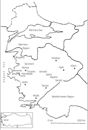
The soil, grape and sultana raisin samples were taken from 62 different vineyards in Manisa and ‹zmir provinces (Figure 1) in 1998 and 1999. In the isolation of moulds from soil samples, the “soil dilution plate method” (Waksman, 1922) was used. In the isolation of moulds from fresh and dried sultanas the “pour plate method” (Brock & Madigan, 1991) was used. Rose-bengal chloramphenicol agar (Oxoid CM549) and dichloran-glycerol (DG18) agar base (Oxoid CM729) were used as isolation media.
The identifications of the isolates were made using Raper & Fennell (1965); Smith (1971), Domsch et al. (1980), Samson et al. (1981), Samson & Pitt (1990), Powell et al. (1994) and Samson & Pitt (2000). Czapex dox agar (CZ) (modified) (Oxoid CM97), malt extract agar (MEA) (Oxoid CM59) and Czapek yeast autolysate agar (CYA) were used as identification media. Citation of the authors of fungal names was performed according to Kirk & Ansell (1992).
Results
As a result of the survey, 772 moulds were isolated from soil, grape and raisin samples. The identifications revealed 39 Aspergillus species and varieties, and among them 2 varieties are new reports for the Turkish mycoflora. The descriptions of the most abundant 5 species and 3 varieties are given below.
Two strains of A. flavofurcatus Bat. & H.Maia were identified, one from the soil and another from dried fruit samples. Two strains of A. foetidus Thom & Raper var. acidus (Nakaz., Simo & A.Watan.) Raper & Fennell, one from grape and another from raisin samples, were found. In A. foetidus Thom & Raper var. pallidus Nakaz., Simo & A.Watan., 29 samples were identified in soil, 79 in fresh grapes and 59 were from raisins, making a total of 167 strains. In A. heteromorphus Bat. & H.Maia a total of 9 strains were identified, 5 from fresh and 4 from dried grapes. For A. nidulans (Eidam) G.Winter. var. acristatus
Marmara Sea Marmara Region Kütahya Uflak Afyon Bergama Saruhanl› Ahmetli Salihli Alaflehir Sar›göl Denizli Ayd›n
Mu¤la Mediterranean Region Manisa Turgutlu Kemalpafla ‹zmir Menemen 0 km 200 km ‹zmir Aegean Sea
Fennell & Raper 4 strains were determined from soil samples only. In A. pulverulentus (McAlpine) Wehmer 1 strain in grapes, in A. unguis (Emile-Weil & L.Gaudin) Thom & Raper 3 strains in raisins and in A. viridinutans Ducker & Thrower 2 strains from soil and raisin samples were found.
Aspergillus flavofurcatus Bat. & H.Maia, Anais Soc Biol Pernambuco 13: 94-96 (1955).
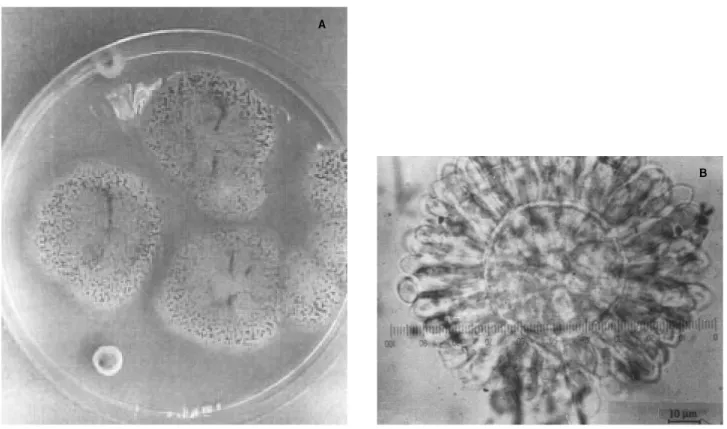
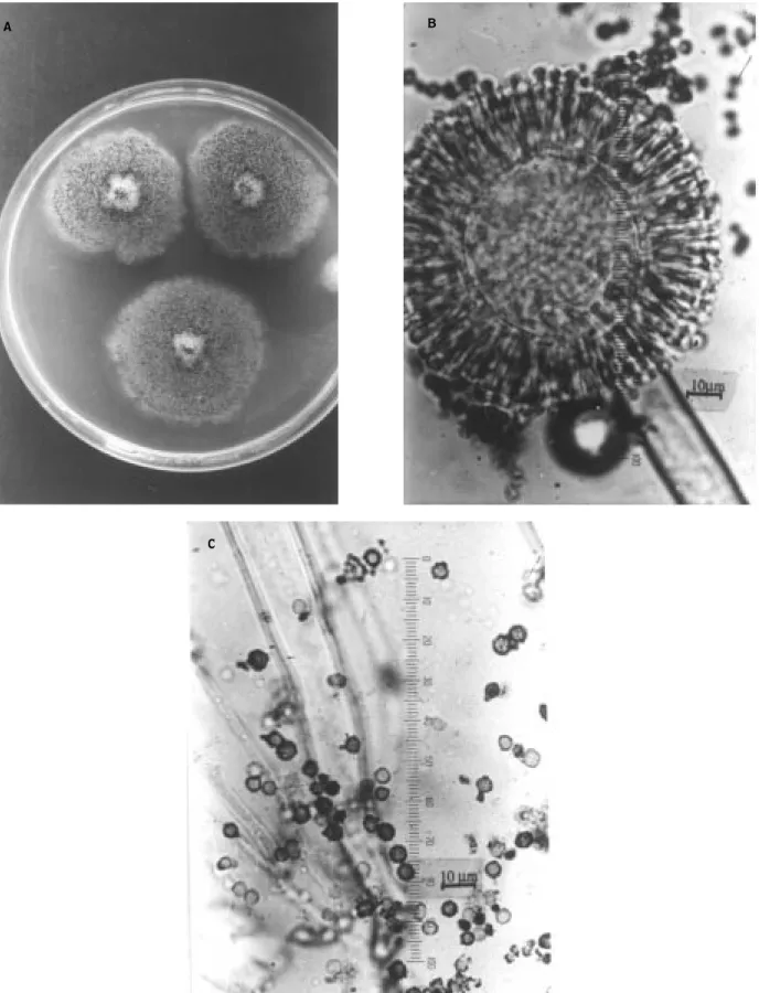
Colony Characteristics: This species, in 10 days, at 25 ºC on CZ, produced colonies 4 cm in diameter. Colony surface is green at margins and brown in the centre and around, the reverse is colourless. There are colourless exudates. The colony is 2.0-3.0 mm deep because of long stipes. Conidial heads are globose formerly, then radiate and 200-400 µm in diameter. Colonies on MEA are 5.5-6.0 cm in diameter and dark brown-black. More or less zonate, basal mycelium thin. On the colony, there are white hyphae.
Microscopic Characteristics: Vesicles are globose or subglobose and 20-70 µm in diameter. Phialide biseriate, metulae 10-12 x 4.0-6.5 µm, phialide 9.0-20 x 3.0-6.5 µm. Stipes are smooth and pigmented, 10-20 µm wide, 1.5-2.1 mm long. Conidia are slightly rough, globose, yellow-brown and 5.0-8.0 µm in diameter.
Aspergillus foetidus Thom & Raper var. acidus (Nakaz., Simo & A.Watan.) Raper & Fennell, Gen. Aspergillus 326 (1965).
Colony Characteristics: On CZ, at 27 ºC, in 2 weeks, colonies are 4.5-6.0 cm in diameter. Texture is lanose, margin white, centre yellowish. On basal mycelium sporulation is not dense. Sporulation is more at colony margin and centre. Conidial heads are blackish brown. Reverse bright yellow. Odour not distinct. On MEA at the same physical conditions, colonies are 4.0-5.0 cm in diameter. There is zonation and mycelium is golden yellow.
Microscopic Characteristics: On CZ conidial heads are 550-650 µm, stipes are 700-1000 x 15-27.5 µm, vesicles are 40-70 µm, phialide biseriate, metulae 18-22 x 4.5-5.0 µm, phialide 7.0-10 x 2.0-3.0 µm. Conidia are globose, 4.0-4.5 µm and wavy.
Aspergillus foetidus Thom & Raper var. pallidus (Nakaz., Simo & A.Watan.), Raper & Fennell, Gen. Aspergillus 325 (1965).
Synonym: Aspergillus aureus var. pallidus Nakaz, Simo & A.Watan. J Agr Chem Soc Japan 12: 961-962 (1936).
Figure 2. A. flavofurcatus on MEA (A), microscopic appearance of conidial head (B). A
Figure 3.A. foetidus var. acidus. on MEA (A), microscopic appearance of conidial head (B) and phialide (C).
A B
Colony Characteristics: This species develops colonies 2.5-4.0 cm in diameter on CZ. Colony surface is white or cream formerly, then turns to dark olive green-brown. Reverse colour of colony is colourless or cream, changes brown; later odour is mouldy, no zonation. There are colourless exudates on colony surface. Conidal heads are in olive green, radiate and 200-600 µm in diameter.
Microscopic Characteristics: Stipes are smooth, brown pigmented, 1.0-2.5 µm x 10-20 µm. Vesicles are globose generally 50-80 µm in diameter. Phialide biseriate, metulae 10-20 x 3-5 µm, phialide 10-20 x 3-5 µm. Conidia are globose or subglobose, obvious and rough, 2.5-5.0 µm in diameter.
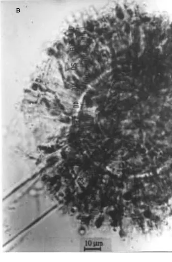
Aspergillus heteromorphus Bat. & H.Maia in Anais Soc Biol Pernambuco 15: 200 (1957).
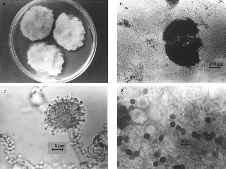
Colony characteristics: On CZ in 2 weeks, at 25 ºC, colonies are 3.0-3.5 cm in diameter. Greenish black, at
the margins there is submerged mycelium 2.0-3.0 mm in width. Conidial heads at margin are small and yellow, thin, smooth, richly sporulated, in centre more sporulated. Reverse is colourless, no distinct odour, no exudate. On MEA under the same physical conditions, colonies are 6.0-7.0 cm in diameter, plane, radiate, velvety; there are brown, globose or subglobose sclerotia 100-150 µm in diameter.
Microscopic Characteristics: On CZ, at the marginal and submarginal areas stipes are 800-900 x 8.0-12 µm, conidial heads are dark green, globose and radiate, 180-200 µm in diameter becoming columnar with age. Vesicles are reddish brown, 35-45 µm, phialide biseriate, metulae 10-12 x 3.5-4.0 µm, phialide 6.0-8.0 x 2.5-3.0 µm. Conidia 3.0-3.5 µm, verruculose, sclerotia are 300-500 µm in diameter. Heads are radiate, 100-3000 µm, colony surface granular, more or less zonate, heads are darker in centre and margins, odour not distinct, no exudate, reverse colourless.
Figure 4. A. foetidus var. pallidus on CZ (A), microscopic appearance of phialide and conidia (B).
Figure 5. A. heteromorphus on CZ (A), microscopic appearance of conidial head (B) and conidia (C). B
A
Aspergillus nidulans (Eidam) G.Winter var. acristatus Fennell & Raper, Mycologia 47 (1): 79 (1955).
Synonym: Emericella acristata (Fennell & Raper) Y.Horie in Trans Brit Mycol Soc Japan 21: 491 (1980).
Colony Characteristics: Colony diameter is 2.5-3.0 cm in 10 days at 25 ºC on CZ. Texture lanose, at the beginning of development colony surface is white then ochre, centre of colony lightly raised. Reverse is dark purple like eggplant. Colourless exudates on the surface. Conidial heads are short and columnar.
Microscopic Characteristics: Stipes are brown and smooth, 75-150 x 4.0-5.0 µm. Vesicles are subglobose and 8.0-11 µm in diameter. Phialide biseriate, metulae 5.0-6.0 x 2.0-3.0 µm, phialide 5.0-6.0 x 2.0-2.5 µm. Conidia are globose, smooth, 3.0-4.0 µm in diameter. Cleistothecia are globose, 60-180 µm. Ascospores are
orange-red, 4.0-5.0 x 3.0-4.0 µm in diameter. Hulle cells are globose, 25-30 µm.
Aspergillus pulverulentus (McAlpine) Wehmer, Centralbl Bacteriol 2. Abth. 18: 394 (1907).
Basionym: Sterigmatocystis pulverulenta McAlpine, Agric Gaz New South Wales 7: 302 (1897).
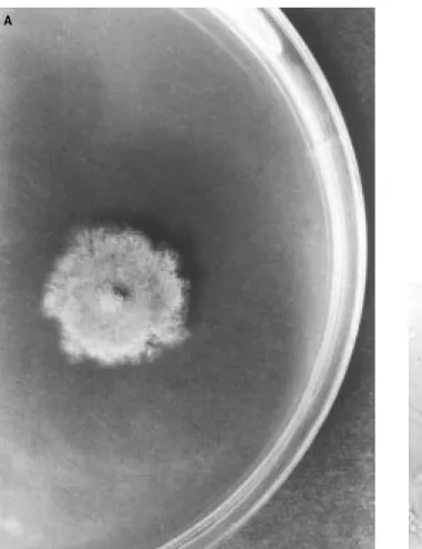
Colony Characteristics: This species developes quickly on CZ in 7 days, at 25 ºC, produces colonies 2.5-4.0 cm in diameter. Texture is velvety, surface grey-black, reverse white-cream. Basal mycelium is white, odour mouldy, exudates are small and colourless. Conidial heads are blackish grey, characteristically radiate, usually 500-600 µm in diameter. Colonies on MEA developed rapidly and reached 5.0 cm in diameter at the same temperature in 7 days. Texture is velvety, surface brown and black, reverse colour white; colourless exudates and zonation are present.
Figure 6. A. nidulans var. acristatus on CZ (A), cleistothecium (B), microscopic appearance of conidial head (C) and ascospores and hulle cells (D).
A B
D C
Microscopic Characteristics: Stipes are smooth, colourless, upper parts are light brown, 15-20 µm in width, 1000-3000 µm sometimes 5000 µm in length. Foot cells are 35-45 µm in length. Vesicles are globose, 60-75 µm in diameter. Phialide biseriate, metulae 20-30 x 3.0-3.5 µm in young individuals and 40-50 x 3.0-3.5 µm in adults. Conidia are globose-subglobose, verruculose or granular and 4.5-5.0 µm in diameter. Measurements of colonies on MEA are similar to those on CZ, but conidial heads are more columnar.
Aspergillus unguis (Emile-Weil & L.Gaudin) Thom & Raper, Mycologia 31: 667 (1939).
Basionym: Sterigmatocystis unguis Emile-Weil & L.Gaudin, Arch Med Exp Anat Pathol 28: 463 (1918).
Teleomorph: Emericella unguis Malloch & Cain, Can J Bot 50: 62 (1972).
Colony Characteristics: On CZ in 10 days at 25 ºC bright yellow in centre, darker and green around. Colony centre is raised and after day 13 becomes wrinkled.
Reverse is nearly orange. At first conidial heads radiate, but later turn columnar. On the MEA at the same physical conditions, colonies are sage green and 3.0-4.0 cm in diameter.
Microscopic Characteristics: On CZ, stipes are smooth and yellow-brown, 45-60 x 3.0-3.5 µm at the foot cells, walls are thick and roughened, there are sterile spicular hyphes. Vesicles are hemispherical and 7.0-12 µm in diameter. Phialide biseriate. Metulae 5.0-6.0 x 2.5-3.0 µm, phialide 6.0-7.0 x 2.5-3.0 µm. Conidia are globose or subglobose in shape and rough, 2.5-3.0 mm in diameter. Conidia chains are 150 µm in length. On MEA, conidial structures are similar to those on CZ.
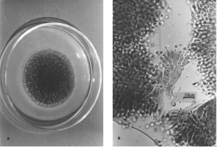
Aspergillus viridinutans Ducker & Thrower, Aust J Bot 2: 355 (1954).
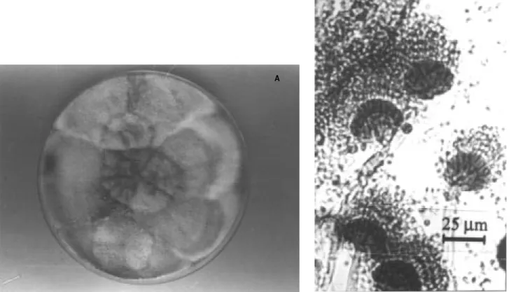
Colony Characteristics: This species produces colonies which are 3.0-4.5 cm in diameter in 14 days at 25 ºC on CZ. Colony is white at first and then turns green in centre and cream along margins. Reverse is light
Figure 7. A. pulverulentus on CZ (A), microscopic appearance of conidial head (B). B
Figure 8. A. unguis on CZ (A), microscopic appearance of conidial head (B) and sterile hyphae (C). B
A
brown. Exudates are small and colourless with no zonation. Conidal heads are columnar and dark, 50-60 x 20-25 µm. Colonies on MEA 6.0-6.5 cm in diameter under the same conditions. Surface is light green-white. Colourless exudates are present. Texture is funiculose-floccose. Reverse is colourless.
Microscopic Characteristics: On CZ, vesicles are globose or hemispherical and 3/4 of it is fertile. Diameter of vesicles is 8.0-13 x 9.0-12 µm. Vesicles are bent. Phialide uniseriate and 5.0-8.0 x 2.5-3.0 µm. Stipes are 80-100 µm in length, 3.0-4.0 µm in width and smooth. Conidia are globose or subglobose, rough, 2.5-3.0 µm in diameter.
Discussion
In studies carried out in Turkey on mycoflora, Aspergillus and Penicillium have been the dominant genera isolated from soil, agricultural and food commodities. In one examination of mycoflora in Turkey 82 Aspergillus species and varieties were determined (Asan, 2000). A. flavofurcatus, A. pulverulentus, A. unguis,A. viridinutans and A. foetidus var. pallidus were
cited for the first time in Turkey by Eltem et al. (2001). In addition, A. heteromorphus was cited for the first time by Azaz & Pekel (2002). In our work 2 varieties (A. foetidus var. acidus and A. nidulans var. acristatus) are reported for the first time increasing the total number to 91.
In Turkey, the most widespread species is A. niger van Tieghem which is followed by A. flavus Link, A. fumigatus Fres., A. versicolor (Vuill.) Tiraboschi, A. ochraceus Wilhelm, A. terreus Thom and A. wentii Wehmer. It is thought that these species are more adapted to the prevailing ecological conditions (Asan, 2000).
Newly reported Aspergillus varieties in the Turkish mycoflora as well as other reported species were isolated and identified from the sultana vineyards in Manisa and ‹zmir provinces in the Aegean Region by Eltem et al. (2001).
A morphological examination of species for the purposes of identification was made first with the naked eye or by using a low magnification microscope and thereafter detailed examinations were performed according to Gams et al. (1987) by measuring the
Figure 9. A. viridinutans on MEA (A), microscopic appearance of conidial head (B).
B
dimensions of the microbiological structures, photographing the microscopic structure and using relevant literature as references.
In the examinations, some colony and microscopic characteristics of the species were found to be different from those stated by Raper & Fennell (1965). For example, the colony diameter of A. flavofurcatus on CZ was smaller. It was determined that the colony diameter of A. foetidus var. pallidus ranged between 2.0 and 4.0 cm and the diameter of the vesicles varied up to 80 µm. The most distinguishing property of A. foetidus var. acidus, a golden yellow mycelium in MEA as stated by
Raper & Fennel (1965), was observed in both of the strains.
Öner (1973) and Smith & Moss (1985) report that Aspergillus species are the most dominant in the soil of mild climatic zones, confirming our results. Eltem et al. (2001) reported that the genus Aspergillus is dominant in vineyards and that one of the most widespread fungi species is A. foetidus var. pallidus; 545 Aspergillus isolates and 167 A. foetidus var. pallidus strains were identified. In conclusion, the descriptions of some Aspergillus species are given in this paper.
References
Alperden ‹, Karaali A, Eke D, Topal fi, Aran N & Arkun G (1982). Türkiye gıdalarında küfler ve mikotoksinler. J Kükem 5: 98-99. Aran N & Eke D (1987). Bazı tahıl çeflitleri ve ürünlerindeki küf florası.
J Kükem 10: 41-52.
Asan A (1997a). Trakya bölgesi mısır tarlaları mikrofungus florası üzerinde arafltırmalar-I. Tr J of Biogy 21: 89-101.
Asan A (1997b). Trakya Bölgesi mısır tarlaları mikrofungus florası üzerinde arafltırmalar-II. J Kükem 20: 9-18.
Asan A (2000). Checklist of Aspergillus and Penicillium species reported from Turkey. Tr J of Botany 24: 151-167.
Asan A & Ekmekçi S (1994). The determination of Penicillium and Aspergillus species in Edirne soils and their seasonal distribution. Tr J of Biology 18: 291-303.
Azaz AD & Haseneko¤lu ‹ (1997). An investigation into the microfungal flora of field soils in the GAP (Southeastern Anatolia Project) irrigation area of Harran Plain. Tr J of Botany 21: 165-172. Azaz AD & Pekel O (2002). Comparision of soil fungi in burnt and
unburnt forest soils in the vicinity of Kargıcak (Alanya, Turkey). Tr J of Botany 26: 409-416.
Birbir M, Ilgaz A, Yurdun T & Çilo¤lu F (1995). Piyasada satılmakta olan hazır çorbalardan küflerin ayırımı ve tanımlanması. Pendik Vet Mikrobiyoloji Dergisi 26: 163-174.
Brock TD & Madigan MT (1991). Biology of Microorganisms. Englewood Cliffs (NJ): Prentice-Hall International.
Çolako¤lu G (1987). Erzurum ili ve ilçelerindeki bu¤day ve arpa depolarından izole edilen küf mantarları üzerinde arafltırmalar. J Kükem 10: 60-69.
Çolako¤lu G (1991). Erzurum yöresinde so¤an hastalı¤ı etmeni fungusların tesbiti ve 1985-1986 yılları arasındaki da¤ılıflları. Do¤a-Tr J of Botany 15: 110-114.
Domsch KH, Gams W & Anderson TH (1980). Compendium of Soil Fungi. Vol. 1-2. London: Academic Press.
Ekmekçi S (1975). Güney yarı Ege Bölgesi topraklarından izole edilen Penicillium ve Aspergillus türleri. Bitki 2: 19-29.
Eltem R & Öner M (1995). Salamura tipi siyah zeytinlerin küf florasının incelenmesi. Tr J of Botany 19: 11- 17.
Eltem R, Özkale E, Sarıgül N, Efendiler H, Karaboz ‹ & Tamer AÜ (2001). Manisa ve ‹zmir ‹llerinde çeflitli sultaniye ba¤larında yetiflen üzümlerin küf florasının incelenmesi. XII. Biyoteknoloji Kongresi 17-21 Eylül, 2001 Bildiriler Kitabı, Ayvalık-Balıkesir, pp. 43-46.
Gams W, van der Aa HA, van der Plaats-Niterink AJ, Samson RA & Stalpers JA (1987). CBS Course of Mycology. 3rd edition. pp 136: Baarn. Institute of Royal Netherlands Academy of Arts and Sciences, Centraalbureau voor Schimmelcultures.
Gür K (1991). Mufl ve Van topraklarından mikrofungus da¤ılımı üzerine bir arafltırma. J Kükem 14: 68-69.
Güven K, Kıvanç M, Karakafl N & Asan A (1997). Eskiflehir’de tüketilen kültür mantarı (Agaricus bisporus (Lange) Imb.) mikroflorasının belirlenmesi. J Kükem 20: 31-36.
Haliki A & Dizbay M (1997). Bergama yöresindeki bazı tarımsal alanlardan mezofilik toprak mikrofunguslarının izolasyonu ve mevsimsel da¤ılımları. Tr J of Biolgy 21: 329-341.
Haseneko¤lu ‹ (1985). Sarıkamıfl civarı orman, çayır ve tarla topraklarının mikrofungus florası. J Kükem 8: 40-46.
Haseneko¤lu ‹ (1987). Do¤u I¤dır ovası çorak topraklarının mikrofungus populasyonu üzerine ön bir arafltırma. J Kükem 10: 53-59.
Haseneko¤lu ‹ (1988a). Erzurum ve çevresinde üretilen küflü peynirlerin mikrofungus florası üzerine bir arafltırma. J Kükem 11: 35-42.
Haseneko¤lu ‹ (1988b). Türkiye’nin Karadeniz bölgesinde depolanmıfl fındıkların mikroflorası üzerinde bir arafltırma. J Kükem 11: 9-20.
Kirk PM & Ansell AE (1992). Authors of Fungal Names. Index of Fungi Supplement. International Mycological Institute. Latimer Kew, Surrey (UK): Trend & Co. Ltd.
Öner M (1970). Soil microfungi of Turkey. Mycopathol Mycol Appl 42: 81-87.
Öner M (1973). Atatürk Üniversitesi Erzurum çiftli¤i E¤erli da¤ı kuzey yamacı ve Trabzon-Hopa sahil fleridi mikrofungus florası ile ilgili bir arafltırma. Erzurum: Atatürk Üniv. Yay. No 21, Arafltırma serisi No: 17.
Öner M (1974). Seasonal distribution of some Fungi Imperfecti in the soils of Western part of Anatolia. Mycopathol Mycol Appl 52: 267-268.
Öner M, Ekmekçi S & Dizbay M (1977). Plant succesion and development of fungi in the soil. Ege Unv J Fac Sci Seri B 1: 57-63.
Powell KA, Renwick A & Pederby JF (1994). The Genus Aspergillus from Taxonomy and Genetics to Industrial Application. New York: Plenum Press.
Raper KB & Fennell DI (1965). The Genus Aspergillus, pp 686 Baltimore: Williams & Wilkins Company.
Samson RA, Hoekstra ES & Oorschut V (1981). Introduction to food-borne fungi. Central Bureau voor Schimmelcultures, Baarn.
Samson RA & Pitt JI (2000). Integration of Modern Taxonomic for Penicillium and Aspergillus Classification. Amsterdam: Harwood Academic Publishers, pp 9-79.
Samson RA & Pitt JI (Eds.) (1990). Modern Concepts in Penicillium and Aspergillus Classification., NATO ASI Series, pp 478. New York: Plenum Press.
Smith G (1971). An Introduction to Industrial Mycology. London: Edward Arnold Ltd.
Smith JE & Moss MO (1985) Mycotoxin Formation, Analysis and Significance. Chichester: John Wiley & Sons.
Sülün Y & Haseneko¤lu ? (1993). A study on Aspergillus Mich ex Fr. and Penicillium Link ex Gray flora of the soils of Northeast Anatolia, Türkiye. Tr J of Botany 17: 49-60.
Topal fi (1984). Gıda maddelerinden ayrılan (izole edilen) ve tanınan (identifiye edilen) küfler üzerinde arafltırmalar. Gıda 9: 253-264. Ulukufl ‹ & Sa¤ır A (1982). Elazı¤ ve Diyarbakır illerinde biber kurumaları ve hastalı?ın fungal etmenleri üzerinde ön çalıflmalar. Plant Protect Bul 22: 13-20.
Waksman SA (1922). A method of counting the number of fungi in the soil. J Bacteriol 7: 339-341.