Yazışma Adresi/Address for Correspondence: Dr. Alev Arslan, Cukurova Universıty Faculty of Medicine, Department of Pediatrics, Adana, Turkey E-mail: [email protected], aarslan@baskent. edu.tr
Geliş tarihi/Received: 04.05.2019 Kabul tarihi/Accepted: 30.07.2019 Çevrimiçi yayın/Published online: 16.09.2019
ARAŞTIRMA / RESEARCH
Serum Fas and Fas Ligand levels in childhood acute lymphoblastic
leukemias
Çocukluk çağı akut lenfoblastik lösemilerde serum Fas ve Fas Ligand düzeyleri
Alev Arslan
1, Bülent Antmen
21Baskent University Faculty of Medicine, Adana Turgut Noyan Application and Research Center, Department of Pediatrics, Department of Pediatric Cardiology Adana, Turkey
2Acıbadem Hospital,Department of Pediatrics, Divison of Pediatric Hematology, Adana, Turkey Cukurova Medical Journal 2019;44(4):1400-1404.
Abstract Öz
Purpose: The aim of this study was to investigate the Fas and Fas Ligand levels of cell membrane receptors that play a role in understanding the mechanism of apoptosis in childhood acute lymphoblastic leukemia (ALL). Materials and Methods: We investigated serum Fas and Fas Ligand levels by using ELISA method in childhood acute leukemias. Twenty-nine cases with acute lymphoblastic leukemia and twenty-seven healthy children included the study. We investigated serum Fas and Fas Ligand levels at the time of diagnosis from peripheral blood samples.
Results: Serum Fas levels of patients were median 63 pg/mL (min 15- max 690 pg/mL) and in the control group were median 37 pg/mL (min 8- max 67 pg/mL). Fas Ligand levels of patients were median 0.18 ng/mL (min 0- max 2.1 ng/mL) and in the control group median 0.1 ng/mL (min 0- max 4 ng/mL). The comparison of the mean values of Fas levels in acute leukemia patients groups and control group have shown important difference as statistically, but Fas Ligand levels were similar. Fas levels in T-ALL patients according to immunophenotypes were statistically higher than B-ALL . The Fas levels of patients who had relapsed were statistically higher than patients in remission.
Conclusion: Serum Fas levels could be a useful prognostic marker in ALL patients follow up.
Amaç: Bu çalışmada, çocukluk çağı akut lenfoblastik lösemilerinde (ALL), apoptozis mekanizmasının anlaşılmasında rol oynayan hücre membran reseptörlerinden serum Fas ve Fas Ligand düzeyinin araştırılması amaçlanmıştır.
Gereç ve Yöntem: Çocukluk çağı akut lenfoblastik lösemi tanısı (1-18 yaş) almış 29 hastada tanı anında periferik kandan elde edilen serum Fas ve Fas Ligand düzeyleri ELISA yöntemi ile çalışıldı. Yirmiyedi çocuktan oluşan kontrol grubunun yaş dağılımı 1-15 yıl arasındaydı.
Bulgular: ALL‟li hastalarda serum Fas düzeyi ortanca 63 pg/mL (15-690 pg/mL) kontrol grubunda ortalama serum Fas düzeyi ortanca 37 pg/mL (8-67 pg/mL) saptandı. ALL‟li hastalarda serum Fas Ligand düzeyi ortanca 0,18 ng/mL (0-2,1 ng/mL), kontrol grubunda ise ortanca 0,1 ng/mL (0-4 ng/mL) saptandı. Akut lenfoblastik lösemi tanılı hastalar ile, kontrol grubu arasında ortalama serum Fas düzeyleri açısından istatistiksel olarak anlamlı fark varken, Fas Ligand düzeyleri arasında fark saptanmadı. Akut Lenfoblastik Lösemili hastalar immunfenotiplemeye göre serum Fas düzeyi karşılaştırıldığında T-ALL hastalarında B-ALL’li hastalara göre istatistiksel olarak anlamlı fark bulundu. Relaps olan hastalarda remisyondaki hastalara göre Fas düzeyleri anlamlı olarak yüksek saptandı.
Sonuç: Bu bulgular sonucunda serum Fas düzeylerinin ALL hastalarda prognostik faktör olarak değerlendirilebileceği düşünüldü.
Key words: ALL, Fas, Fas Ligand Anahtar kelimeler: ALL, Fas, Fas Ligand
INTRODUCTION
Acute lymphoblastic leukemia (ALL) is the most common hematological malignant disease of childhood, occurring in a specific stage of
hematopoiesis of normal lymphoid progenitor cells, resulting in clonal expansion. ALL is important in this age group not only in terms of being the most common malignant disease in childhood but also in terms of success in treatment1.
The regulation of cell death, such as regulation of cell proliferation, is considered to be a complex event. All differentiated cells of the multicellular organism are capable of self-death by activation of the internally coded suicide program. When this program is activated it initiates a complex form of cell death called apoptosis2-4. The most well-defined
death receptor Fas (CD95) functions in the regulation of apoptosis was reported in many tumor types5,6. Fas initiate apoptosis by binding its apoptotic signal to the ligand on cytotoxic T cells. The naturally soluble Fas Ligand form (sFasL) is normally inactive and can act as a Fas Ligand on the surface of cytotoxic T cells by activation. Many defects in Fas-associated apoptosis may cause accumulation and prolonged survival in leukemic cells. Numerous research studies have shown that hematopoietic are dependent on antiapoptotic proteins for survival7-8.
Hence the significance of apoptosis as a prognostic marker is less well studied in paediatric ALL we aimed to investigate the Fas and Fas Ligand levels which play a very important role in the balance of apoptotic and antiapoptotic stimuli related to blastic cells in childhood ALL and evaluate their effects on prognosis and overall survival.
MATERIAL AND METHODS
This cross-sectional study was conducted between October 2005 and August 2008. Medical records of all newly diagnosed pediatric ALL patients younger than 18 years (N=35) who were admitted to the Divison of Pediatric Hematology and Oncology Clinic at the Çukurova University were evaluated. The diagnosis of ALL was confirmed by bone marrow biopsy on all patients, and cytogenetic analysis and flow cytometry were available in all. Patients received treatment with standard BFM TR-ALL 2000 chemotherapy for TR-ALL. Six patients were excluded from the study because of irregular follow up and lack of knowledge about the progress of the disease. Thus, twenty-nine children who were newly diagnosed and regularly followed up at least two year after diagnosis were included the study. The control group included 27 healthy children who were selected from general pediatric outpatient clinic. The study group demographics, relaps and survival outcomes were noted. Patients with ALL were subgrouped according to immunophenotyping.
CALLA (+) B-ALL, CALLA (-) B-ALL and T-ALL. Serum samples were taken from patients by taking 2 mL of venous blood during the diagnosis and stored in the deep-freezer at -80 °C until the day of the study. Bender Medsystems Human sApo-1 Fas Elisa kit
BMS245 (Austria) and Biosource immunoassay kit
Human sFas Ligand KHS9521 (California, USA) according to the manufacturer's specifications were used. The absorbance values were measured via
Medispec ESR 200 Elisa Plate Reader. The ranges of the kits for sCD95 and sCD95L were 0.23–15 ng/mL and 0.72–12.92 ng/mL, respectively. The study is approved by the ethical committee of Cukurova University Medical Faculty (12th April 2005, meeting number 6) and each subject was informed about the study and asked to fill in the signed informed consent form
Statistical analysis
Statistical analysis was performed using the statistical package SPSS (Version 17.0, SPSS Inc., Chicago, IL, USA). If continuous variables were normal, they were describle as the mean±standard deviation (p>0.05 in Kolmogorov-Smirnov test or Shapira-Wilk (n<30)), and if the continuous variables were not normal, they were described as the median. Comparisons between groups were applied using one way ANOVA test or Steudent t test for normally distrubited data and Kruskal Wallis test or Mann Whitney U test were used for the data not normally distrubited. The catagorical variables between the groups was analyzed by using the Chi square test. Correlations between measures were tested by Spearman’s coefficients test.
Overall survival was analyzed using the Wald test, and the log-rank test was used to examine their relationship when different parameters were applied. The survival curve was plotted using the standard Kaplan-Meier methodology.
RESULTS
Patients were 13 male, 16 female with ages mean 7.5±4.8 years (min 1.8- max 17.5 years). The median follow up period for study group was 18 months. Control group was 15 male, 12 female with ages mean 7.5±3.7 years (min 1.5- max 14.5 years). The patients and the control group in terms of age and gender distribution were similar. Blood leukocyte, hemoglobin and platelet levels of the control group
1402 Figure 1. Serum Fas levels according to immunophenotypic classification.
According to the prognostic criteria of the BFM protocol, 4 of the patients were in the standard risk group (14%), 15 were in the moderate risk group (52%) and 10 were in the high-risk group (HRG) (34%). Immunophenotypic examination with flow cytometry revealed that 19 (66%) of the patients with ALL had CALLA (+) B-ALL, 2 (7%) CALLA (-) B-ALL, and 8 (% 27) classified as T-ALL. Five patient were experienced early relapse.
Four of these 5 relapsed patients and 2 of the patients on remission died due to neutropenic sepsis. The mean follow up period in the study group was 18 months (minimum 12- maximum 48 months).
The patients’ median serum Fas level was 63 pg/mL (15-690 pg/mL). No significant correlation was found between serum Fas level, age, sex or Hgb levels (p>0.05) in the study group. There was a statistically significant relationship between leukocyte count and serum Fas level (p<0.001). The patients’ serum Fas and FasL levels were summarised according to subgroups in Table 2. Serum Fas level was significantly higher in T-ALL patients when subgrouped according to immunophenotyping (p<0.001) (Figure 1). The median serum Fas Ligand levels were 0.18 ng/mL (min 0-max 2.1 ng/mL) in the study group and serum Fas Ligand levels were similar between subgroups of immunophenotyping. The Fas and Fas Ligand levels of patient who have died were similar to survivors. Serum Fas levels were statistically higher in relapsed patients when compared with the patient on remission (p<0.004).
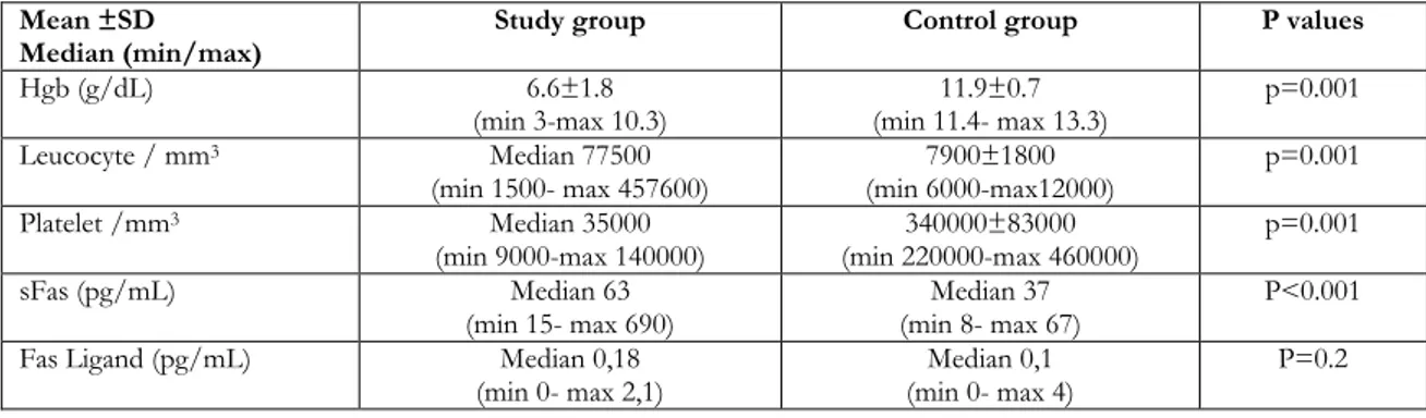
Table 1. Laboratory results of study group and healthy controls Mean ±SD
Median (min/max)
Study group Control group P values
Hgb (g/dL) 6.6±1.8 (min 3-max 10.3) 11.9±0.7 (min 11.4- max 13.3) p=0.001 Leucocyte / mm3 Median 77500 (min 1500- max 457600) 7900±1800 (min 6000-max12000) p=0.001 Platelet /mm3 Median 35000 (min 9000-max 140000) 340000±83000 (min 220000-max 460000) p=0.001 sFas (pg/mL) Median 63 (min 15- max 690) Median 37 (min 8- max 67) P<0.001
Fas Ligand (pg/mL) Median 0,18
(min 0- max 2,1)
Median 0,1 (min 0- max 4)
P=0.2
Tablo 2. sFas and FasL levels according to subclassifications of patients with ALL
Median (min-max) Fas levels (pg/mL) Fas Ligand levels Immunophenotypic classification CALLA (+) B-ALL 54 (15-90) 200 (0-2100) CALLA (-) B-ALL 93 (18-168) 90 (0-180) T-ALL 174 (59-690) 165 (0-1350) Risk classification SRG 63 (15-64) 350 (0-2100) MRG 63 (43-215) 160 (0-1300) HRG 64 (18-690) 400 (0-1350)
DISCUSSION
The relationship between apoptotic signaling pathways and dysregulation in cancer progression and treatment has attracted the interest of many researchers. In the etiopathogenesis of tumors, cell proliferation and inhibition of apoptosis also play an important role and significantly affect prognosis. Apoptosis inhibition and anti-apoptotic proteins are also involved in the development of chemotherapy-resistant cases. The targeted treatment of leukemic cells is about induction of apoptosis. These strategies should focus on targeting and killing leukemia cells without damaging normal hematopoietic progenitor cells9. Kaparou et al reported significantly higher expression of the FAS at remission compared to diagnosis, which might reflect enhanced sensitivity of the leukemic clone to apoptosis and response to treatment10.
Hazar and colleagues investigated the clinical significance of circulating and cellular Fas and Fas ligand expression in lymphoproliferative malignancies in pediatric patients. sFas and FasL expression were investigated in 25 patients with newly diagnosed ALL and NHL and on remission after chemotherapy. There was no significant difference in sFas expression between the control group and newly diagnosed lymphoproliferative malignancy, whereas FasL expression was found to be significantly higher in malignancies 11. Fathi et al. reported that 48 patients who were diagnosed with acute lymphoblastic leukemia had no statistically significant difference in sFas levels compared to the control group12. Although they defended as the sFas may not have a key role in pediatric ALL patients, our study showed a significantly higher level of sFas in the patient group and a significantly higher rate in patients with T-ALL. These differences in other studies suggested further larger series with homogen
patient distribution according to
immunophenotypes.
Karawajew et al. evaluated the expression of CD95 antigen and Bcl-2 protein in leukemic blasts and functionality of the antigen in 42 newly diagnosed
B-ALL and 53 T-ALL patients. In
immunofluorescence method, CD95 expression was found in most of ALL patients and it was higher in T-ALL than B-ALL (p<0.01) 13. Similar results were found in our study.
and Bcl-2 expression in 16 newly diagnosed ALL and 24 AML patients 14. Significant high expression of Fas was detected in the AML and ALL subjects compared to the control group. Both ALL and AML patients reported higher bcl-2 and Fas expression in relapse, compared to those in remission. There was no statistically significant difference between the bcl-2 and Fas expressions when compared between the patients who survived or died.
In the study sFas and Fas Ligand levels were similar between the patient who have died and survivors. Serum Fas levels were statistically higher in relapsed patients. Higher sFas levels at the diagnosis should be a prognostic marker especially early relapses. Studies on larger group, especially with longer follow up or study of relapse cases, will help draw conclusions regarding apoptosis assessment in paediatric ALL.
Our study has some limitations. First, the number of patient in the subgroups was insufficient the distribution by immunophenotyping was not homogeneous. Second, this is a 3-year period cross-sectional study, further prospective longer studies is needed for late relapses and to discuss sFas effects on survival.
In this study, we demonstrated serum Fas and Fas Ligand levels in childhood acute lymphoblastic leukemia, which have been shown to be an important key role in the apoptosis-related mechanism. Our results showed that the level of serum in the diagnosis was higher in lymphoblasts than in healthy controls. However, we didn't detect that serum FasL level was high. When the patients were classified, higher sFas levels in patients with T-ALL showed that it could be a useful prognostic marker especially in the follow-up of these patients.
Yazar Katkıları: Çalışma konsepti/Tasarımı: BA; Veri toplama: AA;
Veri analizi ve yorumlama: AA; Yazı taslağı: AA; İçeriğin eleştirel incelenmesi: BA; Son onay ve sorumluluk: AA, BA; Teknik ve malzeme desteği: AA; Süpervizyon: BA; Fon sağlama (mevcut ise): yok.
Hakem Değerlendirmesi: Dış bağımsız.
Çıkar Çatışması: Yazarlar çıkar çatışması beyan etmemişlerdir. Finansal Destek: Yazarlar finansal destek beyan etmemişlerdir. Author Contributions: Concept/Design : BA; Data acquisition: AA;
Data analysis and interpretation: AA; Drafting manuscript: AA; Critical revision of manuscript: BA; Final approval and accountability: AA, BA; Technical or material support: AA; Supervision: BA; Securing funding (if available): n/a.
Peer-review: Externally peer-reviewed.
1404
REFERENCES
1. Margolin JF, Steuber CP, Poplack DG. Acute lymphoblastic leukemia. In: Principles and Practice of Pediatric Oncology, 4th ed. (Eds Pizzo PA, Poplack DG):4890-544. Philadelphia: Lippincott Williams & Wilkins. 2002:489-544
2. Iannolo G, Conticello C, Memeo L, De Maria R. Apoptosis in normal and cancer stem cells. Crit Rev Oncol Hematol. 2008;66:42–5.
3. Yolcu ES, Ash S, Kaminitz A, Sagiv Y, Askenasy N, Yarkoni S. Apoptosis as a mechanism of T– regulatory cell homeostasis and suppression apoptosis in T–regulatory cell homeostasis. Immunol Cell Biol. 2008;86:650–8.
4. Wickremasinghe RG, Hoffbrand AG. Biochemical and genetic control of apoptosis: Relevance to normal hematopoiesis and hematological malignancies. Blood, 1999;93:3587-600.
5. Verlekar D, Wei1 SJ, Cho H, Yang S, Kang MH. Ceramide synthase-6 confers resistance to chemotherapy by binding to CD95/Fas in T-cell acute lymphoblastic leukemia. Cell Death Dis. 2018;9:925.
6. Vitagliano O, Addeo R, D’Angelo V, Indolfi C, Casale F. The Bcl-2/Bax and Ras/Raf/MEK/ERK signaling pathways: implications in pediatric leukemia pathogenesis and new prospects for therapeutic approaches. Expert Rev. Hematol. 2013;6:587-97. 7. Letai A, Sorcinelli M, Beard C, Korsmeyer SJ.
Antiapoptotic BCL-2 is required for maintenance of a model leukemia. Cancer Cell. 2004;6:241–9.
8. Del Gaizo Moore V, Schlis KD, Sallan SE, Armstrong SA, Letai A. BCL-2 dependence and ABT-737 sensitivity in acute lymphoblastic leukemia. Blood. 2008;111:2300–9.
9. Singh A, Bhatia P, Trehan A, Bansal D, Singh A, Bhatia A. Low spontaneous apoptosis index at diagnosis predicts a high-risk phenotype in paediatric acute lymphoblastic leukemia. Indian J Med Res. 2018;147:248-550.
10. Kaparou M, Choumerianou D, Perdikogianni C, Martimianaki G, Kalmanti M, Stiakak E. Enhanced levels of the apoptotic BAX/BCL-2 ratio in children with acute lymphoblastic leukemia and high-risk features. Genet Mol Biol. 2013;36:7-11.
11. Hazar V, Berber Z, Pestereli E, Coskun M, Yesilipek A, Karpuzoglu G et al. Clinical importance of circulating and cellular expression levels of Fas and Fas Ligand in pediatric patients with lymphoproliferative malignancies. Pediatr Hematol Oncol. 2005;22:247-56.
12. Fathi M, Amirghofran Z, Shahriari. Soluble Fas and Fas ligand and prognosis in children with acute lymphoblastic leukemia. Med Oncol. 2002;29:2046-52.
13. Karawajew L, Wuchter C, Ruppert V, Drexler H, Gruss HJ, DörkenB. Differential CD95 expression and function in T and B lineage acute lymphoblastic leukemia cells. Leukemia.1997;11:1245-52.
14. Aref S, Salama O, Al-tonbary Y,Mansour A. Assesment of Bcl-2 expression as modulator of Fas mediated apoptosis in acute leukemia. Hematology. 2004;9:113-21.