86
Corresponding author Gürhan Gökçe
Selcuk University Shcool of Medicine Meram, Konya, Turkey Tel : +90 (532) 206 37 73
Fax : +90 (332) 323 26 41 E-mail adress : [email protected] Received: 31.08.2005 • Accepted: 26.10.2005
Ankara Üniversitesi Tıp Fakültesi Mecmuası 2006; 59:86-88 CERRAHİ BİLİMLER / SURGICAL SCIENCES
Olgu Bildirisi / Case Report
P
rimary MM of the penis and male urethra are uncommon neoplasms. MM of the urethral meatus, the glans penis, the prepuce and the pe-nile shaft are cutaneous forms, while melanomas of the rest of the urethra are mucosal forms. The diagnosis is often delayed by the patient’s reluctance to consult a physician and by the intrinsic difficulty of such a rare neoplasm. We present a case of malignant melanoma of glans penis and urethral meatus.Case report
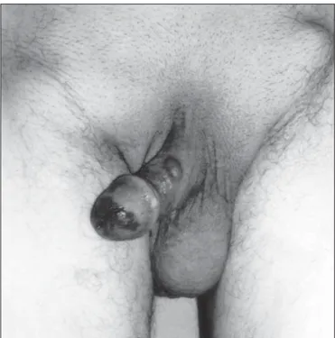
The patient, 73 years old, presented to the urology department of our hospital with pigmented plaque on his glans penis, which had been present for 1 year and rapidly increasing in the last 4 months. Physical examination revealed an approximately 1 cm diameter, unevenly black pigmented, poorly circumscribed, flat lesion on glans penis situated around the meatus externus and merges in the distal part of the urethra. Bilateral palpable inguinal lym-phadenopathy was present. There was an approximately 0.5 cm diameter, soft nodule on the left side of the penile shaft (Figure 1). Chest radiography, sonography of the liver did not show evidence of metastatic disease, but bilateral inguinal lymph node metastases showed by computerized tomog-raphy of the lower abdomen. Incisinal biopsy was done to penile lesions and inguinal lenf node. The histologic diagnosis was: cutaneus lentiginous MM of the glans (Figure 2a, 2b), 5.64 mm in thickness, with multiple con-glomeruleted lenf node metastasis. Patient refuse the all treatment modali-ties, and died six months later.
Malignant melanoma (MM) of the penis is rare and accounts for a small percentage of malig-nant melanomas and of maligmalig-nant penile lesions. The problem of the therapeutic approach is still unsolved. In general prognosis is poor and most patients die within a few years becouse of the distant metastasis.
Key words: Malignant melanoma, penis, urethra
Bu makalede glans penis ve eksternal meatusunda MM’u olan 73 yaşındaki erkek hasta yayın-landı. Penis’in MM’u oldukça nadirdir ve MM’ların ve penisin malign lezyonlarının oldukça az bir kısmını kapsar. Tedavi yaklaşımındaki sorun hala giderilememiştir. Genelde prognozu kötüdür ve hastaların çoğu uzak metastas nedeniyle birkaç yıl içinde ölmektedir.
Anahtar sözcükler: Malign melanom, penis, üretra
Malignant melanoma of the glans penis and
male urethra
Glans penis ve erkek üretrasının primer malign melanomu
Mehmet Arslan, Gürhan Gökçe, Mehmet Mesut Pişkin, Mehmet Kılınç,
Salim Güngör, Selçuk Güven
Department of Urology, University of Selçuk, School of Medicine, Konya, Turkey
Journal of Ankara University Faculty of Medicine 2006; 59(2)
87
M. Arslan, G. Gökçe, M. M. Pişkin et al.
Discussion
MM of the penis accounts for less than 1% of all prima-ry penile malignant lesions (1) and for less than 0,2% of all malignant melonamas in men (2-3). It is usually localized in the glans penis (82%), flowed by the prepuce, the ure-thral meatus and the penile shaft (3). The peak incidence is seen in the sixth to seventh decade (4,5). In advanced stages of the dissease, patients may present symptoms of dysuria, obstruction, haematuria, discharge and occasion-ally melanuria and fistula formation. As happens with oth-er neoplasms of the penis, thoth-ere is often a long intoth-erval be-tween first symptoms and the definitive diagnosis. In fact, the diagnosis is often delayed by the patient’s reluctance to consult a physician and by the intrinsic difıculty in clinical diagnosis of such a rare neoplasm (2,3).
MM present clinically as approximately 1 cm. in di-ameter, blue-black to reddish-brown pigmented papule, plaque or ulceration. The clinical differential diagnoses includes junctional melanocytic naevus, penile melanosis, penile lentigo and atypical pigmented penile macules (6). These completely benign lesions are usually clinically in-distinguishable from MM. Therefore, in order to establish a definitive diagnosis and to avoid large and useless surgical removal in any case of suspected pigmented genital lesion a incisinal biopsy with subsequent histologic examination should be performed.
In cases of penile MM, stage I disease is confined to the penis, stage II is metastatic to regional lymph nodes and
stage III is dissemenated disease (7).At presentation, 43-60% of the cases have lymph node involment (1-4).
The best treatment for penile melanoma is unclear. Whereas some authors recommended an aggresive surgical approach with total amputation of the penis, perineal ure-throstomy and radical inguinal, iliac and obturator lymph node dissection (8,9,10).But Stillwell et al, belive that conservative penile surgery (local excision with 3 to 5 cm margin or distal partial penectomy) with an appropriate Figure 1. The malinant melanoma of the glans penis and urethra with
inguinal lyphadenopathies.
Figure 2a. malignant melanoma of penis 5.64mm in thickness
(H.E.magnification 100x).
Ankara Üniversitesi Tıp Fakültesi Mecmuası 2006; 59(2)
88 Malignant melanoma of the glans penis and male urethra
margin when inguinal nodes are nonpalpable in a patient with thin lesions (less than 1.5 mm) and prophylactic su-perficial inguinal node dissection for those greather than 1.5 mm3 thickand most authors agree with these
treat-ment (11,12,13). Some others reported that sentinel lym-phadenectomy using radiocolloid mapping and dye locali-zation avoided potential morbidity of bilateral superficial inguinal node dissections and allowed accurate staging for further treatment and prognosis (14,15).
Chemotherapy is indicated for disseminated melano-ma. The combination chemotherapy consisting of six cy-cles DTIC, BCNU, cisplatin and tamoxifen gives the best result. The respond rate changes between %15 to %45 (11-16). There has been reported other treatment options; such as radiotherapy, immunotherapy with BCG, endol-ymphatic iodine- iodized oil infusion but the result are not superior to chemotherapy (2).
References
1. Johnson DE, Ayala AG. Primary melanoma of the penis. Urology 1973; 2:174-7.
2. Stillwell TJ, Zincke H, Gaffey TA et al. Malignant melanoma of the penis. J Urol 1988; 140:72-5.
3. Oldbring J, Mikulowiski P. Malignant melanoma of the penis and the male urethra. Cancer 1987; 59:581-7.
4. Begun FP, Grossmann HB, Diokno AC et al. Malignant melanoma of the penis and male urethra. J. Urol 1984; 132:123-5.
5. Weiss J, Elder D, Hamilton R. Melanoma of the male urethra: surgical approach and pathological analysis. J. Urol. 1982; 128:382-5.
6. Primus G, Soyer HP, Smolle J et al. Early ‘invasive’ malignant melanoma of the glans penis and the male urethra. Eur Urol 1990; 18:156-9.
7. DeCosse JJ, McNeer G. Superficial melanoma. A clinical study. Arch. Surg., 1969; 99:531.
8. Pack GT. The pigmented mole and the malignant melanoma. CA Cansen J Clin 1962; 12:11-26.
9. Bracken RB, Diokno AC: Melanoma of the penis and urethra. 2 case reports and review of the liteerature. J.Urol 1974; 111:198-200.
10. Schneiderman C, Simon M, and Levine RM: Malignant melanoma of the penis. J Urol 1965; 93:615-7.
11. Zurrida S, Bartoli C, Clemente C et al.Malignant melanoma of the penis. A report of four cases. Tumori 1990; 76:599-602. 12. Milton GW, Shaw HM. Rare variants of malignant melanoma.
World J Surg 1992; 16:173-8.
13. Myskow MW, Going JJ, McLaren KM et al. Malignant melanoma of the penis J Urol 1988; 139: 817-8.
14. Han KR, Brogle BN, Goydos J et al. Lymphatic mapping and intraoperative lymphoscintigraphy for identifying the sentinel node in penile tumors. Urology 2000; 55:582-5.
15. Southwick A, Rigby O, Daıly M et al. Malıgnant melanoma of the penis and sentınel lymph node bıopsy. J Urol 2001; 166:1833. 16. Bree E, Sanidas E, Tzardi M et al: Malignant melonoma of the