Abstract
©Copyright 2020 by Turkish Ophthalmological Association
Turkish Journal of Ophthalmology, published by Galenos Publishing House.
Introduction
Retinopathy of prematurity (ROP) is a proliferating retinal vascular disorder that can result in poor vision in premature infants.1 The frequency of ROP-associated blindness is low in developed countries. However, in developing countries, the incidence of ROP-associated blindness is higher due to the increased survival of premature infants, a lack of standardized neonatal intensive care unit (NICU) conditions, and limited fundoscopic follow-up evaluations.2,3 The crucial risk factors for ROP development are low birth weight (BW) and low gestational age (GA).4,5 The other risk factors are oxygen therapy needs, sex, sepsis, patent ductus arteriosus (PDA),
intraventricular hemorrhage, neonatal infections, necrotizing enterocolitis (NEC), and blood transfusion needs.4,6,7,8,9,10
Several studies have focused on the role of platelets in angiogenesis and hypothesized that thrombocytopenia might be a possible factor for developing ROP.11,12 Platelets accumulate, carry, and deliver distinct key regulators of angiogenesis such as vascular endothelial growth factor (VEGF), insulin-like growth factor-1 (IGF-1), and platelet-derived growth factor (PDGF).13 Low platelet count may cause delay in normal retinal vascularization and lead to subsequent unregulated retinal neovascularization due to lack of VEGF, IGF-1, and PDGF.11,12 Lundgren et al.14 noted that aggressive posterior ROP is associated with multiple infectious episodes and thrombocytopenia.
Objectives: The aim of the study was to investigate the risk factors for retinopathy of prematurity (ROP), including platelet count. Materials and Methods: This retrospective study analyzed 137 infants in 3 subgroups: no ROP; mild ROP, and severe ROP requiring
laser treatment (type 1 ROP). A retrospective review of records was performed and statistical analysis of possible risk factors for ROP including platelet count was evaluated by using logistic regression.
Results: Birth weight (BW), gestational age (GA), and low platelet count in the first week after birth were significant risk factors
for developing ROP (p=0.038, 0.02, and 0.004, respectively). BW, GA, ventilation, and lower platelet count were associated with progression to type 1 ROP (p=0.004; 0.027, and 0.021, respectively).
Conclusion: Lower platelet count in the first week after birth is a risk factor for ROP development in addition to the previously
established factors of ventilation need, low BW, and low GA.
Keywords: Birth weight, gestational age, retinopathy of prematurity, risk factors, platelet
Cite this article as: Şahinoğlu Keşkek N, Gülcan H, Yılmaz G, Akkoyun İ. Impact of Platelet Count in Retinopathy of Prematurity.
Turk J Ophthalmol. 2020;50:351-355
Address for Correspondence: Nedime Şahinoğlu Keşkek, Başkent University Faculty of Medicine, Adana Research and Training Center, Department of Ophthalmology,
Adana, Turkey E-mail: [email protected] ORCID-ID: orcid.org/0000-0001-8544-103X
Received: 14.01.2020 Accepted: 11.05.2020
*Başkent University Faculty of Medicine, Adana Research and Training Center, Department of Ophthalmology, Adana, Turkey
**Başkent University Faculty of Medicine, Adana Research and Training Center, Department of Pediatrics, Division of Neonatology, Adana, Turkey ***Başkent University Faculty of Medicine, Department of Ophthalmology, Ankara, Turkey
Nedime Şahinoğlu Keşkek*, Hande Gülcan**, Gürsel Yılmaz***, İmren Akkoyun***
Impact of Platelet Count in Retinopathy of
The purpose of this study was to investigate the impact of platelet count in ROP development beyond other known risk factors.
Materials and Methods
The study included 137 infants with a GA of up to 34 weeks who were screened for ROP in the NICU of Başkent University in Adana, Turkey between July 2014 and July 2017. All infants with a GA of up to 34 weeks that were followed up until at least 43 weeks postconception were enrolled in the study (n=137). All the babies included in the study were born at the study site. Exclusion criteria were GA of more than 34 weeks (n=20) and lack of regular follow-up examinations at our institution until 43 weeks postconception (n=18). The Institutional Review Board of Başkent University Faculty of Medicine approved the study (KA:17/308). Informed consent was obtained from the parents of all infants included in the study.
ROP screening was performed by an experienced ophthalmologist (N.S.) using an indirect ophthalmoscope at the postnatal age of 4 weeks or PMA of 31 weeks according to screening guidelines.15 Follow-up examinations were conducted until retinal vascularization reached the ora serrata for 360°. Follow-up intervals were scheduled according to ROP severity. Infants with ROP were examined more often, according to the severity of the disorder. Phenylephrine 2.5% and tropicamide 0.5% were used for dilatation of the pupils. Fundus examination was conducted using an indirect ophthalmoscope and 28 D lens. Fundus findings were noted, and ROP was categorized according to the International Classification of Retinopathy of Prematurity.15
The infants were divided into 3 groups: Group A (no ROP) included babies without retinopathy, Group B (mild ROP) included babies diagnosed with stage 1 or stage 2 ROP but regressed, and Group C (severe ROP) included infants that progressed to Type 1 ROP and underwent laser treatment.16 We noted no noticeable asymmetry in any patient, and no eye progressed to stage 4 or 5 ROP.
We retrospectively reviewed the patient medical records from birth to 43 weeks of age for data such as GA, BW, sex, neonatal morbidities, respiratory distress syndrome (RDS), NEC, intraventricular hemorrhage, PDA, sepsis, red blood cell (RBC) transfusion, apnea, multiple pregnancy of the mother, ventilation need, and surfactant use for RDS. All available laboratory measurements of platelet counts were recorded. The values in the first postnatal week and within 1 week of ROP diagnosis were noted.
Univariate analysis was performed to reveal the significant risk factors for ROP, and the risk factors were included in the logistic regression. Ten potential risk factors (BW, GA, multiple pregnancy, ventilation need, RDS, NEC, PDA, RBC transfusion, apnea, sepsis, surfactant use, and platelet count in the first postnatal week) were analyzed with logistic regression to determine relationships between the variables and identify independent risk factors for ROP. Our criteria for dropping
variables during backward stepwise logistic regression was p=0.05.
Results
We evaluated 137 neonates in our NICU with GA ≤34 weeks during the study period. ROP was diagnosed in 47 cases (34.3%). Severe ROP was detected and treated in 15 cases (10.9%).
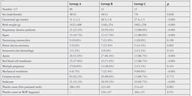
Univariate analysis showed that, in order of significance, BW, GA, PDA, RDS, RBC transfusion, apnea, low platelet count in the first postnatal week, ventilation need, surfactant, and sepsis were associated with ROP based on the p values. Risk factors are listed in Table 1.
With subsequent logistic regression analysis, BW, GA, and low platelet count in the first postnatal week were shown to be independent risk factors for ROP development. Also, logistic regression analysis demonstrated that GA, ventilation, and low platelet count in the first postnatal week were independently related to severe ROP. Low platelet count in the first postnatal week was shown to be an independent risk factor for both ROP development and progression.
The mean GA was 31.3±2.2 weeks for Group A, 28.5±1.8 weeks for Group B, and 27.4±1.5 weeks for Group C. The mean GA of Group A was significantly different from Group B and Group C (p<0.05 for each). Group B and C were not statistically significantly different (p=0.17).
Mean platelet count in the first postnatal week was 280±103x103/µL in Group A, 222±69x103/µL in Group B, and 214±62x103/µL in Group C. According to the post hoc analysis, Group A had a significantly higher mean platelet count than Groups B and C (p=0.002). Group B and C were not statistically significantly different.
The mean platelet count in the week of ROP diagnosis was 339±147x103/µL in Group B and 366±121x103/µL in Group C. Group B and C were not statistically significantly different (p=0.5).
Discussion
The incidence of ROP varies among countries due to different socioeconomic development and variability in study designs and survival rates. In the current study, the overall ROP incidence was 34.3%, while severe ROP was recorded in 10.9% of the infants. These results were similar to the rates of developing countries.17,18,19
In developing countries, infants with higher BW and GA are at risk for ROP development.20,21 Therefore, the current study consisted of infants with a GA ≤34 weeks. In a recent ROP study, fundus examinations of infants with GA ≤34 weeks or BW <1,700 g was recommended for Turkey.22
In our study, low GA, low BW, and low platelet count in the first week after birth were independent risk factors for developing ROP. However, low GA, ventilation need, and low platelet count in the first week after birth arose as independent risk factors for ROP progression.
Both low GA and low BW are associated with incomplete vascular and retinal neural development at birth, given the vulnerable structure of the retina.4 In our study, according to the logistic regression, GA was an independent risk factor for developing mild and severe ROP. However, BW was not statistically significant as a risk factor of severe ROP. This result may indicate the importance of weight gain to prevent the progression of ROP.23,24,25
The association between ROP and blood transfusion is well documented.26 The number of blood transfusions received by premature infants has been a major indicator of ROP in addition to GA and BW. Stutchfield et al.26 hypothesized that changing fetal hemoglobin to adult hemoglobin during transfusion may lead to ROP development by rapidly increasing oxygen accessibility to the retina. In our NICU, RBC transfusion was performed rather than whole blood transfusion when necessary. Therefore, RBC transfusion and platelet count were considered independent risk factors in our study. RBC transfusion was not found to be an independent risk factor for ROP, but this result may be due to the existence of many other risk factors in these infants.
Several pro- and antiangiogenic regulators were shown to be accumulated and carried in platelets.11,12 Platelet alpha granules have been shown to include IGF-1, IGF-binding protein 3 (the primary serum binding protein for IGF-1), VEGF, and platelet-derived growth factor. IGF-1 and VEGF levels are critical for ROP development.27 Our first hypothesis about the mechanism
linking low platelet count and ROP development implies the delivery of IGF-1 by platelets. While IGF-1 is needed for VEGF-induced vessel growth, low platelet count at an early gestational week slows down vasculogenesis and leads to development of subsequent type 1 ROP.
ROP is a disorder with pathological angiogenesis in the inner retina and preretinal space.28 The newly formed blood vessels are not mature, which may lead to vascular leakage.28 Pericytes have a crucial role in angiogenesis by contributing survival signals for endothelial cells.29 PDGF is essential for pericyte viability.30 Moreover, PDGF is fundamental for both proliferation and migration of endothelial cells.30 A lack of pericytes is connected with endothelial hyperplasia, dilated capillaries, irregularly shaped endothelial cells, and increased transendothelial permeability.30 Hammes et al.31 indicated that PDGF-deficient mice had fewer pericytes compared to wild-type mice during the early postnatal phase of the growing retina. They studied a PDGF-receptor β-deficient mice model of oxygen-induced proliferative retinopathy (resembling ROP) to investigate the proliferative phase of diabetic retinopathy. PDGF-receptor β-deficient mice had significantly lower pericyte numbers and significantly higher numbers of acellular capillaries compared with wild-type. After exposure to a high-oxygen environment, the neovascular response to hypoxia nearly doubled in PDGF-receptor β-deficient mice. They also noted the degeneration of endothelial cells (indicated by narrow vessels) and obstructive occlusion in the absence of the PDGF-β
Table 1. Comparison of the demographic characteristics and morbidities of infants with and without retinopathy of prematurity (ROP)
Group A Group B Group C p
Number: 137 90 32 15
Sex (male/female) 48/42 18/14 7/8 0.828 Gestational age (weeks) 31.3±2.2 28.5±1.8 27.4±1.5 <0.001 Birth weight (g) 1622±408 1166±254 1002±258 <0.001 Respiratory distress syndrome 21 (23.3%) 19 (59.4%) 12 (80.0%) <0.001 Sepsis 15 (16.7%) 12 (37.5%) 12 (80.0%) <0.001 Necrotizing enterocolitis 9 (10.0%) 7 (21.9%) 3 (20.0%) 0.190 Patent ductus arteriosus 5 (5.6%) 7 (21.9%) 5 (33.3%) 0.002 Intraventricular hemorrhage 3 (3.3%) 3 (9.4%) 2 (13.3%) 0.193 Apnea 26 (31.0%) 27 (84.4%) 13 (86.7%) <0.001 Red blood cell transfusion 25 (27.8%) 23 (71.9%) 13 (86.7%) <0.001 Multiple pregnancy 27(30.0%) 13 (40.6%) 2 (13.3%) 0.163 Mechanical ventilation 6 (6.7%) 7 (21.9%) 9 (60.0%) <0.001 Cesarean section 83 (92.2%) 29 (90.6%) 13 (86.7%) 0.772 Surfactant 21 (23.3%) 18 (56.3%) 10 (66.7%) <0.001 Platelet count (first postnatal week) 280±103 222±69 214±62 0.002 Platelet count (at ROP diagnosis) -- 339±147 366±121 0.551
Values are presented as number of neonates (with the percentage in brackets) or mean ± standard deviation. Group A: No retinopathy; Group B: Mild ROP; Group C: Prethreshold or threshold ROP, receiving laser treatment
receptor.31 Pericytes likely have a role in promoting endothelial cell survival and limiting endothelial hyperplasia. Our second hypothesis about the mechanism linking low platelet count and ROP development is the lack of PDGF. Our results and data from the literature demonstrate that at high VEGF levels (e.g., ROP), the deficiency of pericyte coverage due to low levels of circulating PDGF may lead to an increased neovascular response.
In ROP models, the introduction of hyperoxia to the retinas of newborn rats decreased VEGF levels and weakens retinal angiogenesis.32,33 Relative hypoxia of room air during the second week led to increased VEGF synthesis and pathological angiogenesis.34 During this proliferative phase of ROP, VEGF levels increase locally and systemically.35
VEGF induces endothelial cell migration and proliferation after hypoxia.36 During that period, thrombocytopenia may deepen the PDGF deficiency which is necessary for pericyte viability. PDGF deficiency may result in pathological angiogenesis.
Vinekar et al.12 presented a case of aggressive posterior ROP with severe thrombocytopenia regressing after serum platelet transfusions. Jensen et al.11 showed a relation between thrombocytopenia and the existence of type 1 ROP in zone 1 cases. The results of these studies suggest thrombocytopenia is a risk factor for zone 1 ROP. Cakir et al.37 showed that any episode of thrombocytopenia at ≥30 weeks postmenstrual age (PMA), was associated with severe ROP in a mouse model of ROP. The researchers evaluated mean weekly platelet count of mice and found a statistically significant difference between the severe ROP group and the no or less severe ROP group. On the contrary, Jensen et al.38 demonstrated that thrombocytopenia from birth to 34 weeks of PMA was related to severe ROP. In the current study, we evaluated the platelet count of the infants on the week of delivery and found lower platet count as a risk factor for ROP development. Our result is compatible with study by Jensen et al.38
The study group of the current study included all ROP cases classified in zone 1 and zone 2. Although platelet counts did not reach the level of thrombocytopenia and none of the infants needed a platelet transfusion, there was a significant difference in platelet count between infants that developed ROP and those who did not. Platelets are major regulators of angiogenic regulatory proteins such as VEGF and PDGF, which are stored, transported, and delivered by platelets.13 Our findings suggest that the growth factors in circulating platelets have a potential protective role against ROP and are necessary for retinal vascular maturation.
Conclusion
The infants with lower platelet counts may have a higher risk for developing ROP. Our findings further contribute to the body of work producing a predictive model to estimate the likelihood for an infant to develop ROP. Further large-scale studies are required to define the potential relation between thrombocytopenia and ROP.
Acknowledgements
I would like to express my special thanks to Çağla Sarıtürk for her assistance with the statistics used in this research.
Ethics
Ethics Committee Approval: There was no funding for
the study. All procedures performed in studies involving human participants were in accordance with the ethical standards of the institutional committee and with the 1964 Declaration of Helsinki and its later amendments or comparable ethical standards.
Informed Consent: Informed consent was obtained from
the parents of all infants included in the study.
Peer-review: Externally peer reviewed. Authorship Contributions
Concept: N.Ş.K., H.G., G.Y., İ.A., Design: N.Ş.K., H.G., G.Y., İ.A., Data Collection or Processing: N.Ş.K., H.G., G.Y., İ.A., Analysis or Interpretation: N.Ş.K., H.G., G.Y., İ.A., Literature Search: N.Ş.K., H.G., G.Y., İ.A., Writing: N.Ş.K., H.G., G.Y., İ.A.
Conflict of Interest: No conflict of interest was declared by
the authors.
Financial Disclosure: The authors declared that this study
received no financial support.
References
1. Good WV, Hardy RJ, Dobson V, Palmer EA, Phelps DL, Quintos M, Tung B, Early Treatment for Retinopathy of Prematurity Cooperative Group. The incidence and course of retinopathy of prematurity: findings from the early treatment for retinopathy of prematurity study. Pediatrics. 2005;116:15-23. 2. Blencowe H, Lawn JE, Vazquez T, Fielder A, Gilbert C. Preterm-associated
visual impairment and estimates of retinopathy of prematurity at regional and global levels for 2010. Pediatr Res. 2013;74(Suppl 1):35-49.
3. Austeng D, Källen KB, Ewald UW, Jakobsson PG, Holmström GE. Incidence of retinopathy of prematurity in infants born before 27 weeks’ gestation in Sweden. Arch Ophthalmol. 2009;127:1315-1319.
4. Kocabeyoglu S, Kadayifcilar S, Eldem B. Retinopathy of Prematurity; Risk Factors, Prognosis and Treatment. Turk J Ophthalmol. 2011;41:128-132. 5. Aclimandos W. Seventy years of retinopathy of prematurity. Br J Ophthalmol.
2011;95:899-900.
6. Brooks SE, Marcus DM, Gillis D, Pirie E, Johnson MH, Bhatia J. The effect of blood transfusion protocol on retinopathy of prematurity: a prospective, randomized study. Pediatrics. 1999;104(3 Pt 1):514-518.
7. Gupta VP, Dhaliwal U, Sharma R, Gupta P, Rohatgi J. Retinopathy of prematurity-risk factors. Indian J Pediatr. 2004;71:887-892.
8. Ali AA, Gomaa NAS, Awadein AR, Al-Hayouti HH, Hegazy AI. Retrospective cohort study shows that the risks for retinopathy of prematurity included birth age and weight, medical conditions and treatment. Acta Paediatr. 2017;106:1919-1927.
9. Mitsiakos G, Papageorgiou A. Incidence and factors predisposing to retinopathy of prematurity in inborn infants less than 32 weeks of gestation. Hippokratia. 2016;20:121-126.
10. Akkoyun I, Oto S, Yilmaz G, Gurakan B, Tarcan A, Anuk D, Akgun S, Akova YA. Risk factors in the development of mild and severe retinopathy of prematurity. J AAPOS. 2006;10:449-453.
11. Jensen AK, Ying GS, Huang J, Karp K, Quinn GE, Binenbaum G. Thrombocytopenia and retinopathy of prematurity. J AAPOS. 2011;15:e3-e4. 12. Vinekar A, Hegde K, Gilbert C, Braganza S, Pradeep M, Shetty R, Shetty KB.
Do platelets have a role in the pathogenesis of aggressive posterior retinopathy of prematurity? Retina. 2010;30(4 Suppl):S20-S23.
13. Italiano JE Jr, Richardson JL, Patel-Hett S, Battinelli E, Zaslavsky A, Short S, Ryeom S, Folkman J, Klement GL. Angiogenesis is regulated by a novel mechanism: pro- and antiangiogenic proteins are organized into separate platelet alpha granules and differentially released. Blood. 2008;111:1227-1233.
14. Lundgren P, Lundberg L, Hellgren G, Holmström G, Hård AL, Smith LE, Wallin A, Hallberg B, Hellström A. Aggressive posterior retinopathy of prematurity is associated with multiple infectious episodes and thrombocytopenia. Neonatology. 2017;111:79-85.
15. Fierson WM, American Academy of Pediatrics Section on Ophthalmology, American Academy of Ophthalmology, American Association for Pediatric Ophthalmology and Strabismus, American Association of Certified Orthoptists. Screening examination of premature infants for retinopathy of prematurity. Pediatrics. 2013;131:189-195.
16. Early Treatment for Retinopathy of Prematurity (ETROP) Cooperative Group. Revised indications for the treatment of retinopathy of prematurity: results of the early treatment for retinopathy of prematurity randomized trial. Arch Ophthalmol. 2003;121:1684-1694.
17. Tavosnanska J, Carreras IM, Fariña D, Luchtenberg G, Celadilla ML, Celotto M, Ferreiro N, González J, Grois I, Lamelas J, Mardyks M, Menalled A, Panzitta M, Petruccelli N, Solana C, Somaruga L, Caparrós S, Capriata A, Gangi D, Goyeneche N, Molina P, Nagel D, Orsini J, Piñón AL, Rojas E, Roldán L, Rubio C, Russmann M, Siniscalco M. Mortality and morbidity of very low birth weight newborn infants assisted in Buenos Aires public hospitals. Arch Argent Pediatr. 2012;110:394-403.
18. Hungi B, Vinekar A, Datti N, Kariyappa P, Braganza S, Chinnaiah S, Donthi K, Shetty B. Retinopathy of prematurity in a rural neonatal intensive care unit in south India-a prospective study. Indian J Pediatr. 2012;79:911-915. 19. Vătavu I, Nascutzy C, Ciomârtan T, Brezan F, Anca I, Stoicescu S. Retinopathy
of prematurity-screening results. Oftalmologia. 2010;54:110-117.
20. Tabarez-Carvajal AC, Montes-Cantillo M, Unkrich KH, Trivedi RH, Peterseim MMW. Retinopathy ofprematurity: screening and treatment in Costa Rica. Br J Ophthalmol. 2017;101:1709-1713.
21. Esen E, Erdem E, Yar K, Demircan N, Soylu M. Results of Screening for Retinopathy of Prematurity: How the Ideal Screening Program Should Be? Turk J Ophthalmol. 2014;44:42-46.
22. Bas AY, Demirel N, Koc E, Ulubas Isik D, Hirfanoglu İM, Tunc T, TR-ROP Study Group. Incidence, risk factors and severity of retinopathy of prematurity in Turkey (TR-ROP study): a prospective, multicentre study in 69 neonatal intensive care units. Br J Ophthalmol. 2018;102:1711-1716. 23. Wang ZH, Gao PF, Bai H, Li YY. Postnatal weight gain in very low birth
weight infants in Beijing and the risk of retinopathy of prematurity. Int J Ophthalmol. 2015;8:1207-1210.
24. Anuk İnce D, Gülcan H, Hanta D, Ecevit A, Akkoyun I, Kurt A, Tarcan A. Poor postnatal weight gain predicts stage 3+ retinopathy of prematurity in very low birth weight infants. Turk J Pediatr. 2013;55:304-308.
25. Jung JL, Wagner BD, McCourt EA, Palestine AG, Cerda A, Cao JH, Enzenauer RW, Singh JK, Braverman RS, Wymore E, Lynch AM. Validation of WINROP for detecting retinopathy of prematurity in a North American cohort of preterm infants. J AAPOS. 2017;21:229-233.
26. Stutchfield CJ, Jain A, Odd D, Williams C, Markham R. Foetal haemoglobin, blood transfusion, and retinopathy of prematurity in very preterm infants: a pilot prospective cohort study. Eye (Lond). 2017;31:1451-1455.
27. Yenice Ö, Ashour A, Çerman E, Firat R, Haklar G, Sirikçi Ö, Akman I, Kazokoglu H. Serum Erythropoietin, Insulin-Like Growth Factor 1 and Vascular Endothelial Growt Factor in Ethiopathogenesis of Retinopathy of Prematurity. Turk J Ophthalmol. 2012;42:423-428.
28. Reynaud X, Dorey CK. Extraretinal neovascularization induced by hypoxic episodes in the neonatal rat. Invest Ophthalmol Vis Sci. 1994;35:3169-3177. 29. Verbeek MM, Otte-Höller I, Wesseling P, Ruiter DJ, de Waal RM. Induction
of α-smooth muscle actin expression in cultured human brain pericytes by transforming growth factor-β1. Am J Pathol. 1994;144:372-382.
30. Hellström M, Gerhardt H, Kalén M, Li X, Eriksson U, Wolburg H, Betsholtz C. Lack of pericytes leads to endothelial hyperplasia and abnormal vascular morphogenesis. J Cell Biol. 2001;153:543-554.
31. Hammes HP, Lin J, Renner O, Shani M, Lundqvist A, Betsholtz C, Brownlee M, Deutsch U. Pericytes and the pathogenesis of diabetic retinopathy. Diabetes. 2002;51:3107-3112.
32. Benjamin LE, Hemo I, Keshet E. A plasticity window for blood vessel remodeling is defined by pericyte coverage of the preformed endothelial network and is regulated by PDGF-B and VEGF. Development. 1998;125:1591-1598.
33. Akkoyun I. Pathophysiology of Retinopathy of Prematurity. Turk J Ophthalmol. 2012;42:63-67.
34. Moravski CJ, Kelly DJ, Cooper ME, Gilbert RE, Bertram JF, Shahinfar S, Skinner SL, Wilkinson-Berka JL. Retinal neovascularization is prevented by blockade of the renin-angiotensin system. Hypertension. 2000;36:1099-1104.
35. Hellgren G, Löfqvist C, Hård AL, Hansen-Pupp I, Gram M, Ley D, Smith LE, Hellström A. Serum concentrations of vascular endothelial growth factor in relation to retinopathy of prematurity. Pediatr Res. 2016;79:70-75. 36. Bressler SB. Introduction: understanding the role of angiogenesis and
antiangiogenic agents in age related macular degeneration. Ophthalmology. 2009;116(10 Suppl):S1-7.
37. Cakir B, Liegl R, Hellgren G, Lundgren P, Sun Y, Klevebro S, Löfqvist C, Mannheimer C, Cho S, Poblete A, Duran R, Hallberg B, Canas J, Lorenz V, Liu ZJ, Sola-Visner MC, Smith LE, Hellström A. Thrombocytopenia is associated with severe retinopathy of prematurity. JCI Insight. 2018;3:e99448. 38. Jensen AK, Ying GS, Huang J, Quinn GE, Binenbaum G. Longitudinal study
of the association between thrombocytopenia and retinopathy of prematurity. J AAPOS. 2018;22:119-123.