RESEARCH ARTICLE
The effectiveness of two different exercise
approaches in adolescent idiopathic scoliosis:
A single-blind, randomized-controlled trial
Hikmet KocamanID1*, Nilgu¨ n Bek2, Mehmet Hanifi KayaID3, Buket Bu¨ yu¨ kturan4, Mehmet Yetiş5, O¨ znur Bu¨yu¨kturan41 Department of Physiotherapy and Rehabilitation, Prosthetics-Orthotics Physiotherapy, Karamanoglu Mehmetbey University, Karaman, Turkey, 2 Department of Physiotherapy and Rehabilitation, Faculty of Health Sciences, Lokman Hekim University, Ankara, Turkey, 3 Faculty of Medicine, Ahi Evran University, Kırşehir, Turkey, 4 School of Physical Therapy and Rehabilitation, Ahi Evran University, Kırşehir, Turkey, 5 Department of Orthopedics and Traumatology, Faculty of Medicine, Ahi Evran University, Kırşehir, Turkey *kcmnhikmet@gmail.com
Abstract
Objectives
The purpose of this study was to compare the efficacy of two different types of exercise methods in patients with adolescent idiopathic scoliosis.
Methods
In total, 28 subjects with adolescent idiopathic scoliosis with a mild curve magnitude (10˚– 26˚) were randomly divided into two groups: the Schroth group (n = 14) and the core group (n = 14). The patients in the Schroth group were treated with supervised Schroth exercises, and the patients in the core group were treated with supervised core stabilization exercises; both groups performed the exercises for three days per week for a total of 10 weeks, and both were given additional traditional exercises to perform. Assessment included Cobb angle (Radiography), trunk rotation (Adam’s test), cosmetic trunk deformity (Walter Reed Visual Assessment Scale), spinal mobility (Spinal Mouse), peripheral muscle strength (Bio-dex System 4-Pro), and quality of life (Scoliosis Research Society-22 questionnaire).
Results
It was found that patients in the Schroth group showed greater improvement in Cobb angles, thoracic trunk rotation angle, cosmetic trunk deformity, spinal mobility, and quality of life than those in the core group (p<0.05), except for in lumbar trunk rotation angle. Peripheral muscle strength improvement was greater in the core group than in the Schroth group (p<0.05).
Conclusion
Schroth exercises are more effective than core stabilization exercises in the correction of scoliosis and related problems in mild adolescent idiopathic scoliosis, and core stabilization
a1111111111 a1111111111 a1111111111 a1111111111 a1111111111 OPEN ACCESS
Citation: Kocaman H, Bek N, Kaya MH, Bu¨yu¨kturan B, Yetiş M, Bu¨yu¨kturan O¨ (2021) The effectiveness of two different exercise approaches in adolescent idiopathic scoliosis: A single-blind, randomized-controlled trial. PLoS ONE 16(4): e0249492.
https://doi.org/10.1371/journal.pone.0249492
Editor: Walid Kamal Abdelbasset, Prince Sattam Bin Abdulaziz University, College of Applied Medical Sciences, SAUDI ARABIA
Received: November 25, 2020 Accepted: March 16, 2021 Published: April 15, 2021
Copyright:© 2021 Kocaman et al. This is an open access article distributed under the terms of the
Creative Commons Attribution License, which permits unrestricted use, distribution, and reproduction in any medium, provided the original author and source are credited.
Data Availability Statement: All revelant data are within the paper.
Funding: The authors received no financial support for the research, authorship, and/or publication of this article.
Competing interests: The authors have declared that no competing interests exist.
exercises are more effective than Schroth exercises in the improvement of peripheral mus-cle strength.
Trial registration
NCT04421157
Introduction
Adolescent idiopathic scoliosis (AIS) is a three-dimensional deformity of the spine with an unknown etiology; it is clinically described as a curvature of the spine in the coronal plane greater than 10 degrees. It presents in 2–2.5% of adolescents and is often accompanied by a rotation of the spine in the axial plan and an alteration in sagittal physiological curvature [1]. AIS affects body alignment, the spinal column, and soft tissue structures, leading to physical problems such as postural abnormality, cosmetic trunk deformity, degraded flexibility of the spinal column, change in the characteristics of the erector spine muscle, back pain, and, in severe cases, reduced respiratory function [2]. Various treatment approaches have been pro-posed to deal with these complications and others [3,4].
Depending on the severity of the curve, treatment approaches consist of exercises, bracing, and surgery to prevent, correct, or halt the progression of the deformity caused by AIS [4]. A review of the literature revealed that exercises are often recommended to decrease progression, correct postural behavior, increase neuro-motor control of the spine, and to improve spine and thoracic flexibility, muscle strength, and elasticity [5]. Exercise is almost always a part of the treatment plan for patients with AIS. In mild cases, exercise may be the main treatment, and it may serve as an adjunct in more severe cases [3].
There are several approaches to exercise designed to treat scoliosis, including the Schroth method [6]. The Schroth method is a scoliosis-specific exercise approach commonly per-formed in scoliosis rehabilitation that uses postural, scoliosis-specific sensorimotor and breathing exercises [6,7]. The treatment program consists of scoliotic posture correction with the help of exteroceptive and proprioceptive stimulations and mirrors, isometrics, and other exercises to lengthen or strengthen the asymmetrical muscles while maintaining a specific breathing pattern. Auto-correction is a basic component of the Schroth method, described as the patient’s ability to decrease the spinal deformity using active postural realignment of the spine in three dimensions. The Schroth method is intended to improve the patient’s motor control of their posture through the repetition of corrective movements with progressively less feedback [6,8]. In several studies, the Schroth method was demonstrated to improve Cobb angles, slow curve progression, reduce the need for surgery, increase back muscle strength, and boost breathing function [7,9,10]. However, although the Schroth method is widely used in AIS rehabilitation, there are few randomized controlled studies on the effectiveness of Schroth exercises [7].
Other recently introduced stabilizing physiotherapeutic exercises used in the conservative treatment of AIS include yoga, Pilates, and core stabilization (CS). These exercises focus on core strength training and spinal stability [11–13]. CS exercises are commonly used for improving neuromuscular control, strength, and the endurance of different muscles around the spine to correct and maintain the alignment of the spine [14]. One of the major therapeutic aims of CS exercises used in the treatment of scoliosis is to improve spinal stability [13]. Previ-ously, limited studies have demonstrated that CS exercises are effective in the treatment of sco-liosis and in the improvement of posture in patients with AIS [13,15,16]. For example, Gu¨r
et al. [15] found CS exercises to be more effective than traditional exercises (breathing exer-cises, posture training, spinal flexibility exerexer-cises, stretching exercises) for the reduction of pain and the correction of vertebral rotation in patients with moderate AIS. Other studies have concluded that CS exercises decrease Cobb angle, increase lumbar muscle strength, and improve sitting balance in patients with AIS [13,16].
The Scientific Society on Scoliosis Orthopaedic Rehabilitation and Treatment (SOSORT) has stated that evidence on the effectiveness of conservative treatment in AIS is scarce and that the isolated effects of therapeutic and corrective exercises on AIS patients have not been clearly defined [4]. Recently, a systematic review identified the positive effects of therapeutic exercises based on the Schroth method or CS exercises in the management of AIS. The review also sug-gested that further studies of better methodological quality are required to confirm the effec-tiveness of such exercises and to determine the best therapeutic exercise intervention for AIS [17]. To the best of our knowledge, no research has previously been done comparing the effects of the Schroth method with those of CS exercises in patients with AIS. Thus, there is a need for randomized controlled studies on the effectiveness of different methods of exercise in the treatment of AIS. This study was done to compare the effects of Schroth exercises and CS exer-cises on Cobb angle, spinal mobility, trunk rotation, peripheral muscle strength, cosmetic deformity, and health-related quality of life in patients with AIS.
Materials and methods
Study design
The study design was a randomized, single-blind 1:1 parallel-group study and was conducted at Kırşehir Ahi Evran University Faculty of Medicine, Departments of Orthopedics and Trau-matology and the School of Physical Therapy and Rehabilitation between October 2019 and March 2020. The study proposal was approved by the Research Ethics Board of Kırşehir Ahi Evran University Faculty of Medicine (08/10/2019). Prior to the study, written and oral con-sent was given by all participants and their families. The study was conducted in accordance with the Declaration of Helsinki principles. The authors confirm that all ongoing and related trials for this study were registered. Due to an error of omission, the trial was registered retrospectively on May 12, 2020, before the data was analyzed (ClinicalTrials.gov Identifier: NCT04421157). We hereby state that all future trials will be registered prospectively.
Participants
Participants in this study were adolescents diagnosed with AIS by a physician according to Lenke criteria and referred to receive an exercise treatment at the School of Physical Therapy and Rehabilitation of Kırşehir Ahi Evran University. The inclusion criteria were as follows: diagnosed with AIS, aged between 10–18 years, Lenke curve type 1 [18], Risser stage � 3 [19], and a Cobb angle of 10–30˚. Participants with a history of neuromuscular, cardiovascular, pul-monary, vestibular, or rheumatological diseases were excluded. Patients were also excluded if they had been prescribed brace treatment, had non-idiopathic scoliosis, had taken medicine periodically, had received any previous surgical or conservative treatment of the spine, were unable to participate, or were reluctant to receive treatment.
Interventions
Supervised exercise treatment programs were performed 90 minutes three times per week for 10 weeks with patients in both groups. The Schroth group (SG) performed Schroth exercises, and the core group (CG) performed CS exercises.
Both groups also performed supervised traditional exercises. These traditional exercises consisted of stretching exercises (particularly for the muscles on the concave side of the curve), posture training, breathing exercises, and spinal flexibility exercises. The exercise program gradually increased in intensity in accordance with each participant’s functional improvement.
The CS training principles, demonstrated in previous studies [13,14,20]. The CS training consisted of three phases. The primary purpose of the first phase was to activate the core mus-cles to improve proprioception and muscular coordination in spinal areas. Thus, the training program focused on local muscle stability training (transversus abdominis, diaphragm, and multifidus) in static positions. In the second and third phases, exercises were made more intense to improve muscular stability and endurance [20].
The CS exercises were explained and assisted by a certified and experienced physiotherapist (B.B.). In the CG, participants were taught isolated activation of the transversus abdominis in the first session while performing an abdominal drawing-in maneuver. Once isolated activation of the transversus abdominis was achieved, the participants were asked to perform the maneu-ver during all stability exercises and in different positions (quadruped, sitting, and standing). The first and second phases each lasted three weeks, and the third phase took place over four weeks. Each training session began with 10 minutes of warm-up exercises and finished with 10 minutes of cool-down exercises; both warm-up and cool-down exercises included breathing and stretching exercises. The number of repetitions was adjusted according to the participant’s exercise tolerance. During the first week of each phase, the number of repetitions of each exer-cise was 7–10, and this progressed to 10–15 based on the patient’s physical tolerance. Training progression was achieved by adding movements to the extremities, working in different posi-tions, the use of TheraBands, the use of exercise balls, and by using the weight of the patient’s body’. The rules and improvements of CS exercises are shown inS1 Appendix.
Schroth exercises consist of passive and active postural auto-correction exercises done repeatedly and based on kinesthetic and sensorimotor principles. The ultimate goal of the Schroth method is to enable the patient to consciously maintain correct posture in their daily living activities [8]. To achieve this requires repeating corrective movements performed to improve postural motor control. Schroth exercises also include strength and endurance train-ing of postural muscles in order to improve the curve, raise the patient’s self-image, and reduce pain [6]. In this study, the Schroth exercises progressed from more to less passive support, from more to less feedback, and from lying to sitting or standing positions, according to the patient’s ability to perform the specific exercise [6,9].
The Schroth exercises were demonstrated and supervised by a certified and experienced physiotherapist (H.K.). Patients were placed in an asymmetric position to maximize correction in trunk symmetry. The Schroth program includes exercises for rotational breathing, spinal elongation, de-flexion, stretching, de-rotation, and strengthening, and these exercises were per-formed to improve the curvature, muscle strength, and endurance of postural muscles. During the Schroth exercises, rice bags, foam blocks, a stool, and long sticks were used to adjust the pos-ture and give passive support. The intensity of the Schroth exercises was gradually increased depending on the patient’s improvement in exercise performance by decreasing the amount or degree of passive support, changing the patient’s position, and adjusting the sets and repetitions of exercises. The rules and improvements of Schroth exercises are shown inS2 Appendix.
Outcome measurements
Socio-demographic data (age, gender, weight, height, body mass index) were collected in an in-person interview. Cobb angle, trunk rotation angle, spinal mobility, cosmetic trunk
deformity, peripheral muscle strength, and quality of life of the participants were assessed by the same researcher who performed the interview.
Primary outcome. Cobb angle. Curve magnitude was evaluated using the Cobb method,
which is considered the gold standard for monitoring scoliosis progression. Cobb angles, in degrees, were obtained using a standard anterior-posterior standing full spine radiograph [21].
Secondary outcomes. Trunk rotation angle. Trunk rotation angle (ATR) was evaluated
using Bunnell’s scoliometer and Adam’s forward bend test. The patients were asked to bend forward, and the angle of trunk rotation (the angle between the horizontal plane and a plane across the posterior aspect of the trunk) was measured using the apical vertebrae of the curve. In order to have clinical significance, the change in ATR must be >4˚. This measurement has been proven to be sensitive, specific, and reliable [22].
Cosmetic trunk deformity. The cosmetic trunk deformity was evaluated using the Walter
Reed Visual Assessment Scale (WRVAS). The WRVAS was designed to assess the perceived physical deformity of patients with idiopathic scoliosis. The test allows patients to describe their perception of their deformity. The WRVAS demonstrates seven visible aspects of spinal deformity, including shoulder level, body curve, head pelvis, flank prominence, rib promi-nence, scapular rotation, and head rib pelvis. Scores for each catagory range from 1 (no defor-mity) to 5 (the worst defordefor-mity), and the total score is generated from the sum of the scores from the seven domains [23]. The WRVAS was found to have high reliability and validity for AIS patients to assess their perception of deformity [24].
Spinal mobility. Spinal mobility was evaluated using a skin-surface computer-aided device
(The Spinal Mouse1System, Idiag, Fehraltorf, Switzerland). Spinal Mouse is a non-invasive external measurement method developed to evaluate segmental and global thoracal and lum-bar mobility on multiple planes [25]. It has been found to be a practical, valid, and reliable device for the clinical assessment of AIS patients [26]. In this study, measurements were taken from the spinous process of C7 to the top of the anal crease (roughly S3). Maximal flexion and maximal extension were measured in the sagittal plane (SP), and maximal right and left lateral flexion positions were measured in the frontal plane (FP). Total SP and FP movements were recorded [27].
SRS-22 questionnaire. In this study, the Scoliosis Research Society-22 (SRS-22)
question-naire was used to assess health-related quality of life. The SRS-22 questionquestion-naire is a valid self-reported instrument for the assessment of quality of life related to scoliosis. It includes five domains: self-image, function, pain, mental health (five questions each), and satisfaction with treatment (two questions). The questionnaire has a total of 22 items that are scored from 1 (worst) to 5 (best) for each item. The final score is the average of these five domains. This instrument has been found to have good validity and test-retest reliability [28].
Muscle strength. A Biodex System 4-Pro dynamometer (Biodex, Inc., Shirley, New York)
was used to assess patients’ strength of knee flexion-extension, flexion-abduction-external rotation (FAE), extension-adduction-internal rotation (EAI) upper extremity patterns in both extremities. All test protocols provided by the manufacturers were strictly followed. The angu-lar velocity was adjusted to 60˚/sec and 120˚/sec, as recommended in previous studies and as can be tolerated by AIS patients [29,30]. To assess the flexion-extension muscle strength of both knees, the patients were placed in a sitting position with arms lying against the body, hands holding lateral fixed handles, and the tested leg stabilized by a belt on the thigh. During testing of the strength of the upper extremity patterns, patients were placed in a sitting position with one arm lying against the body, one hand holding a lateral fixed handle, and the trunk sta-bilized by belts. Three repetitive adaptation periods were applied at an angular velocity of 60˚/ sec in order to increase the adaptation of the participants to the device before measurements were taken at both 60˚/sec and 120˚/sec angular velocity. Five minutes after the adaptation
period, muscle strength tests were performed using five repetitions at an angular velocity of 60˚/sec and 10 repetitions at an angular velocity of 120˚/sec. Rest periods of five minutes were given between the tests of each muscle group and the velocity trials to prevent fatigue [30]. The patients were asked to perform the exercise as forcefully and fast as possible, and they were ver-bally encouraged during the process. Peak torque/body weight (Nm/kg) was recorded as an outcome parameter. Studies have shown that isokinetic muscle testing is useful for assessing scoliosis and should be included in the determination of treatment strategies [29,30].
During the initial period of our study, the gait parameters of most of the participants were evaluated. Unfortunately, the device broke down before all participants could be assessed. Despite all efforts, the device could not be repaired within the required timeframe. Thus, this parameter was omitted because no similar device was found in the province where the study was conducted. We subsequently informed the ethics committee about this issue.
Sample size
To determine the sample of the study, version 3.1.9.4 of the G�Power program (Heinrich-Heine-Universita¨t Du¨sseldorf, Germany) was used [31]. According to previous studies, the effects of exercises on the Cobb angle of the main curve were determined to be from small to moderate (0.16–0.38) [32,33]. To obtain 80% statistical power (1− β error probability) with anα error level probability of 0.05, we performed repeated measure analysis of variance (ANOVA) within and between interactions, used a medium effect size of 0.30 to consider the two groups, and used two measurements for the primary outcome, generating a sample size of 24 participants. Considering the drop-out rate of 15% and aiming to increase the statistical power of the results, a total of 28 participants (14 for each group) were recruited into the study.
Randomization and blinding
A randomization process was performed for the 28 AIS patients. The participants were ran-domly divided into two groups as SG and CG using matched pairs randomization based on their Cobb angle, age, and gender. Matched pairs randomization was performed with numbers sorted using the Research Randomizer program on therandomizer.org website [34]. At base-line and after the 10-week treatment period, all assessments were evaluated by the investigator, who was blinded to the groups throughout the study (M.H.K.).
Statistical analysis
Statistical analysis was performed using IBM1SPSS (Version 21.0 software, IBM Corp., Armonk, NY, USA). According to analytical methods (Kolmogorov–Smirnov and Shapiro– Wilk tests), the data were normally distributed. Thus, parametric tests were used for the statis-tical analysis. Descriptive statistics were presented as mean± standard deviation (SD) or num-ber and frequency. For comparison of demographic and subject characteristics between the two groups, the chi-square test was used for categorical variables, and the t-test was used for continuous variables. To analyze the changes within the groups over time and group–time interactions for continuous variables, a two-way mixed design repeated measures analysis of variance was performed. Based on Box’s M test, the assumption of the equality of covariance matrices, which is an important assumption in this test, was not violated. Also, pairwise com-parisons, known as Bonferroni corrections, were performed for subsequent multiple compari-sons. Partial eta squared was considered as the effect size. The effect size values considered were 0.10 = small, 0.25 = medium, and 0.40 = large [35]. Statistical significance was deter-mined at a p value of < 0.05.
Results
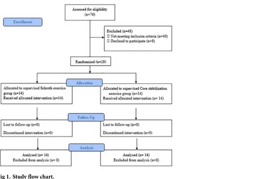
Seventy-six scoliosis patients were admitted to the department, and 28 (21 females, 7 males) satisfied the inclusion criteria. The patient distributions were n = 14 for the SG and n = 14 for the CG after randomization. The study was completed with 100% attendance compliance. The flow chart of the study is shown inFig 1.
Baseline clinical and demographic characteristics of the patients are shown inTable 1. There was no significant difference between the two groups in terms of age, gender, body mass index, Risser sign, curve type, or dominant sides (p>0.05).
Fig 1. Study flow chart.
https://doi.org/10.1371/journal.pone.0249492.g001
Table 1. Baseline clinical and demographic characteristics of groups.
SG CG p (n = 14) (n = 14) Mean±SD Mean±SD Age (years) 14.07±2.37 14.21±2.19 0.87 BMI (kg/m2) 19.77±4.12 19.73±2.36 0.98 Risser sign 1.64±1.34 1.78±1.19 0.77 n n Gender Female 10 (71.4%) 11 (78.6%) 0.66 Male 4 (28.6%) 3 (21.4%)
Dominant side (upper extremity) Right 14 (100.0%) 12 (85.7%) 0.14
Left 0 (0.0%) 2 (14.3%)
Dominant side (lower extremity) Right 13 (92.9%) 13 (92.9%) 1.00
Left 1 (7.1%) 1 (7.1%)
Curve type Right Thoracic 3 (21.4%) 3 (21.4%) 1.00
Left Thoracic 5 (35.7%) 5 (35.7%)
Right Thoracic-Left lumbar 6 (42.9%) 6 (42.9%) SG: Schroth group, CG: Core group, SD: Standard deviation, BMI: Body-mass index.
There were significant group-by-time interactions for Cobb angles (thoracic: F = 32.39, p
<0.001,η2
= 0.55 and lumbar: F = 19.47, p = 0.002,η2
= 0.68), WRVAS (F = 20.47, p<0.001, η2
= 0.44), thoracic angle of trunk rotation (ATR-T) (F = 35.61, p<0.001,η2= 0.57), SRS-22 (F = 17.44, p<0.001,η2
= 0.40), total FP (F = 17.19, p<0.001,η2
= 0.39), and SP motion (F = 27.26, p<0.001,η2= 0.51). There were no significant group-by-time interactions between the groups for the lumbar angle of trunk rotation (ATR-L) (F = 1.10, p = 0.302,η2
= 0.04). Within and between-group differences for the baseline and final treatment measurements for these parameters are shown inTable 2. Patients in the SG demonstrated a greater improve-ment in these parameters than those in the CG, except for in ATR-L.
There were significant group-by-time interactions for the FAE upper extremity pattern in both extremities at angular velocities of 60˚/sec and 120˚/sec (left 60˚/sec: F = 9.47, p = 0.005, η2
: 0.26 and 120˚/sec: F = 4.76, p = 0.038,η2
: 0.15; right 60˚/sec: F = 11.71, p = 0.002,η2
: 0.31 and 120˚/sec: F = 14.24, p = 0.001,η2: 0.35). There were no significant group-by-time interac-tions for the EAI upper extremity pattern in both extremities at both angular velocities (left 60˚/sec: F = 2.15, p = 0.154,η2: 0.07 and 120˚/sec: F = 3.55, p = 0.070,η2: 0.12; right 60˚/sec: F = 4.25, p = 0.051,η2
:0.14 and 120˚/sec: F = 3.51, p = 0.072,η2
:0.11). As indicated by mean difference change scores, patients who received CS exercises showed significantly greater improvements in FAE upper extremity pattern in both extremities compared to those who received the Schroth exercises (Table 3).
There were significant group-by-time interactions for the muscle strength of the left knee extensor (F = 28.76, p = <0.001,η2:0.52) and the right knee flexor (F = 15.92, p<0.001,η2: 0.38) at an angular velocity of 60˚/sec. There were significant group-by-time interactions for the muscle strengths of the knee flexor (left: F = 21.15, p = <0.001,η2:0.44; right: F = 9.84,
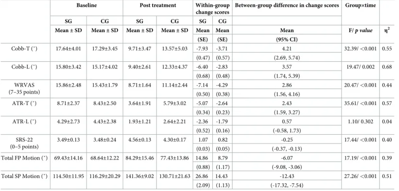
Table 2. Baseline, post-ıntervention and change scores for the Cobb angle, WRVAS, ATR, SRS-22, total FP and SP motion. Baseline Post treatment Within-group
change scores
Between-group difference in change scores Group×time
SG CG SG CG SG CG
Mean± SD Mean± SD Mean± SD Mean± SD Mean Mean Mean F/p value η2
(SE) (SE) (95% CI)
Cobb-T (˚) 17.64±4.01 17.29±3.45 9.71±3.47 13.57±5.03 -7.93 -3.71 4.21 32.39/ <0.001 0.55 (0.47) (0.57) (2.69, 5.74) Cobb-L (˚) 15.80±3.42 15.17±4.02 9.40±2.61 12.33±4.37 -6.40 -2.83 3.57 19.47/ 0.002 0.68 (0.68) (0.48) (1.74, 5.39) WRVAS (7–35 points) 15.86±2.48 15.43±1.79 8.71±1.64 11.14±2.44 -7.14 -4.29 2.86 20.47/ <0.001 0.44 (0.50) (0.38) (1.56, 4.16) ATR-T (˚) 8.71±2.37 8.43±2.50 3.64±1.91 5.79±3.02 -5.07 -2.64 2.43 35.61/ <0.001 0.57 (0.34) (0.23) (1.59, 3.27) ATR-L (˚) 4.29±2.73 4.43±2.38 1.93±1.21 2.64±2.21 -2.36 -1.79 0.57 1.10/ 0.302 0.04 (0.52) (0.16) (-0.58, 1.73) SRS-22 (0–5 points) 3.49±0.13 3.48±0.24 4.56±0.13 4.30±0.17 1.07 0.82 -0.25 17.44/ <0.001 0.40 (0.03) (0.05) (-0.37, -0.13) Total FP Motion (˚) 69.43±14.16 68.64±12.22 84.29±15.46 77.43±13.86 14.86 8.79 -6.07 17.19/ <0.001 0.39 (0.88) (1.17) (-9.08, -3.06) Total SP Motion (˚) 114.50±11.95 116.29±20.29 141.36±9.02 130.71±21.63 26.86 14.43 -12.43 27.26/ <0.001 0.51 (2.09) (1.13) (-17.32, -7.54)
SG: Schroth group, CG: Core group, SD: Standard deviation, SE: Standard error, CI: Confidence interval,η2: Effect size, Cobb-T: Cobb angle of thoracic, Cobb-L: Cobb
angle of lumbar, ATR-T: Thoracic angle of trunk rotation, ATR-L: Lumbar angle of trunk rotation, FP: Frontal plane, SP: Sagittal plane.
p<0.001,η2:0.27) and extensor (left: F = 26.79, p<0.001,η2:0.50; right: F = 39.46, p<0.001,η2: 0.60) in both extremities at an angular velocity of 120˚/sec. There were no significant group-by-time interactions for the muscle strength of the left knee flexor (F = 2.89, p = 0.101,η2:0.10) and right knee extensor (F = 1.00, p = 0.326,η2
:0.03) at an angular velocity of 60˚/sec. As indi-cated by mean difference change scores, patients treated with CS exercises showed significantly greater improvements in knee flexor-extensor muscle strength in both extremities than those who were treated with the Schroth exercises, except in muscle strength of the left knee flexor (angular velocity of 60˚/sec) and right knee extensor (angular velocity of 60˚/sec) (Table 4).
In addition, pairwise comparisons revealed significant differences between pretreatment and posttreatment for Cobb-T (p<0.001), Cobb-L (p = 0.001 in SG; p = 0.002 in CG), WRVAS (p<0.001), ATR-T (p<0.001), ATR-L (p = 0.001 in SG; p<0.001 in CG), SRS-22 (p<0.001), total FP and SP motion (p<0.001), left and right EAI and FAE upper extremity patterns’ mus-cle strengths (p<0.001), and left and right knee flexor-extensor musmus-cle strengths (p<0.001) in both intervention groups.
Discussion
To the best of our knowledge, the present study is the first to compare these two exercise meth-ods used in the conservative treatment of AIS. This single-blind, randomized controlled study indicated that the Schroth exercises are superior in improving Cobb angles, ATR-T, spinal mobility, cosmetic trunk deformity, and quality of life compared to CS exercises in patients with AIS after a 10-week treatment. Improvement in the ATR-L was similar in both groups, and CS exercises were superior in the improvement of peripheral muscle strength.
Table 3. Baseline, post-ıntervention and change scores for the EAI and FAE upper extremity patterns’ muscle strengths. Baseline After treatment Within-group
change scores
Between-group difference in change scores
Group×time
SG CG SG CG SG CG
Mean± SD Mean± SD Mean± SD Mean± SD Mean Mean Mean F/p value η2
(SE) (SE) (95% CI) Left EAI-UEP PT/BW 60˚/sec 41.49
±11.38 41.98 ±10.55 57.91 ±16.05 63.18 ±11.29 16.42 21.20 4.79 2.15/ 0.154 0.07 (2.81) (1.66) (-1.92, 11.49) Left FAE- UEP PT/BW 60˚/
sec 43.39±7.56 39.67 ±10.20 58.86 ±13.69 67.06 ±12.81 15.48 27.39 11.92 9.47/ 0.005 0.26 (3.22) (2.15) (3.96, 19.87) Left EAI- UEP PT/BW 120˚/
sec 45.73 ±14.79 42.51 ±11.23 60.80 ±15.84 63.25 ±10.96 15.08 20.73 3.47 3.55/ 0.070 0.12 (2.63) (1.44) (-5.46, 12.39) Left FAE- UEP PT/BW 120˚/
sec 41.89±7.98 40.86±8.73 60.82 ±11.73 68.00 ±13.85 18.93 27.14 8.20 4.76/ 0.038 0.15 (3.01) (2.25) (0.47, 15.93) Right EAI- UEP PT/BW 60˚/
sec 47.13±8.77 44.16 ±13.38 64.26 ±12.67 67.01 ±12.25 17.13 22.86 5.73 4.25/ 0.051 0.14 (2.22) (1.67) (0.02, 11.44) Right FAE- UEP PT/BW 60˚/
sec 46.93 ±10.74 39.92±7.89 64.63 ±16.21 70.71 ±14.86 17.70 30.79 13.08 11.71/ 0.002 0.31 (3.03) (2.33) (5.23, 20.94) Right EAI- UEP PT/BW 120˚/
sec 47.11 ±13.34 44.23 ±12.99 64.02 ±15.81 67.17 ±12.81 16.91 22.95 6.04 3.51/ 0.072 0.11 (2.76) (1.66) (-0.59, 12.66) Right FAE- UEP PT/BW 120˚/
sec 44.52 ±10.84 41.82 ±10.19 61.41 ±12.46 70.06 ±13.86 16.89 28.24 11.35 14.24/ 0.001 0.35 (2.28) (1.96) (5.17, 17.53)
SG: Schroth group, CG: Core group, SD: Standard deviation, SE: Standard error, CI: Confidence interval,η2: Effect size, EAI: Extension-adduction-internal rotation,
FAE: Flexion-abduction-external rotation, UEP: Upper extremity pattern, PT/BW: Peak torque/body weight.
In the study of AIS, Cobb angle and ATR are major prognostic and clinical indications of curve progression [36]. Strategies that slow the progression of scoliosis and decrease the need for surgery are important in the treatment of AIS [37]. In the present study, the Cobb angle and ATR of the scoliotic curve decreased in all participants. However, patients in the SG had a greater decrease in Cobb angle and ATR than those in the CG, except in lumbar trunk rotation angle. These findings suggest that the Schroth exercise program is superior to the CS exercise program in reducing Cobb angle or ATR. Similar to the results presented here, a systematic review reported that corrective, therapeutic exercise based on the Schroth method or involving CS exercises can reduce vertebral angles and body asymmetries [17]. Kuru et al. [32] found that the Schroth exercise program applied in the clinic under the supervision of a physiothera-pist decreased the Cobb angle and ATR, and Gu¨r et al. [15] found that a 10-week CS exercise program decreased Cobb angles (thoracic and lumbar) and ATR in patients with AIS. It has been established that ATR is associated with curve magnitude and directly affects Cobb angle [38]. In the present study, the reduction in the ATR, as it relates to the decrease in Cobb angle, is consistent with the literature.
A main concern of scoliosis patients is anxiety developed due to three-dimensional defor-mity [39]. Correction of cosmetic deformity is a primary goal of treatment, as reported in a consensus by SOSORT [4]. Therefore, measuring the perception of cosmetic problems is important for patients, their families, and clinicians in order to evaluate the outcome of treat-ment [23]. WRVAS has been found to be sensitive to improvement or worsening of the defor-mity of scoliosis [23,24]. A study on treatments using CS exercise or the scientific exercise approach to scoliosis (SEAS) found both approaches considerably improve cosmetic deformity in moderate AIS [40]. Another study reported that when the Schroth exercise program was
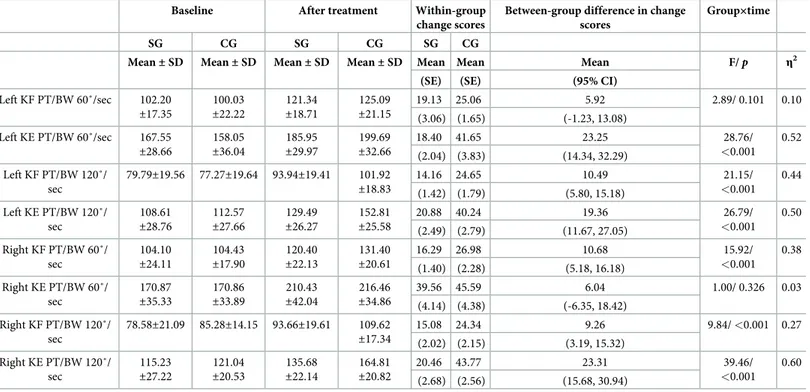
Table 4. Baseline, post-intervention and change scores for the knee flexor-extensor muscle strengths. Baseline After treatment Within-group
change scores
Between-group difference in change scores
Group×time
SG CG SG CG SG CG
Mean± SD Mean± SD Mean± SD Mean± SD Mean Mean Mean F/p η2
(SE) (SE) (95% CI) Left KF PT/BW 60˚/sec 102.20 ±17.35 100.03 ±22.22 121.34 ±18.71 125.09 ±21.15 19.13 25.06 5.92 2.89/ 0.101 0.10 (3.06) (1.65) (-1.23, 13.08) Left KE PT/BW 60˚/sec 167.55 ±28.66 158.05 ±36.04 185.95 ±29.97 199.69 ±32.66 18.40 41.65 23.25 28.76/ <0.001 0.52 (2.04) (3.83) (14.34, 32.29) Left KF PT/BW 120˚/ sec 79.79±19.56 77.27±19.64 93.94±19.41 101.92 ±18.83 14.16 24.65 10.49 21.15/ <0.001 0.44 (1.42) (1.79) (5.80, 15.18) Left KE PT/BW 120˚/ sec 108.61 ±28.76 112.57 ±27.66 129.49 ±26.27 152.81 ±25.58 20.88 40.24 19.36 26.79/ <0.001 0.50 (2.49) (2.79) (11.67, 27.05) Right KF PT/BW 60˚/ sec 104.10 ±24.11 104.43 ±17.90 120.40 ±22.13 131.40 ±20.61 16.29 26.98 10.68 15.92/ <0.001 0.38 (1.40) (2.28) (5.18, 16.18) Right KE PT/BW 60˚/ sec 170.87 ±35.33 170.86 ±33.89 210.43 ±42.04 216.46 ±34.86 39.56 45.59 6.04 1.00/ 0.326 0.03 (4.14) (4.38) (-6.35, 18.42) Right KF PT/BW 120˚/ sec 78.58±21.09 85.28±14.15 93.66±19.61 109.62 ±17.34 15.08 24.34 9.26 9.84/ <0.001 0.27 (2.02) (2.15) (3.19, 15.32) Right KE PT/BW 120˚/ sec 115.23 ±27.22 121.04 ±20.53 135.68 ±22.14 164.81 ±20.82 20.46 43.77 23.31 39.46/ <0.001 0.60 (2.68) (2.56) (15.68, 30.94)
SG: Schroth group, CG: Core group, SD: Standard deviation, SD: Standard error, CI: Confidence interval,η2: Effect size, KF: Knee flexor, KE: Knee extensor, PT/BW:
Peak torque/body weight.
added to standard care, the body image of patients with AIS improved [9]. A systematic review on this subject reported that corrective, therapeutic exercises, such as the Schroth method or CS exercises, improved body symmetry in patients with AIS [17]. In the present study, cos-metic deformity was evaluated using WRVAS, and the coscos-metic deformities of both groups improved considerably. However, patients in the SG had greater cosmetic improvement than those in the CG. The greater cosmetic deformity improvement in the SG may be the result of greater improvement in body symmetry and greater reduction in curve magnitudes.
Spinal flexibility and mobility decrease in AIS patients due to structural deformity of the spine, which can become more rigid over time [41]. Kao et al. [42] found a negative correlation between Cobb angle, vertebral rotation, and results of the sit-and-reach test in individuals with AIS and determined that a greater Cobb angle and vertebral rotation lead to greater restriction of lumbar flexion. In conservative treatment, increasing the flexibility and mobility of the spine is important to correct the curve of the spine in scoliosis [43]. The effect of the Schroth method on the flexibility and mobility of the spine has been documented in previous studies [7,44,45]. Malaj et al. [44] reported an improvement in trunk flexion after treatment with combined Schroth and Pilates exercises in patients with AIS. Another study examined the effects of CS exercise on flexibility in scoliosis and showed that lumbar flexibility increased sig-nificantly after CS exercise in patients with AIS [13]. Consistent with the previous studies, FP and SP mobility increased in both groups in our study. However, the Schroth exercise program produced more improvement in FP and SP mobility compared to the CS exercise program. This result may be attributed to a greater increase in the flexibility of the curve with the Schroth exercise program.
Depending on the severity of the curve, physical and psychological problems can arise in patients with AIS [36,39]. Different results have been reported in the literature regarding the effect of exercise on quality of life in scoliosis patients. Some studies have reported that differ-ent exercise protocols have positive effects on quality of life, while some studies have not found any effect from exercise on improving the quality of life of patients with AIS [32,46–48]. The findings of the present study support the hypothesis that exercise promotes a positive quality of life. However, in our study, patients in the SG showed more improvement in quality of life than those in the CG.
Generalized muscle dysfunction is common in patients with AIS and contributes to limita-tions in their exercise capacity, even in the absence of severe ventilatory impairment [39,49,
50]. Several studies have reported on the effect of Schroth or CS exercise on the strength of trunk muscles in patients with AIS [10,13,45,51]. Screiber et al. [9] demonstrated that adding the Schroth exercise program to standard care improves back muscle endurance in AIS patients, and Otman et al. [10] found that trunk muscle strength increased significantly after a six-week Schroth exercise program. Another study reported that lumbar muscle strength improved after 12 weeks of CS exercise in AIS patients [13], and a recent meta-analysis deter-mined that the Schroth exercise program mostly influences core muscle strength [7]. In the present study, peripheral muscle strength increased in both groups. However, the peripheral muscle strength improvement of patients in the CG was greater than that of those in the SG. We could not find a study that examined the effect of exercise therapies on peripheral muscle strength in AIS with which we could compare our results. To the best of our knowledge, the present study is the first to compare the effect of two different exercise methods on peripheral muscle strength in patients with AIS.
This study has several limitations. First, only the patients who had Lenke curve type 1 were recruited for the study. Second, the findings of this study are only for a 10-week treatment program. Third, although the inclusion criterion for Cobb angle was 10–30˚, most of the patients included in the study had mild curve magnitude. Therefore, this study could not be
generalized to other types of scoliotic curves, different curve magnitudes, and different treat-ment periods. Future studies should take these into consideration.
Conclusion
The present study indicated that Schroth exercises are more effective in reducing Cobb angle and ATR (main curve) and in improving spinal mobility and quality of life in patients with mild AIS than are CS exercises, while CS exercises are more effective than Schroth exercises in the improvement of peripheral muscle strength. Both exercise methods can be used in the con-servative treatment of mild AIS, depending on the treatment’s purpose. Further studies are needed with long-term follow-up periods and patients with different curve types, curve magni-tudes, and other scoliosis types (neuromuscular, early-onset, adult) and conditions.
Supporting information
S1 Appendix. Program of core stabilization exercises.
(DOCX)
S2 Appendix. Program of Schroth exercises.
(DOCX)
S1 File. Study protocol.
(DOCX)
S2 File. Study protocol-Turkish.
(DOC)
S1 Checklist. CONSORT 2010 checklist of information to include when reporting a rando-mised trial.
(DOC)
Author Contributions
Conceptualization: Hikmet Kocaman, Nilgu¨n Bek, O¨ znur Bu¨yu¨kturan.
Formal analysis: Nilgu¨n Bek, Buket Bu¨yu¨kturan, O¨ znur Bu¨yu¨kturan.
Investigation: Hikmet Kocaman, Mehmet Hanifi Kaya, Mehmet Yetiş.
Methodology: Hikmet Kocaman, Nilgu¨n Bek, Mehmet Hanifi Kaya, Buket Bu¨yu¨kturan, Meh-met Yetiş, O¨ znur Bu¨yu¨kturan.
Project administration: Mehmet Yetiş.
Supervision: Nilgu¨n Bek, O¨ znur Bu¨yu¨kturan.
Visualization: Hikmet Kocaman.
Writing – original draft: Hikmet Kocaman.
Writing – review & editing: Hikmet Kocaman, Nilgu¨n Bek, Mehmet Hanifi Kaya, Buket Bu¨yu¨kturan, Mehmet Yetiş, O¨ znur Bu¨yu¨kturan.
References
1. Parent EC, Wong D, Hill D, Mahood J, Moreau M, Raso VJ, et al. The association between Scoliosis Research Society-22 scores and scoliosis severity changes at a clinically relevant threshold. Spine. 2010; 35(3):315–22.https://doi.org/10.1097/BRS.0b013e3181cabe75PMID:20075764
2. Miller NH. Cause and natural history of adolescent idiopathic scoliosis. Orthopedic Clinics. 1999; 30 (3):343–52.https://doi.org/10.1016/s0030-5898(05)70091-2PMID:10393760
3. Romano M, Minozzi S, Zaina F, Saltikov JB, Chockalingam N, Kotwicki T, et al. Exercises for adoles-cent idiopathic scoliosis: a Cochrane systematic review. Spine. 2013; 38(14):883–93.https://doi.org/10. 1097/BRS.0b013e31829459f8PMID:23558442
4. Negrini S, Aulisa AG, Aulisa L, Circo AB, de Mauroy JC, Durmala J, et al. 2011 SOSORT guidelines: orthopaedic and rehabilitation treatment of idiopathic scoliosis during growth. Scoliosis. 2012; 7(1):1– 35.
5. Negrini S, Fusco C, Minozzi S, Atanasio S, Zaina F, Romano M. Exercises reduce the progression rate of adolescent idiopathic scoliosis: results of a comprehensive systematic review of the literature. Dis-ability and rehabilitation. 2008; 30(10):772–85.https://doi.org/10.1080/09638280801889568PMID:
18432435
6. Berdishevsky H, Lebel VA, Bettany-Saltikov J, Rigo M, Lebel A, Hennes A, et al. Physiotherapy scolio-sis-specific exercises–a comprehensive review of seven major schools. Scoliosis and spinal disorders. 2016; 11(1):1–52.https://doi.org/10.1186/s13013-016-0076-9PMID:27525315
7. Park g, Jeon H-S, Park H-W. Effects of the Schroth exercise on idiopathic scoliosis: a meta-analysis. European journal of physical and rehabilitation medicine. 2017; 54(3):440–9.https://doi.org/10.23736/ S1973-9087.17.04461-6PMID:28976171
8. Lehnert-Schroth C. Introduction to the three-dimensional scoliosis treatment according to Schroth. Physiotherapy. 1992; 78(11):810–5.
9. Schreiber S, Parent EC, Moez EK, Hedden DM, Hill D, Moreau MJ, et al. The effect of Schroth exercises added to the standard of care on the quality of life and muscle endurance in adolescents with idiopathic scoliosis—an assessor and statistician blinded randomized controlled trial:“SOSORT 2015 Award Win-ner”. Scoliosis. 2015; 10(1):1–12.
10. Otman S, Kose N, Yakut Y. The efficacy of Schroth’s 3-dimensional exercise therapy in the treatment of adolescent idiopathic scoliosis in Turkey. Neurosciences (Riyadh). 2005; 10(4):277–83.
11. Fishman LM, Groessl EJ, Bernstein P. Two isometric yoga poses reduce the curves in degenerative and adolescent idiopathic scoliosis. Topics in Geriatric Rehabilitation. 2017; 33(4):231–7.
12. Kim G, HwangBo P-n. Effects of Schroth and Pilates exercises on the Cobb angle and weight distribu-tion of patients with scoliosis. Journal of physical therapy science. 2016; 28(3):1012–5.https://doi.org/ 10.1589/jpts.28.1012PMID:27134403
13. Ko K-J, Kang S-J. Effects of 12-week core stabilization exercise on the Cobb angle and lumbar muscle strength of adolescents with idiopathic scoliosis. Journal of exercise rehabilitation. 2017; 13(2):244–49.
https://doi.org/10.12965/jer.1734952.476PMID:28503541
14. Akuthota V, Nadler SF. Core strengthening. Archives of physical medicine and rehabilitation. 2004; 85:86–92.https://doi.org/10.1053/j.apmr.2003.12.005PMID:15034861
15. Gu¨r G, Ayhan C, Yakut Y. The effectiveness of core stabilization exercise in adolescent idiopathic scoli-osis: A randomized controlled trial. Prosthetics and orthotics international. 2017; 41(3):303–10.https:// doi.org/10.1177/0309364616664151PMID:27625122
16. Shin SS, Lee YW, Song CH. Effects of lumbar stabilization exercise on postural sway of patients with adolescent idiopathic scoliosis during quiet sitting. Journal of physical therapy science. 2012; 24 (2):211–5.
17. Laita LC, Kinesiologista CTC, Kinesiologistb TMG, del Barrio SJ. Effects of corrective, therapeutic exer-cise techniques on adolescent idiopathic scoliosis. A systematic review. Archivos argentinos de pedia-tria. 2018; 116(4):582–9.
18. Slattery C, Verma K. Classifications in Brief: The Lenke Classification for Adolescent Idiopathic Scolio-sis. Clinical orthopaedics and related research. 2018; 476(11):2271–76.https://doi.org/10.1097/CORR. 0000000000000405PMID:30179943
19. Risser JC. The iliac apophysis: an invaluable sign in the management of scoliosis. Clinical Orthopaedics and Related Research®. 1958; 11:111–9. PMID:13561591
20. Akuthota V, Ferreiro A, Moore T, Fredericson M. Core stability exercise principles. Current sports medi-cine reports. 2008; 7(1):39–44.https://doi.org/10.1097/01.CSMR.0000308663.13278.69PMID:
18296944
21. Deacon P, Flood B, Dickson R. Idiopathic scoliosis in three dimensions. A radiographic and morphomet-ric analysis. The Journal of bone and joint surgery British volume. 1984; 66(4):509–12.https://doi.org/ 10.1302/0301-620X.66B4.6746683PMID:6746683
22. Amendt LE, Ause-Ellias KL, Eybers JL, Wadsworth CT, Nielsen DH, Weinstein SL. Validity and reliabil-ity testing of the Scoliometer®. Physical therapy. 1990; 70(2):108–17.https://doi.org/10.1093/ptj/70.2. 108PMID:2296610
23. Sanders JO, Polly DW Jr, Cats-Baril W, Jones J, Lenke LG, O’Brien MF, et al. Analysis of patient and parent assessment of deformity in idiopathic scoliosis using the Walter Reed Visual Assessment Scale. Spine. 2003; 28(18):2158–63.https://doi.org/10.1097/01.BRS.0000084629.97042.0BPMID:14501929
24. C¸ olak I, C¸ olak TK. A Study of the Reliability and Validity of the Turkish Version of the Walter Reed Visual Assessment Scale in Adolescents with Idiopathic Scoliosis. The Journal of Turkish Spinal Surgery. 2020; 31(3):125–9.
25. Post R, Leferink V. Spinal mobility: sagittal range of motion measured with the SpinalMouse, a new non-invasive device. Archives of Orthopaedic and Trauma Surgery. 2004; 124(3):187–92.https://doi. org/10.1007/s00402-004-0641-1PMID:14968367
26. Livanelioglu A, Kaya F, Nabiyev V, Demirkiran G, Firat T. The validity and reliability of "Spinal Mouse" assessment of spinal curvatures in the frontal plane in pediatric adolescent idiopathic thoraco-lumbar curves. Eur Spine J. 2016; 25(2):476–82.https://doi.org/10.1007/s00586-015-3945-7PMID:25900295
27. Demir E, Guzel NA, Cobanoglu G, Kafa N. The reliability of measurements with the spinal mouse device in frontal and sagittal planes in asymptomatic female adolescents. Age. 2020; 16:1–8.
28. Alanay A, Cil A, Berk H, Acaroglu RE, Yazici M, Akcali O, et al. Reliability and validity of adapted Turkish Version of Scoliosis Research Society-22 (SRS-22) questionnaire. Spine. 2005; 30(21):2464–8.https:// doi.org/10.1097/01.brs.0000184366.71761.84PMID:16261127
29. Tsai Y-T, Leong C-P, Huang Y-C, Kuo S-H, Wang H-C, Yeh H-C, et al. The electromyographic responses of paraspinal muscles during isokinetic exercise in adolescents with idiopathic scoliosis with a Cobb’s angle less than fifty degrees. Chang Gung Med J. 2010; 33(5):540–50. PMID:20979705
30. Blay GL, Atamaz F, Biot B, Calmels P, Mouilleseaux B. Isokinetic findings in scoliosis: Their relationship to clinical measurements and reliability studies. Isokinetics and Exercise Science. 2007; 15(1):23–8. 31. Faul F, Erdfelder E, Lang A-G, Buchner A. G*Power 3: A flexible statistical power analysis program for
the social, behavioral, and biomedical sciences. Behavior research methods. 2007; 39(2):175–91.
https://doi.org/10.3758/bf03193146PMID:17695343
32. Kuru T, Yeldan I, Dereli EE, Ozdincler AR, Dikici F, Colak I. The efficacy of three-dimensional Schroth exercises in adolescent idiopathic scoliosis: a randomised controlled clinical trial. Clinical rehabilitation. 2016; 30(2):181–90.https://doi.org/10.1177/0269215515575745PMID:25780260
33. Białek M. Conservative treatment of idiopathic scoliosis according to FITS concept: presentation of the method and preliminary, short term radiological and clinical results based on SOSORT and SRS crite-ria. Scoliosis. 2011; 6(1):1–19.
34. Research Randomizer.https://www.randomizer.org/. (accessed 15 October 2019).
35. Maher JM, Markey JC, Ebert-May D. The other half of the story: effect size analysis in quantitative research. CBE—Life Sciences Education. 2013; 12(3):345–51.https://doi.org/10.1187/cbe.13-04-0082
PMID:24006382
36. Horne JP, Flannery R, Usman S. Adolescent idiopathic scoliosis: diagnosis and management. Ameri-can family physician. 2014; 89(3):193–8. PMID:24506121
37. Nachemson AL, Peterson L-E, Bradford D, Burwell R, Duhaime M, Edgar M, et al. Effectiveness of treatment with a brace in girls who have adolescent idiopathic scoliosis. A prospective, controlled study based on data from the Brace Study of the Scoliosis Research Society. Journal of Bone and Joint Sur-gery-Series A. 1995; 77(6):815–22.https://doi.org/10.2106/00004623-199506000-00001PMID:
7782353
38. Ma H-H, Tai C-L, Chen L-H, Niu C-C, Chen W-J, Lai P-L. Application of two-parameter scoliometer val-ues for predicting scoliotic Cobb angle. Biomedical engineering online. 2017; 16(1):1–13.
39. Negrini S, Grivas TB, Kotwicki T, Maruyama T, Rigo M, Weiss HR. Why do we treat adolescent idio-pathic scoliosis? What we want to obtain and to avoid for our patients. SOSORT 2005 Consensus paper. Scoliosis. 2006; 1(1):1–14.
40. Yagci G, Yakut Y. Core stabilization exercises versus scoliosis-specific exercises in moderate idiopathic scoliosis treatment. Prosthetics and orthotics international. 2019; 43(3):301–8.https://doi.org/10.1177/ 0309364618820144PMID:30628526
41. Deviren V, Berven S, Kleinstueck F, Antinnes J, Smith JA, Hu SS. Predictors of flexibility and pain pat-terns in thoracolumbar and lumbar idiopathic scoliosis. Spine. 2002; 27(21):2346–9.https://doi.org/10. 1097/00007632-200211010-00007PMID:12438982
42. Kao F-C, Lai P-L, Chang C-H, Tsai T-T, Fu T-S, Niu C-C, et al. Influence of lumbar curvature and rota-tion on forward flexibility in idiopathic scoliosis. Biomedical Journal. 2014; 37(2):78–83.https://doi.org/ 10.4103/2319-4170.113182PMID:24732662
43. Hawes MC. The use of exercises in the treatment of scoliosis: an evidence-based critical review of the literature. Pediatric rehabilitation. 2003; 6(3–4):171–82.https://doi.org/10.1080/
44. Rrecaj-Malaj S, Beqaj S, Krasniqi V, Qorolli M, Tufekcievski A. Outcome of 24 Weeks of Combined Schroth and Pilates Exercises on Cobb Angle, Angle of Trunk Rotation, Chest Expansion, Flexibility and Quality of Life in Adolescents with Idiopathic Scoliosis. Medical Science Monitor Basic Research. 2020; 26:1–10.https://doi.org/10.12659/MSMBR.920449PMID:32280133
45. Duangkeaw R, Laddawong T, Rattanapongbundit N, Polmang B. Effects of Three-Dimension Schroth Exercises and Kinesio Taping on General Mobility of Vertebrae, Angle of Trunk Rotation, Muscle Strength and Endurance of Trunk, and Inspiratory and Expiratory Muscle Strength in Children with Idio-pathic Scoliosis. Walailak Journal of Science and Technology (WJST). 2019; 16(12):965–73.
46. Kwan KYH, Cheng AC, Koh HY, Chiu AY, Cheung KMC. Effectiveness of Schroth exercises during bracing in adolescent idiopathic scoliosis: results from a preliminary study—SOSORT Award 2017 Win-ner. Scoliosis and spinal disorders. 2017; 12(1):1–7.https://doi.org/10.1186/s13013-017-0139-6PMID:
29051921
47. Monticone M, Ambrosini E, Cazzaniga D, Rocca B, Ferrante S. Active self-correction and task-oriented exercises reduce spinal deformity and improve quality of life in subjects with mild adolescent idiopathic scoliosis. Results of a randomised controlled trial. European Spine Journal. 2014; 23(6):1204–14.
https://doi.org/10.1007/s00586-014-3241-yPMID:24682356
48. Anwer S, Alghadir A, Shaphe A, Anwar D. Effects of exercise on spinal deformities and quality of life in patients with adolescent idiopathic scoliosis. BioMed research international. 2015; 2015:123848.
https://doi.org/10.1155/2015/123848PMID:26583083
49. Jiang H, Meng Y, Jin X, Zhang C, Zhao J, Wang C, et al. Volumetric and fatty infiltration imbalance of deep paravertebral muscles in adolescent idiopathic scoliosis. Medical Science Monitor: International Medical Journal of Experimental and Clinical Research. 2017; 23:2089–95.
50. Martı´nez-Llorens J, Ramı´rez M, Colomina M, Bago´ J, Molina A, Ca´ceres E, et al. Muscle dysfunction and exercise limitation in adolescent idiopathic scoliosis. European Respiratory Journal. 2010; 36 (2):393–400.https://doi.org/10.1183/09031936.00025509PMID:20032022
51. Kim J-H, Oh D-H, Zhang S-A, Lee J-K. Effects of Schroth 3-Dimensional Exercise on Cubb’s Angle, Abdominal Endurance, Flexibility and Balance in Adolescents with Idiopathic Scoliosis. Journal of the Korea Academia-Industrial cooperation Society. 2015; 16(6):4098–107.