Medical Journal of Western Black Sea
Batı Karadeniz Tıp Dergisi
Med J West Black Sea 2020;4(2): 78-85DOI: 10.29058/mjwbs.2020.2.6Estrogen Receptor-Alpha Expression and The
Correlation Between Pathologic and Clinical Features
in Parathyroid Adenomas
Corresponding Address Mehmet Sözen E-mail [email protected] Received 22.06.2020 Revision 12.08.2020 Accepted 14.08.2020Paratiroid Adenomlarında Östrojen Reseptör-Alfa Ekspresyonu ve
Patolojik ve Klinik Özellikler Arasındaki Korelasyon
Funda ÖZTÜRK
1, Berrin ÇETINARSLAN
2, Emre GEZER
2, Yeşim Saliha GÜRBÜZ
3,
Alev SELEK
2, Mehmet SÖZEN
2, Zeynep CANTÜRK
2, Ilhan TARKUN
41 Department of Endocrinology and Metabolism, Acıbadem Healthcare Group Kozyatağı Hospital, Istanbul, Turkey 2 Department of Endocrinology and Metabolism, Kocaeli University College of Medicine, Kocaeli, Turkey
3 Department of Medical Pathology, Istinye University College of Medicine, Istanbul, Turkey 4 Department of Endocrinology and Metabolism, Anadolu Medical Center, Gebze, Kocaeli
ORCID ID: Funda Öztürk 0000-0003-4621-9334, Berrin Çetinarslan 0000-0002-8041-8161, Emre Gezer 0000-0002-5340-6106, Yeşim Saliha Gürbüz 0000-0002-4278-8241, Alev Selek 0000-0002-0646-8697, Mehmet Sözen 0000-0002-8428-1115, Zeynep Cantürk 0000-0001-7114-2565, İlhan Tarkun 0000-0002-3529-7495
Cite this article as: Öztürk F et al. Estrogen Receptor-Alpha Expression and The Correlation Between Pathologic and Clinical Features in Parathyroid Adenomas. 2020;4(2):78-85.
ABSTRACT
Aim: Primary hyperparathyroidism is the most common cause of hypercalcemia. Primary hyperparathyroidism shows a marked increase in postmenopausal women.In this study, we aimed to evaluate the estrogen receptors alpha (ERα) expression in parathyroid adenomas and the association of preoperative serum calcium and parathyroid hormone (PTH) levels with adenoma weight of patients diagnosed with primary hyperparathyroidism.
Material and Methods: Total of 50 patients were included in the study, after excluding the patients diagnosed with tertiary hyperparathyroidism, double adenoma, parathyroid hyperplasia and parathyroid carcinoma. The diagnosis of parathyroid adenoma was confirmed by pathologist. After validation of parathyroid adenoma, ERα expression was evaluated with immunohistochemical method. Pathological features of the parathyroid adenomas were recorded.
Results: The patients included in the study are mostly women and are in the postmenopausal period. Parathyroid adenoma hasn’t shown ERα expression in immunohistochemical staining. Male patients had higher mean pre-operative calcium and vitamin D levels than women (p=0.026, p=0.009 respectively). There was no correlation between menopause and pre-postoperative calcium, vitamin D and PTH. Correlation was found between adenoma diameter and adenoma weight with pre-operative serum calcium and PTH levels.
Conclusion: In our study, statistically significant positive correlation was detected between the diameter of parathyroid adenoma and preoperative serum calcium together with PTH levels. This finding suggests the preoperative clinical features of the patients may be useful in predicting the adenoma diameter. However, it is important to determine the cut-off values of serum calcium and PTH level which might have a significant association with the adenoma diameter.
INTRODUCTION
Primary hyperparathyroidism is a common endocrine disorder and the most common cause of hypercalcemia. Hypercalcemia develops due to autonomous release of parathyroid hormone (PTH) from one or more parathyroid glands. Increased PTH secretion usually occurs due to solitary parathyroid adenoma (80%) or less often parathyroid hyperplasia (10-15%) (1). Parathyroid cancer is rare and accounts for less than 1% of all cases (2). Risk factors thought to be effective in the development of primary hyperparathyroidism are age, gender, radiotherapy and long-term use of lithium (3).
Primary hyperparathyroidism shows a marked increase in postmenopausal women. The increase in the incidence of postmenopausal women suggests that female hormones and receptors may be effective in the pathogenesis of the disease. Postmenopausal reduction of estrogen, which is involved in tumorigenesis and calcium homeostasis, has been the primary suspect in postmenopausal hyperparathyroidism (4). Studies have shown the ability of estrogens and tamoxifen to alter PTH secretion. It has been even recommended as a treatment option for patients who cannot be operated (5,6). The estrogen signal affects cellular processes such as cell death, proliferation, metabolism, and inflammation. Estrogen receptors are members of the nuclear hormone receptor superfamily and play a role in many types of tumors such as breast, prostate, endometrium (7).
The relationship between serum calcium, PTH levels and parathyroid adenoma weight is uncertain in primary hyperparathyroidism. The studies had carried out present conflicting results between adenoma weight and preoperative biochemical data (8-10).
In this study, we aimed to evaluate the estrojen receptor alpha (ERα) expression in parathyroid adenomas and the association of preoperative serum calcium and PTH levels with adenoma weight of patients diagnosed with primary hyperparathyroidism.
MATERIALS and METHODS
This study was approved by local ethics committee of Kocaeli University Clinical Research Ethics Committee (KKAEK 2012/137). In this study, the data of patients who underwent parathyroidectomy between January 2009 and December 2012 were analyzed retrospec-tively from the database of Pathology. A total of 50 patients were included in the study, after excluding the patients diagnosed with tertiary hyperparathyroidism, double adenoma, parathyroid hyperplasia and para-thyroid carcinoma. In addition to the demographic data and female patients’ menopausal status, preoperative hypercalcemia symptoms (anorexia, common body pain, nausea, vomiting, abdominal pain), albumin, serum calcium, phosphorus, PTH, vitamin D and postoperative first day serum calcium and PTH levels were recorded. Pathological samples of 50 patients were reevaluated by the pathologist and the diagnosis of parathyroid ÖZ
Amaç: Primer hiperparatiroidizm hiperkalseminin en yaygın nedenidir. Primer hiperparatiroidizm postmenopozal kadınlarda belirgin bir artış gösterir. Bu çalışmada primer hiperparatiroidi tanısı alan hastaların paratiroid adenomlarında östrojen reseptör alfa (ERα) ekspresyonu ile preoperatif serum kalsiyum ve paratiroid hormon (PTH) düzeylerinin adenom ağırlığı arasındaki ilişkiyi değerlendirmeyi amaçladık.
Gereç ve Yöntemler: Tersiyer hiperparatiroidizm, çift adenom, paratiroid hiperplazi ve paratiroid karsinomu tanısı alan hastalar hariç tutulduktan sonra toplam 50 hasta çalışmaya dâhil edildi. Paratiroid adenom tanısı bir patolog tarafından doğrulandı. Paratiroid adenomunun doğrulanmasından sonra immünohistokimyasal yöntemle ERα ekspresyonu değerlendirildi. Paratiroid adenomlarının patolojik özellikleri kaydedildi.
Bulgular: Çalışmaya dâhil edilen hastalar çoğunlukla kadın ve menopoz sonrası dönemdedirler. Hiçbir paratiroid adenomu im-münohistokimyasal boyamada ERα ekspresyonu göstermemiştir. Erkek hastaların ameliyat öncesi ortalama kalsiyum ve D vitamini düzeyleri kadınlara göre daha yüksekti (sırasıyla p = 0.026, p = 0.009). Menopoz ile ameliyat öncesi kalsiyum, D vitamini ve PTH arasında korelasyon yoktu. Ameliyat öncesi serum kalsiyum ve PTH düzeyleri ile adenom çapı ve adenom ağırlığı arasında güçlü bir korelasyon bulundu.
Sonuç: Çalışmamızda PTH düzeyleri ile birlikte paratiroid adenom çapı ve preoperatif serum kalsiyum arasında istatistiksel olarak anlamlı pozitif güçlü korelasyon saptandı. Bu bulgu hastaların ameliyat öncesi klinik özelliklerinin adenom çapının öngörülmesinde yararlı olabileceğini düşündürmektedir. Bununla birlikte, adenom çapı ile anlamlı bir ilişkisi olabilecek serum kalsiyum ve PTH seviyesinin cut-off değerlerini belirlemek önemlidir.
Descriptive statistics were expressed for continuous variables and in the form of mean ± SD (standard deviation) and for cathegorical variables in the form of count (percentages). The relationship between ERα staining intensity and clinicopathological factors was evaluated with Pearson correlation analysis. All p values are 2 sided and values less than 0.05 were considered statistically significant.
RESULTS
Descriptive characteristics of 50 patients included in the study are given in Table 1. The majority of the patients included are women, and most of them are in the postmenopausal period. In immunohistochemical staining performed to evaluate ERα expression, none of the parathyroid adenomas were stained. Therefore, ERα expression was negative (Figure 2).
adenoma was confirmed. After validation of parathyroid adenoma, it was evaluated with immunohistochem-ical method.The presence of normal parathyroid tissue called “rim” surrounding the parathyroid adenoma was evaluated before immunohistochemical staining. Patho-logical features of the parathyroid adenomas [adenoma diameter (largest size specified in the pathology report), weight, presence of rim, ERα expression] were recorded.
Immunohistochemical Analysis
A block representing the parathyroid adenoma was selected for the immunohistochemical study. 4-micron-thick sections were prepared from the paraffin block for the slides coated with Poly-L-Lysine. The sections were first deparaffinized in a 56°C etuve with xylene for one night, and progressively rehydrated by treatment with ethanol solutions. The “antigen retrieval” procedure was performed by keeping the sections in a pressure pot with sodium citrate buffer solution (pH = 6) at 100°C for 10 minutes. Endogenous peroxidase activity was prevented by keeping the sections in H2O2 solution at a concentration of 3% for 15 minutes. The sections were then incubated with 1/200 diluted ERα primary antibody (rabit monoclonal, clone SP1; Thermo Scientific, USA) for 60 minutes. After incubation, the primary antibody was made visible using secondary antibody conjugated with streptavidin biotin complex (UltraVision Detection SystemLarge Volume anti-Polyvalent, HRP, LabVision, Fremont, CA, USA) and chromogen (AEC Substrate System, LabVision, CA, USA). Slides counterstained with Mayer H-E and covered with a water-based mounting medium. Human breast tissue was used as positive control (Figure 1).
Since ERα is expressed in nucleus, only nuclear staining was counted and ERα expression was evaluated as follows:
0; no painting,
1; Less than 10% nuclear painting, 2; Nuclear staining between 10 - 50%, 3; More than 50% nuclear staining,
0 and 1 was accepted as negative staining (due to the importance of staining less than 10% is uncertain), 2 and 3 evaluated as positive staining (10 - 50% staining was defined as focal positive, if more than 50% staining was defined as diffuse positive)
Statistical analysis
In this study, all statistical analyzes were performed using SPSS 20.0 (SPSS Inc., Chicago, IL, USA) version.
Figure 1. ERα expression positive control human breast
tissue (Immunohistochemistry ERα x 200).
Figure 2. Negative ERα expression in parathyroid
Evaluation of the relationship between menopause and clinical findings
There was no correlation between menopause and pre-postoperative calcium, vitamin D and PTH. Patho-logical features (diameter, weight, presence of rim) of adenomas were similar between premenopausal and postmenopausal patients (p>0.05).
Evaluation of the relationship between the pathological features of parathyroid adenoma and biochemical parameters
A correlation was found between adenoma diameter and pre-operative serum calcium and PTH levels. A
Evaluation of clinicopathological findings between genders
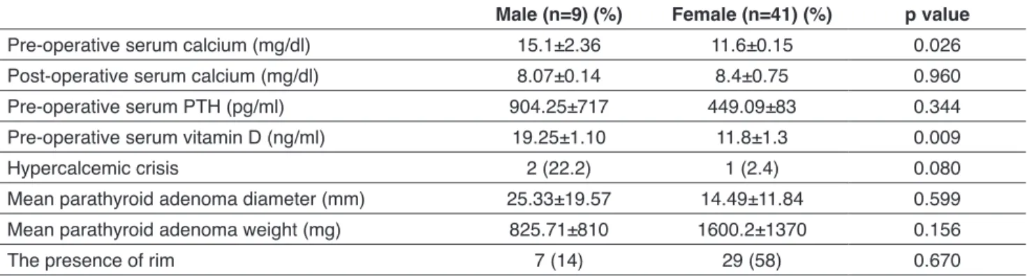
Male patients had higher pre-operative calcium and vitamin D levels than women (p=0.026, p=0.009 respectively), but post-operative serum calcium and pre-operative PTH levels were similar. The pre-operative hypercalcemic crisis incidence was similar in both groups. There was no difference between the two groups in terms of adenoma diameter, weight and presence of rim in the adenoma (Table 2). ERα staining intensity was evaluated as “zero” in all patient’s preparations. Therefore, ERα expression was considered negative in all patients (Table 1).
Table 1. General clinicopathological features of patients
n (%)
Gender Female Male 41 (87.5)9 (24.5)
Menopause status PostmenopausalPremenopausal 31 (75.5)10 (22.5)
Hypercalcemic crisis Yes No 47 (94)3 (6)
The presence of rim Yes No 36 (72)14 (28)
ERα expression No 50 (100)
mean±SD
Average age (year) 57.72±13.16
Average age by gender (year) Female Male 57.63±12.5158.11±16.69
Pre-operative serum calcium (mg/dl) 11.91±1.7
Post-operative serum calcium (mg/dl) 10.75±0.88
Pre-operative serum PTH (pg/ml) 498.46±600.79
Pre-operative serum vitamin D (ng/ml) 12.62±6.55
Parathyroid adenoma diameter (mm) 23.82±13.32
Parathyroid adenoma weight (mg) 1910.21±2400.4
Table 2. Comparison of clinical features in male and female patient with parathyroid adenoma
Male (n=9) (%) Female (n=41) (%) p value
Pre-operative serum calcium (mg/dl) 15.1±2.36 11.6±0.15 0.026
Post-operative serum calcium (mg/dl) 8.07±0.14 8.4±0.75 0.960
Pre-operative serum PTH (pg/ml) 904.25±717 449.09±83 0.344
Pre-operative serum vitamin D (ng/ml) 19.25±1.10 11.8±1.3 0.009
Hypercalcemic crisis 2 (22.2) 1 (2.4) 0.080
Mean parathyroid adenoma diameter (mm) 25.33±19.57 14.49±11.84 0.599
Mean parathyroid adenoma weight (mg) 825.71±810 1600.2±1370 0.156
After excluding two heavy adenomas, it was found that ongoing negative correlation between adenoma weight and PTH level/ weight ratio, which was not statistically significant (r=-0.208, p=0.156).
negative correlation was found between adenoma diameter and post-operative serum calcium level, but the difference was not statistically significant. Similarly, there was a correlation between adenoma diameter and pre-operative serum vitamin D levels, which was not statistically significant (Table 3).
There was a statistically significant correlation between adenoma weight and pre-operative serum calcium and PTH levels, but an insignificant negative correlation with post-operative calcium level. There was also a correlation between adenoma weight and pre-operative serum vitamin D levels, but the difference was not statistically significant (Table 4).
When two parathyroid adenomas (30,000 and 23,000 mg) heavier than the expected average adenoma weight were excluded, the correlation power between adenoma weight and pre-operative PTH and vitamin D levels decreased and lost statistical significance (r=0.243,
p=0.109 and r=0.090, p=0.590 respectively) (Figure 3).
However, a statistically significant correlation between preoperative serum calcium levels and adenoma weight was observed to continue at lower levels (r=0.387,
p=0.007).
A statistically insignificant negative correlation was found between adenoma weight and PTH (ng/ml) adenoma weight (gram) ratio which gave an idea about the amount of PTH secreted by the adenoma weight per gram (r=-0.157, p=0.227) (Figure 4). After two heavy adenomas were excluded from the analysis, it was found that negative correlation between adenoma weight and PTH / weight ratio persisted (r=-0.208, p=0.156)
Table 3. Correlation of adenoma diameter with clinical features
Correlation coefficient p value
Pre-operative serum calcium (mg/dl) r=0.595 p<0.001
Post-operative serum calcium (mg/dl) r=-0.117 p=0.418
Pre-operative serum PTH (pg/ml) r=0.418 p=0.003
Pre-operative serum vitamin D (ng/ml) r=0.023 p=0.887
Table 4. Correlation of adenoma weight with clinical features
Correlation coefficient p value
Pre-operative serum calcium (mg/dl) r=0.607 p<0.001
Post-operative serum calcium (mg/dl) r=-0.130 p=0.369
Pre-operative serum PTH (pg/ml) r=0.503 p<0.001
Pre-operative serum vitamin D (ng/ml) r=0.052 p=0.751
Figure 3. Correlation between adenoma weight and serum
PTH.(•) heavy adenoma.
Figure 4. Correlation between adenoma weight and PTH /
In line with our hypothesis, a study had reported that while ERα expression was present in only 1-3.5% of the patients in that study; ER-β1 and ER-β2 subgroups were expressed in all of 37 patients with parathyroid adenoma. Furthermore, ER-β1 expression has been indicated to correlate with clinical findings (4). For the first time, ESR1 gene -which also encodes ERα and ESR2 gene -which encodes ERβ - expressions had been demonstrated in the same study. It is also been reported that ESR2 gene expression was high in normal parathyroid tissue and parathyroid adenoma. Since demographic data and menopausal status of the patients included in that study were not clearly noted, statistical strength of the study was considered to be weak.
When the clinical features of male and female patients were compared in our study, there was no difference between preoperative PTH levels; however, preoperative calcium levels of male patients were higher than female patients and the difference was statistically significant. The possible reason of male patients having higher serum calcium levels was assumed as consulting a physician too late for examination and treatment. Few studies had reported that male patients have higher levels of calcium and PTH, and also lower vitamin D levels, besides some other studies reporting there was no difference in vitamin D levels between male and female patients (15,16). In our study, vitamin D levels of male patients were higher than those of females; however, the low number of male patients made it difficult to interpret this result. The situation might be explained by traditional dressing style and lack of outdoor activities of females or postmenopausal period in the majority of women included in the study. In some small studies, no significant difference has been found between vitamin D levels of healthy women and men in Turkey (17).
In multiple studies, it has been shown that adenoma weight was heavier in male patients than females (18,19). In our study, no statistically significant difference was found when compared pathological characteristics of parathyroid adenomas in men with those in women. In 2012, Shah et al. had reported that there was no statistically significant difference between adenoma weight and serum calcium, PTH and vitamin D levels in female patients without regard to their menopausal status (16). In our study, in accordance with the results of that study, the clinical features of pre-postmenopausal women and the pathological characteristics of the adenoma were similar; no statistically significant difference was detected.
DISCUSSION
In accordance with the results of several studies in literature, ERα expression could not be demonstrated in the parathyroid adenoma by immunohistochemical technique in our study. In all patient groups, the staining characteristics of normal parathyroid tissue, which suppressed by the parathyroid adenoma, called “rim” was also examined. Any significant nuclear staining wasn’t observed in any patient with detected “rim” to indicate ERα expression at the normal parathyroid tissue in histological assessment.
It is known that primary hyperparathyroidism is more common in women, and its incidence increases during the perimenopausal period, after an average age of 40 years and reaches a peak in the 6th decade
(11). It has been thought that the increased incidence in postmenopausal period may be associated with a decrease in serum estrogen levels. It has been suggested, parathyroid gland may be one of the target tissues affected by estrogen and also various studies have been conducted to identify ER expression in parathyroid gland.
It is quite difficult to determine ER expression in parathyroid tissue due to small size of the normal parathyroid gland and being difficult to acquire the specimen besides autopsies and animal studies. Prince
et al. had reported that ER expression was not present
in 7 human parathyroid adenoma specimens which were analyzed with immunohistochemical method for the first time in 1991 (12). In another study examining 3 parathyroid hyperplasia and 5 parathyroid adenoma specimens, ER expression had not been demonstrated in any of them (13). ER subgroups were not defined within these studies, because ER-β had not been identified in that era, yet.
It has been shown that ERα and ER-β expression was not present in parathyroid gland by using rat models with chronic kidney failure and oophorectomy. However, PTH, vitamin D and phosphate levels have been shown to decrease and also fibroblast growth factor-23 (FGF-23) levels to increase following estrogen therapy (14). These findings have supported that estrogen plays an important role in the pathophysiology of hyperparathyroidism. Without ER expression, the exact mechanism of estrogenic effect on parathyroid gland has not been totally elucidated. For this reason, the expression of ER in the parathyroid gland has gained importance.
expected for small adenomas. Combining these findings, growth in parathyroid adenoma doesn’t always correlate with a precise increasein PTH (21-23).
Limitations of our study, in terms of immunohistochemical examination, are the low number of male and premenopausal female patients; and the absence of a healthy control group with normal parathyroid tissue. It is very difficult to create a control group to examine normal parathyroid tissue, since it is not possible to obtain the material other than autopsy and animal experiments. However, in our study; the absence of ERα expression in normal parathyroid tissue around the adenoma capsule, called “rim”, is a finding that may indirectly indicate the absence of ERα expression in normal parathyroid tissue.
In our study, statistically significant positive correlation was detected between the diameter of parathyroid adenoma and preoperative serum calcium together with PTH levels. In the literature, generally adenoma weight or volume was used in correlation analyses, and the correlation of adenoma diameter with other biochemical studies has not been assessed, yet. This finding suggests the preoperative clinical features of the patients may be useful in predicting the adenoma diameter. However, it is important to determine the cut-off values of serum calcium and PTH level which might have a significant association with the adenoma diameter. In order to achieve that, new larger studies examining the correlation of serum calcium and PTH levels with the adenoma diameter by dividing them into tertiles are necessary.
Acknowledgement
We would like to thank the residents at the Department of Pathology for their contribution to the preparation of pathological specimen.
Ethical Approval
This study was approved by local ethics committee of Kocaeli University Clinical Research Ethics Committee (KKAEK 2012/137).
Conflict of Interest
The authors declare that they have no conflict of interest. Funding
This research did not receive any specific grant from funding agencies in the public, commercial, or not-for-profit sectors.
Author Contributions
Concept: Funda Öztürk, Berrin Çetinarslan, Design: Alev Selek, Supervision: İlhan Tarkun, Zeynep Cantürk, A study conducted by Williams et al. in 1992, in which
40 patients included, had demonstrated a correlation between adenoma weight and PTH, but the researchers had concluded that the result was mainly affected by two parathyroid adenomas much heavier than expected in the cohort group. So, when they excluded these two adenomas and repeated the analysis, they figured out there was no correlation and some of larger adenomas secreted less PTH comparing with small adenomas (20). A statistically significant correlation has been found between preoperative calcium, PTH levels and adenoma diameters in our study (r=0.607, p<0.001; r =0.503,
p<0.001). However, in the repeated analysis when two
adenomas much heavier than expected (30.000mg and 23.000mg) were excluded, it was observed that the correlation between adenoma weight and preoperative serum PTH levels was weakened and statistically significant difference disappeared (r=0.234, p=0.109). The ratio of PTH secretion to adenoma weight which has been suggested as a reasonable predictor for the function of parathyroid adenoma by Williams et al., was also evaluated in our study (20). The high ratio indicates increased adenoma function. In our study, a negative correlation was found between adenoma weight and PTH level (ng/ml) to adenoma weight (gram) ratio (r=-0.157, p=0.278). When two parathyroid adenomas heavier than expected were excluded from the analysis, presented negative but nonsignificant correlation did not change.
Contrary to our results, by using the same formula, Hamidi et al. had reported there was a statistically significant negative correlation (r=-0.46, p<0.001)and also the ratio was shown to be greater in adenomas lighter than 3.9 grams (21). That result supported the hypothesis of unexpected increased PTH secretion in lighter adenomas. In our study, this ratio was found higher in adenomas weighted less than 1.5 grams which was due to the secretion of PTH in the parathyroid gland depended on the number of active cells secreting hormone rather than the total cell number or adenoma weight. There is no relationship between adenoma weight and number of active cells. A large parathyroid adenoma can be heavier due to some fibrotic, calcific, hemorrhagic and cystic degenerated areas. PTH secretion is associated with the capacity of secretory cells and calcium set point, regardless of the macroscopic features of adenoma. Therefore, it is challenging to find a mathematical formula that indicates the relationship between adenoma size and PTH secretion. It should be noted that PTH levels might be much higher than
12. Prince RL, Maclaughlin DT, Gaz RD, Neer RM. Lack of evidence for estrogen receptors in human and bovine parathyroid tissue. J Clin Endocrinol Metab. 1991;72(6):1226-1228.
13. Saxe AW, Gibson GW, Russo IH, Gimotty P. Measurement of estrogen and progesterone receptors in abnormal human parathyroid tissue. Calcif Tissue Int. 1992;51(5):344-347. 14. Carrillo-López N, Román-García P, Rodríguez-Rebollar
A, Fernández-Martín JL, Naves-Díaz M, Cannata-Andía JB. Indirect regulation of PTH by estrogens may require FGF23. J Am Soc Nephrol. 2009;20(9):2009-2017.
15. Mazeh H, Sippel RS, Chen H. The role of gender in primary hyperparathyroidism: Same disease, different presentation. Ann Surg Oncol. 2012;19(9):2958-2962.
16. Shah VN, Bhadada SK, Bhansali A, Behera A, Mittal BR, Bhavin V. Influence of age and gender on presentation of symptomatic primary hyperparathyroidism. J Postgrad Med. 2012;58(2):107-111.
17. Cigerli O, Parildar H, Unal AD, Tarcin O, Erdal R, Guvener Demirag N. Vitamin D deficiency is a problem for adult out-patients? A university hospital sample in Istanbul, Turkey. Public Health Nutr. 2013;16(7):1306-1313.
18. Moretz WH, Watts TL, Virgin FW, Chin E, Gourin CG, Terris DJ. Correlation of intraoperative parathyroid hormone levels with parathyroid gland size. Laryngoscope. 2007;117(11):1957-1960.
19. Kebebew E, Hwang J, Reiff E, Duh QY, Clark OH. Predictors of single-gland vs multigland parathyroid disease in primary hyperparathyroidism: A simple and accurate scoring model. Arch Surg. 2006;141(8):777-782.
20. Williams JG, Wheeler MH, Aston JP, Brown RC,
Woodhead JS. The relationship between adenoma weight and intact (1-84) parathyroid hormone level in primary hyperparathyroidism. Am J Surg. 1992;163(3):301-304. 21. Hamidi S, Aslani A, Nakhjavani M, Pajouhi M, Hedayat
A, Kamalian N. Are biochemical values predictive of adenoma’s weight in primary hyperparathyroidism? ANZ J Surg. 2006;76(10):882-885.
22. Cetani F, Picone A, Cerrai P, Vignali E, Borsari S, Pardi E, et al. Parathyroid expression of calcium-sensing receptor protein and in vivo parathyroid hormone-Ca(2+) set-point in patients with primary hyperparathyroidism. J Clin Endocrinol Metab. 2000;85(12):4789-4794.
23. Mun HC, Conigrave A, Wilkinson M, Delbridge L, Duh QY, Delbridge L. Surgery for hyperparathyroidism: Does morphology or function matter most? Surgery. 2005;138(6):1111-1120.
Resources: Funda Öztürk, Mehmet Sözen, Materials:
Emre Gezer, Yeşim Saliha Gürbüz, Data Collection and/or
Processing: Mehmet Sözen, Yeşim Saliha Gürbüz, Analysis
and/or Interpretation: Funda Öztürk, Berrin Çetinarslan, Alev Selek, Literature Search: İlhan tarkun, Zeynep Cantürk, Emre Gezer, Writing Manuscript: Funda Öztürk, Mehmet Sözen, Emre Gezer, Critical Review: Alev Selek, Berrin Çetinarslan
Peer Review
Extremly peer-reviewed.
REFERENCES
1. O’Connor C., Levine J.A., Hahr A. Primary
Hyperparathyroidism. In: Camacho P. (ed.) Metabolic Bone Diseases. Springer. 2019, 15-25.
2. Cetani F, Marcocci F. Parathyroid Carcinoma. In: John P. Bilezikian (ed.), The Parathyroids (Third Edition), Academic Press, 2015, 409-421.
3. Bilezikian JP, Bandeira L, Khan A, Cusano NE. Hyperparathyroidism. Lancet. 2018 J;391(10116):168-178. 4. Haglund F, Ma R, Huss M, Sulaiman L, Lu M, Nilsson IL, et al. Evidence of a functional estrogen receptor in parathyroid adenomas. J Clin Endocrinol Metab. 2012;97(12):4631-4639.
5. Farford B, Presutti RJ, Moraghan TJ. Nonsurgical management of primary hyperparathyroidism. Mayo Clin Proc. 2007;82(3):351-355.
6. Greenberg C, Kukreja SC, Bowser EN, Hargis GK, Henderson WJ, Williams GA. Parathyroid hormone secretion: Effect of estradiol and progesterone. Metabolism. 1987;36(2):151-154.
7. Nilsson S, Koehler KF, Gustafsson JÅ. Development of subtype-selective oestrogen receptor-based therapeutics. Nat Rev Drug Discov. 2011;10(10):778-792.
8. Bindlish V, Freeman JL, Witterick IJ, Asa SL. Correlation of biochemical parameters with single parathyroid adenoma weight and volume. Head Neck. 2002;24(11):1000-1003. 9. Wagner PK, Rothmund M. Correlation of tumor weight and
typical pathologic laboratory parameters in primary and secondary hyperparathyroidism. Langenbecks Arch Chir. 1983;360(2):133-139.
10. Saadeh G, Licata AA, Esselstyn C, Gupta M. Relationship of parathyroid adenoma volume and biochemical function. Horm Res Paediatr. 1989;32(4):142-144.
11. F. Richard Bringhurst, Demay MB, Kronenberg HM. Hormones and Disorders of Mineral Metabolism. In: Melmed S, Polonsky K, Larsen PR, Kronenberg H (eds). Williams Textbook of Endocrinology. 12th ed. Elsevier Sanders Publications; 2011, 1237-1304.