The size and number is not everything to evaluate the tumor response in
gastrointestinal stromal tumors
Gastrointestinal stromal tümörlerde tümör yanıtı değerlendirilmesinde
boyut ve sayı herşey demek değildir
Erdem Şen1, İrem Öner1, Farise Yılmaz2, Özlem Ata1 1Selçuk Üniversitesi Tıp Fakültesi, Tıbbi Onkoloji Bilimdalı, Konya 2Selçuk Üniversitesi Tıp Fakültesi, Nükleer Tıp Anabilimdalı, Konya Dergiye Ulaşma Tarihi: 12,09,2017 Dergiye Kabul Tarihi: 13,02,2018 Doi: 10.5505/aot.2018.53244
ÖZET
Gastrointestinal stromal tümörler gastrointestinal sistemin en yaygın görülen mezenkimal tümörleridir. Kontrastlı bilgisayarlı tomografi, tedaviye yanıtı değerlendirmede en yaygın kullanılan yöntemdir. Hedefli tedaviler tümör yapısında bazı değişikliklere neden olabilir. Lezyon vaskülaritesinde azalma, kavitasyon ve intratümöral kanama, imatinib tedavisi sonrası hastalarda herhangi bir boyut azalması olmadan bile görülebilen bazı değişikliklerdir. Çelişkili olarak, kistik değişiklik ve intratümöral kanama nedeniyle bazı durumlarda geçici bir boyut artışı görülebilir. Gastrointestinal stromal tümörlerde Choi ve arkadaşları tarafından alternatif tümör yanıt kriterleri geliştirilmiştir. Choi ve arkadaşları Yanıtın değerlendirilmesinde sadece tümör boyutunun değil tümör yoğunluğunun da önemli olduğunu gösterdiler. Bu çalışmada biz gastrointestinal stromal tümör tanısı alan, imatinib ile tedavi edilen 67 yaşında erkek hastayı sunduk. 3 aylık imatinib tedavisinden sonra, yanıt değerlendirilmesinde karaciğer lezyonları progresyon olarak rapor edilmişti.
ABSTRACT
Gastrointestinal stromal tumors are the most common mesenchymal tumors of the gastrointestinal tract. Contrast-enhanced computed tomography is the most commonly used for evaluating response to treatment. Targeted therapies may cause some changes in tumor structure. Reduced lesion vascularity, cavitations, and intratumoral hemorrhage are some changes in patients after imatinib therapy, even without any size reduction. Paradoxically, a transient increase in size may be seen in some cases due to cystic change and intratumoral hemorrhage. Alternative tumor response criteria were developed by Choi et al. in gastrointestinal stromal tumor. Choi et al. showed that not only the tumor size but also the tumor density are important in evaluating the response. In this study, we presented a 67-year-old male patient treated with imatinib receiving a gastrointestinal stromal tumor diagnosis. After 3 months of imatinib treatment, liver lesions were reported as progression in response assessment.
Gastrointestinal stromal tumor (GIST) is one of the most common soft tissue sarcoma subtypes; each year ~3,300–6,000 new cases are diagnosed.1,2 GIST is usually seen in
middle age and elderly patients., with ~60%
located in the stomach, 30% in the small intestine, and 10% in other regions of the gastrointestinal (GI) tract.3 Size, mitotic rate,
and location of the primary lesion are the most
important prognostic factors.4 GIST may be
increased in size during treatment because of intratumoral hemorrhage or myxoid degeneration. Decreasing in tumor density on computed tomography (CT) is an important early clinical marker of antitumor activity. Once tumors become hypodense (cystic), the size of the lesions may decrease slowly and eventually stabilize.5,6 Responses can be
observed within 24 hours of starting therapy on PET-CT. 7
Table 1: Response Evaluation Criteria in Solid
Tumors (RECIST) version 1.1
Response assessment
RECIST, version 1.1 Target lesions
CR Disappearance of all target
lesions and reduction in the short axis measurement of all pathologic lymph nodes to ≤10 mm
PR ≥30 percent decrease in the
sum of the longest diameter of the target lesions compared with baseline
PD ≥20 percent increase of at least
5 mm in the sum of the longest diameters of the target lesions compared with the smallest sum of the longest diameter recorded
OR
The appearance of new lesions, including those detected by FDG-PET
SD Neither PR nor PD
CR: complete response; PR: partial response; PD: progressive disease; FDG-PET: fludeoxyglucose-positron emission tomography; SD: stable disease
The 67-year-old male patient was admitted to our hospital to determine the cause of anemia. Heterogeneously enhancing solid mass (11x17 cm) revealed in the pelvic region and multiple metastatic lesions in the liver were determined on abdominal CT (Figure 1,2). The mass was originated from the ileum. GIST was diagnosed as a result of the biopsy of the abdominal mass and liver. Imatinib therapy (400 mg/day) was started. After 3 months of imatinib treatment thoracic and abdominal CT scans were taken. The mass in the pelvic area measured 7x7.5 cm( Figure 3). However, in the liver parenchyma, metastatic lesions were reported as progression by the department of radiology according to RECIST 1.1 criteria
time imatinib therapy (400 mg/day) is still ongoing. There is no disease progression for 10 months. Stable disease is going on.
Table 2: Choi criteria. Response
assessment
Choi criteria
CR Disappearance of all lesions
No new lesions
PR A decrease in size ≥ 10 or a
decrease in tumor attenuation (HU) ≥ 15 on CT
No new lesions
No obvious progression of non measurable disease
PD An increase in tumor size ≥ 10
and does not meet criteria of PR by tumor attenuation on CT
New lesions
SD Does not meet the above
criteria
CR: complete response; PR: partial response; PD: progressive disease; SD: stable disease
CT:Computed tomography
In metastatic GIST; There are differences for evaluating treatment response compared to other solid tumors. For example, late response may occur who have stable disease. A clinical trial performed by Verweij et al. demonstrated that the median time of objective response is four months, a maximal response may be in six months or even longer.8 In clinical trials
performed that PET-CT is able to show an earlier response than CT.9,10 Treglia et al.,
demonstrated that PET-CT has a significant value in assessing treatment response in GIST. This modality allows an early evaluation of treatment response and is a strong predictor of clinical outcome.11 Alternative response
assessment criteria (Choi criteria) have been developed. in GIST. Investigators at MD
Anderson reported that Choi criteria are a better predictor of response to therapy than standard RECIST criteria. Also, they showed that 10 percent decrease in unidimensional tumor size or a 15 percent decrease in tumor density on contrast-enhanced CT scans (Partial
New ascites or effusions noted on CT scans can be mistakenly regarded as a new finding of peritoneal disease when the disease is otherwise stable or resolving. This is an important consideration in evaluating the response.14
Tumoral lesions may enlarge paradoxically during treatment in GIST. These enlargements can be due to the development of intratumoral hemorrhage or myxoid
degeneration. Enlargement of the tumor does not indicate progression. Also at the beginningi uninvolved tumoral solid lesions may become cystic and may appear as new lesions in the later months of treatment. İn this study, we presented the response evaluation process in our patient with metastatic GIST who received imatinib therapy.
Conflict of interest:None
Figure 1: Multipl metastatic lesions are seen in left and right lobes of the liver. Before
starting treatment of imatinib. A, B: Metastatic lesions in right lobe. C: Metastatic lesions in
right and left lobes.
Figure 2: Heterogeneous mass (11x17 cm) in the pelvic region of abdomen. The mass was
originated from ileum. GIST was diagnosed by biopsy in thic primary lesion. (Before starting
Imatinib treatment)
Figure 3: The primary, heterogeous mass dimensions have decreased (7x7.5 cm). Tumor has
Adress for correspondence:Selçuk Üniversitesi Tıp Fakültesi Hastanesi Aleaddin Keykubat Kampüsü Konya/Türkiye
C: Metastatic lesions in right and left lobes
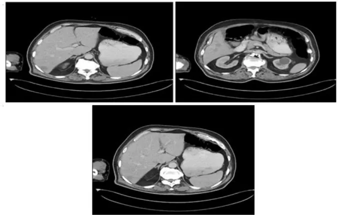
Figure 4: Multipl metastatic lesions are seen in left and right lobes of the liver. Metastatic
lesions number and size were increased. It can be interpreted as progression according to
RECIST criteria. (After three month of the imatinib treatment.) A,C: Metastatic lesions in
right and left lobes. B: Metastatic lesions in right lobe
Figure 5: FDG uptake is not seen in metastatic lesions. PET CT was performed 3 months
after the imatinib therapy
Figure 6: Comparison of tumor densities on primary mass before treatment and after 3
months of imatinib treatment. (Before treatment: 57 Hounsfield Units (HU), After 3 months
of imatinib treatment: 44 HU) A: Tumor density of primary mass before treatment. B: Tumor
density of primary mass after 3 months of imatinib treatment.
Adress for correspondence:Selçuk Üniversitesi Tıp Fakültesi Hastanesi Aleaddin Keykubat Kampüsü Konya/Türkiye Figure 7: Comparison of tumor densities on same
metastatic liver lesionbefore treatmentand after 3 months of imatinib treatment. (Before treatment: 61 HU, After 3 months ofimatinib treatment: 28 HU) A: Tumor density of metastatic liver lesion in right lobe before treatment. B: Tumor density of
metastatic liver lesion in right lobe after 3 months of imatinib treatment
Figure 8 : Comparison of tumor densities on same
metastatic liver lesion before treatment and after 3 months of imatinib treatment. (Before treatment: 75 HU, After 3 months of imatinib treatment: 40 HU) A: Tumor density of metastatic liver lesion in right lobe before treatment. B: Tumor density metastatic liver lesion in right lobe after 3 months of imatinib treatment.
REFERENCES
1. Corless CL, Fletcher JA, Heinrich MC. Biology of gastrointestinal stromal tumors. J Clin Oncol. 2004;22(18):3813–3825.
2. Plesec TP. Gastrointestinal mesenchymal neoplasms other than gastrointestinal stromal tumors: focusing on their molecular aspects. Patholog Res Int. 2011;2011:952569.
3. Miettinen M, Lasota J. Gastrointestinal stromal tumors. Gastroenterol Clin North Am.
2013;42(2):399–415.
4. Rajendra R, Pollack SM, Jones RL. Management of gastrointestinal stromal tumors. Future Oncol. 2013;9(2):193–206.
5. Bechtold RE, Chen MY, Stanton CA, et al. Cystic changes in hepatic and peritoneal metastases from gastrointestinal stromal tumors treated with Gleevec. Abdom Imaging. 2003; 28:808.
6. Linton KM, Taylor MB, Radford JA. Response evaluation in gastrointestinal stromal tumors treated with imatinib: misdiagnosis of disease progression on CT due to cystic change in liver metastases. Br J Radiol. 2006; 79:e4
7. Stroobants S, Goeminne J, Seegers M, et al. 18FDG-Positron emission tomography for the early prediction of response in advanced soft tissue sarcoma treated with imatinib mesylate (Glivec). Eur J Cancer. 2003; 39:2012.
8. Verweij J, Casali PG, Zalcberg J, et al. Progression-free survival in gastrointestinal stromal tumors with high-dose imatinib: randomized trial. Lancet. 2004; 364:1127.
9. Van den Abbeele AD. The lessons of GIST--PET and PET/CT: a new paradigm for imaging. Oncologist. 2008; 13 Suppl 2:8.
10. Antoch G, Kanja J, Bauer S, et al. Comparison of
PET, CT, and dual-modality PET/CT imaging for monitoring of imatinib (STI571) therapy in patients with gastrointestinal stromal tumors. J Nucl Med. 2004; 45:357.
11. Treglia G, Mirk P, Stefanelli A et al.
18F-Fluorodeoxyglucose positron emission tomography in evaluating treatment response to imatinib or other drugs in gastrointestinal stromal tumors: a systematic review. Clin. Imaging. 2012; 36 (3): 167– 75.
12. Choi H, Charnsangavej C, Faria SC, et al.
Correlation of computed tomography and positron emission tomography in patients with metastatic gastrointestinal stromal tumor treated at a single institution with imatinib mesylate: proposal of new computed tomography response criteria. J Clin Oncol. 2007; 25:1753.
13. Benjamin RS, Choi H, Macapinlac HA, et al. We
should desist using RECIST, at least in GIST. J Clin Oncol. 2007; 25:1760.
14. Demetri GD, Benjamin R, Blanke CD, et al. NCCN
task force report: optimal management of patients with gastrointestinal stromal tumors (GIST)— expansion and update of NCCN clinical practice guidelines. J Natl Compr Cancer Netw. 2004; 2:S1– S26