Investigation of paranasal sinuses based on dissection and multidetector computed
tomography images in New Zealand rabbits
Sema Özkadif1*, Emrullah Eken2 Özet
Özkadif S, Eken E. Yeni Zelanda tavşanlarında sinus paranasales’in diseksiyon bulguları ve multidedektor bilgisayarlı tomografi görüntülerinin araştırılması. Eura-sian J Vet Sci, 2012, 28, 1, 10-14
Amaç: Yeni Zelanda tavşanlarında sinus paranasales ve concha’ların anatomik özelliklerini diseksiyon ve multid-edektör bilgisayarlı tomografi (MDBT) ile ortaya koymaktır. Gereç ve Yöntem: Araştırmada her iki cinsiyetten to-plam 16 yetişkin Yeni Zelanda tavşanı kullanıldı. Sinus paranasales’in yüksek çözünürlüklü MDBT görüntüleri elde edildikten sonra hayvanlar bilinen metodlar ile ötenazi yapıldı ve paranasal sinusları diseke edildi.
Bulgular: MDBT ve diseksiyon bulguları ışığında Yeni Ze-landa tavşanlarının kafatasının her bir yarımında ductus nasolacrimalis vasıtasıyla bağımsız olarak ikiye ayrılan iki bölümlü bir adet sinus maxillaris’in mevcut olduğu tespit edildi. Bu türlerde iyi gelişmiş olan concha’ların concha na-salis dorna-salis, concha nana-salis media, concha nana-salis ventra-lis ve endoturbinalia’dan oluştuğu görüldü. Sinus frontaventra-lis, sinus sphenoidalis ve sinus ethmoidalis’in şekillenmediği saptandı. Diseksiyon ve MDBT görüntülerinden elde edilen verilerin birbirini desteklediği belirlendi.
Öneri: Bilinen metodların yanında görüntüleme yöntemi de kullanan bu çalışmanın sinus paranasales ile ilgili ge-lecek çalışmalara katkı sağlayabileceği ve anatomi eğitimine modern bir açılım sağlayabileceği düşünülmektedir.
Abstract
Ozkadif S, Eken E. Investigation of paranasal sinuses based on dissection and multidetector computed tomography im-ages in New Zealand rabbits. Eurasian J Vet Sci, 2012, 28, 1, 10-14
Aim: The aim of this study is to present anatomic properties of paranasal sinuses and conchae by dissection and multi-detector computed tomography (MDCT) images in New Zealand rabbits.
Materials and Methods: In this study a total of 16 adult New Zealand rabbits of both sexes were used. After obtain-ing high resolution MDCT images of paranasal sinuses, the animals were euthanasied by conventional methods and then dissected their paranasal sinuses.
Results: Based on dissection and MDCT images, it was de-termined that each half of New Zealand rabbit skull had max-illary sinus formed by two compartments that were divided by independently a nasolacrimal duct. We also showed that the well developed conchae were formed by dorsal, middle, ventral nasal conchae as well as endoturbinalia. Interest-ingly, we recorded no frontal, sphenoidal and ethmoidal si-nuses in this species. The data obtained from dissection and MDCT images were in parallel to each other
Conclusion: It has been suggested that this work using imaging technique in addition to common anatomical tec-niques may contribute to future studies on paranasal sinus-es and may add modern dimension to anatomical education.
1Department of Biology Education, Faculty of Ahmet Keleşoğlu Education, 2Department of Anatomy, Faculty of Veterinary Medicine, Selcuk University, Konya, 42075, Turkey Received: 15.11.2011, Accepted: 28.11.2011 *[email protected]
Anahtar kelimeler: Diseksiyon, bilgisayarlı tomografi, sinus paranasales, concha, tavşan
Keywords: Dissection, computed tomography, paranasal sinuses, concha, rabbit
RESEARCH ARTICLE
Journal of Veterinary Sciences
Introduction
The main properties of paranasal sinuses are to have air holes, complex anatomies and individual varia-tions. Genetic diseases, environmental conditions and experienced infections may affect the developmen-tal process of paranasal sinuses (Tos and Stangerup 1985, Kubal 1998). Knowing anatomic variations are important not only for reducing endoscopic sinus sur-gery complications but also for correcting surgically variations that might cause chronic sinusitis (Çağıcı et al 2006).
Head dissection is important to reveal nose and sinus anatomy of human cadaver and for technical applica-tion. It’s mostly difficult for medical students to find enough head of human cadaver (Gardiner et al 1996). At the same time, it’s ethically not possible to perform experiments on human beings for the research of na-sal and paranana-sal sinus diseases. Almost all of stud-ies related with surgical traumas of these regions and regeneration patterns following them have been also performed on animals (Köybaşıoğlu et al 1997). For these reasons, knowing anatomy, biology and behav-ior of animals is highly important in terms of produc-ing models that might be applied on human beproduc-ings (Palumbo et al 2004). Basic information should be presented before transferring techniques and models to the clinical medium. Results of studies with animals can be adapted on human models (Grauer et al 2000). For this purpose, rabbits are mostly used as experi-ment animals in human and veterinary medicines. For example, rabbits can be used in the development of infection similar to paranasal sinus aspergillosis in human beings. In the study of this infection, mice and rats were failed (Chakrabarti et al 1997).
In the literature, although there were few studies re-lated to morphology of rabbit paranasal sinuses dis-section findings of rabbit paranasal sinuses, no stud-ies using imaging tecniques were found. Therefore this study was performed for the aim of filling the deficiency in this field as comparing dissection and MDCT findings of paranasal sinuses and conchae in New Zealand rabbits.
Materials and Methods
In this study, a total of 16 New Zealand rabbits of both sexes aged 1-1.5 and weighing between 3 and 3.5 kg were used. Study protocol was approved by Ethical Committee of Veterinary Faculty, Selcuk University. The rabbits were intravenously anaesthetized with the mixture of 5 mg/kg ketamine-HCl (KetamidorTM RicherPharma AG, Wels, Austria) and 20 mg/kg pro-pofol (Propro-pofolTM Amp., Fresenius Kabi, Austria). Under anesthesia, MDCT images of animals in prone position were obtained. The parameters of MDCT (Somatom Sensation 64;Siemens Medical Solutions, Forchheim, Germany) instrument were adjusted as; physical detector collimation, 32 x 0.6 mm; final sec-tion collimasec-tion, 64 x 0.6 mm; secsec-tion thickness, 0.75
mm; gantry rotation period; 330 msec; kVp; 120; mA, 300; resolution, 512 x 512 pixel; resolution range, 0.92 x 0.92. Dosage parameters and scannings were performed by predicating them on standard protocols and literature (Prokop 2003, Kalra et al 2004). Thus, it was tried to obtain radiometric resolution (MONO-CHROME 2;16 bit) at the lowest radiation level with optimum image quality. The axial images obtained were stocked in DICOM format and then were evalu-ated in a personal computer.
After obtaining high resolution MDCT images of para-nasal sinuses were obtained, the animals were eu-thanasied by conventional methods for dissection. After the skin of the dorsal portion of the nose was trimmed, the skin and periosteum in middle line were elevated carefully to reveal the nasal bone between naso-frontal suture and naso-incisiv suture (Bahadır at al 2008). Osteotomy was performed transversally along the back of nasal and maxillary bones and a cut was formed on upper part of the lateral cartilages. Lateral nasal septum including maxillary sinus ostium was also cut and removed. This cut piece is conjugat-ed with front part of ethmoid (Velwoerd- Velhoef and Verwoerd 2003). Anatomy of lateral nasal septum, conchae and ostium of maxillary sinus were investi-gated within these incisions. Moreover, the inner side of sinus maxillaris was investigated by getting coronal cross sections (Köybaşıoğlu et al 1997). The emerging maxillary sinus and conchae were photographed and named. Nomina Anatomica Veterinaria (NAV 2005) was used as terminology in study.
Results
Dissection of paranasal sinuses
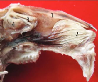
The right and left lateral nasal septums were ob-served after nasal bone and maxilla are removed. Nasal septums were intimate contact with the rostral part of ethmoid bone.We showed the right and left maxillary sinus ostiums at the bottom of both nasal septums. Dorsal, ventral and middle nasal conchae and endoturbinalia were determined on lateral nasal septum. The dorsal nasal concha was on top of cavum nasi, middle nasal concha was the oro-ventral third of the dorsal nasal concha, the ventral nasal concha was aboro-ventral third of the dorsal nasal concha. Endoturbinalia was located ventral to the middle na-sal concha. It was observed that conchae were quite sinuous (Figure 1).
It was recorded that maxillary sinus was in the max-illa ventral to conchae. Maxmax-illary sinus ostium was opened to the narrow field at the back of ethmotur-binal concha. In coronal cross sections, it was ob-served that maxillary sinus was constituted by dorsal and ventral compartments. The nasolacrimal canal was determined to cross between the ventral and dor-sal compartments (Figure 2). The ventral compart-ment of the maxillary sinus was placed at the bace of the nasal cavity. It was observed that the maxillar
si-nus ostium was opened to the nasal cavity at the base of the dorsal compartment (Figure 3). In coronal and lateral cross sections, frontal, sphenoidal and ethmoi-dal sinuses were not seen.
Analysis of MDCT images taken from paranasal si-nuses
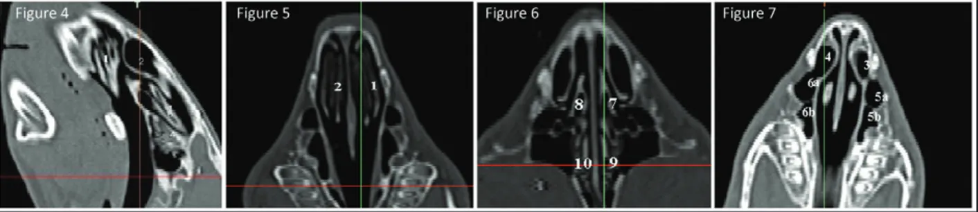
In sagittal sections of paranasal sinuses, it was deter-mined that dorsal nasal concha was dorsal to the na-sal cavity. The ventral nana-sal and middle nana-sal conchae were located aboro-ventral and oro-ventral to the dorsal nasal concha, respectively. Both conchae were seen to be formed by white colored curles whose spaces were black colored. Endoturbinalia was ob-served caudal to the middle nasal concha (Figure 4).
In coronal sections of paranasal sinuses, there was the ventral nasal concha that was located in the right and left sides of the cranial part of nasal septum had grey colored curlings including black colored spaces (Figure 5).
In coronal sections of paranasal sinuses, the mid-dle nasal concha was seen as grey colored line in the right and left side of medial face of the nasal septum. The endoturbinalia was also grey colored in the both side of caudal face of nasal septum. In all sections, the spaces among the middle nasal concha and endotur-binalia were black colored (Figure 6).
In coronal sections of paranasal sinuses, the dorsal nasal concha and maxillary sinus were seen as black colored, and they were bordered by a grey colored septum. The dorsal and ventral compartment could be discernible (Figure 7).
Discussion
The conformations and locations of the nasal sinuses and conchae in New Zeland rabbit were consistent with the Köybaşıoğlu et al (1997) and Özer (2004). We showed that the maxillary sinus was located just ventral to the nasal bone as recorded by Asai et al (2002). New Zealand rabbit had two compartments that the nasolacrimal canal separated one another, which is inconsestent with human being. The con-chaae of New Zealand rabbit were more developted and more curling than human being (Köybaşıoğlu et al 1997). In both dissection and MDCT images the frontal, sphenoidal and ethmoidal sinuses were not seen in this work.
As mentioned by Pinkston et al (1995), the anatomy of nose and maxillary sinus belonging to rabbits prin-cipally was similar to those of human beings. How-ever, the most important difference was that the up-per part and turbinals of maxillary sinus in rabbits were placed in the upwards of lateral nasal wall. Due to this similarity, it was determined in the studies of Chakrabarti et al (1997) and Jyonouchi et al (1998) that rabbit maxilary sinus can be used in the research of paranasal sinus aspergilosis and bacteroids.
Figure 1. View of 1: Dorsal nasal concha, 2: Ventral nasal concha, 3.
Middle nasal concha and 4: Endoturbinalia on lateral nasal septum. Figure 2. In coronal cross section, 1: dorsal compartment of maxillary sinus, 2: ventral compartment of maxillary sinus, 3: Canalis nasolac-rimalis.
As it was specified by Verwoerd-Verhoef and Ver-woerd (2003), maxillary sinus was connected with nasal cavity. Maxillary sinus which is below maxillary sinus turbinale was opened to nasal cavity with a sin-gle hole which is called the maxillary sinus ostium. In maxillary sinus research of Watanabe et al (1999) and in experimental operations of Pinkston et al (1995) on lateral nasal wall, it was determined that rabbit had many advantages as a good model.
It was shown by Peltola et al (2001), Peltola et al (2003), Peltola et al (2006) that the frontal sinus in rabbits was present just in young rabbits and was dis-appeared in the course of time. Since the rabbits used in our study were adult, frontal sinus was not encoun-tered. Although Jacob and Chole (2006) reported that the mouse had a pair of sphenoidal sinus, it was not encountered in New Zealand rabbits.
Conclusions
This study using dissection and MDCT imaging tech-niques revealed that each half of the New Zeland rab-bit skull had one maxillary sinus consisting of two compartments and four conhcae named dorsal, mid-dle, ventral nasal conchae and endoturbinalia. We hoped that the results from this study will shed light on further studies on paranasal sinuses, and we can say that making use of computer technology in chang-ing and developchang-ing anatomy education and research-es is ethically quite important.
Acknowledgements
This study is supported by the Coordinatorship of Selcuk University Scientific Research Projects as the Project number 2009/056.
References
Asai S, Shimizu Y, Ooya K, 2002. Maxillary sinus augmenta-tion model in rabbits: Effect of occluded nasal ostium on new bone formation. Clin Oral Imp Res, 13, 405-409.
Bahadir O, Bahadir A, Kosucu P, Livaoglu M, 2008. The ef-fect of maxillary sinus surgery on its development. Acta Otolaryngol, 128, 551-555.
Çağıcı CA, Yavuz H, Erkan AN, Akkuzu B, Özlüoğlu L, 2006. Paranazal sinüs anatomik varyasyonlarının değerlendirilmesinde bilgisayarlı tomografi. Turk Arch Otolaryngol, 44, 201-210.
Chakrabartı A, Jatana M, Sharma SC, 1997. Rabbit as an ani-mal model of paranasal sinus mycoses. J Med Vet Mycol, 35, 295-297.
Gardiner Q, Oluwole M, Tan L, White PS, 1996. An animal model for training in endoscopic nasal and sinus sur-gery. J Laryngo Otol, 110, 425-428.
Grauer NJ, Erulkar SJ, Patel CT, Panjabi MM, 2000. Biome-chanical evaluation of the New Zeland white rabbit lum-bar spine: a physiologic characterization. Eur Spine J, 9, 250-255.
International Committee on Veterinary Gross Anatomical Nomenclature, 2005. General Assemble of the World As-sociation of Veterinary Anatomists. Nomina Anatomica Veterinaria, 5th edition, Gent, Belgium.
Jacob A, Chole RA, 2006. Survey anatomy of the paranasal sinuses in the normal mouse. Laryngoscope, 116, 558-563.
Jyonouchi H, Sun S, Kennedy CA, Kajander KC, Rimel FL, 1998. Ig Antibody levels in the sinus, ear and airway in a rabbit model of sinusitis with bacteroides. Arch Otolaryngol Head Neck Surgery, 124, 767-772.
Kalra MK, Maher MM, Toth TL, Hamberg LM, Blake MA, Shepard J, Saini S, 2004. Strategies for CT radiation dose optimization. Radiology, 230, 619-628.
Köybaşıoğlu A, İleri F, Beder L, İnal E, 1997. Tavşan mak-siller sinüs anatomisi. KBB ve Baş ve Boyun Cerrahisi Dergisi, 5, 41-44.
Kubal WS, 1998. Sinonasal anatomy. Neuroimaging Clin N Am, 8, 143-156.
Özer A, 2004. Deneysel mide çeriği sıvısının tavşan bu-run ve paranazal sinüslerine etkisi. Süleyman Demirel Üniversitesi Tıp Fakültesi Kulak Burun Boğaz Ana Bilim Dalı, Uzmanlık Tezi, pp: 31-32.
Figure 4. Sagittal MDCT view of paranasal sinuses. 1: Left ventral nasal concha, 2: Left dorsal nasal concha, 3: Left middle nasal concha, 4: Left endoturbinalia.
Figure 5. Coronal MDCT view of paranasal sinuses. 1: Left ventral nasal concha, 2: Right ventral nasal concha.
Figure 6. Coronal MDCT view of paranasal sinuses. 7: Left middle nasal concha, 8: Right middle nasal concha, 9: Left endoturbinalia, 10: Right endoturbinalia.
Figure 7. Coronal MDCT view of paranasal sinuses. 3: Left dorsal nasal concha, 4: Right dorsal nasal concha, 5a: Dorsal compartment of left maxillary sinus, 5b: Ventral compartment of left maxillary sinus, 6a: Dorsal compartment of right maxillary sinus, 6b: Ventral compartment of right maxillary sinus.
Palumbo M, Valdes M, Robertson A, Sheikh S, Lucas P, 2004. Posterolateral intertransverse lumbar arthrodesis in the New Zealand white rabbit model: I Surgical anato-my. Spine J, 4, 287-292.
Peltola MJ, Aitasalo KMJ, Suonpaa JTK, Varpula M, Yli-Urpo A, 2006. Bioactive glass S53P4 in frontal sinus oblitera-tion: a long-term clinical experience. Inc Head Neck, 28, 834-841.
Peltola MJ, Aitasalo KMJ, Suonpaa JTK, Yli-Urpo A, Laippala PJ, Forsback AP, 2003. Frontal sinus and skull bone de-fect obliteration with three synthetic bioactive materi-als. A comparative study. Inc. J Biomed Mater Res Part B: Appl Biomater, 66, 364-372.
Peltola MJ, Aitasalo KMJ, Suonpaa JTK, Yli-Urpo A, Laippala PJ, 2001. In vivo model for frontal sinus and calvarial bone defect obliteration with bioactive glass S53P4 and hydroxyapatite. J Biomed Mater Res Appl Biomater, 58, 261-269.
Pinkston DR, Schubkegel AJ, Zimmerman MB, Smith RJH, 1995. The effects of sinus surgery on midfacial growth in the rabbit. Amer J Rhinol, 9, 115-124.
Prokop M, 2003. General principles of MDCT. Eur J Radiol, 45, 4-10.
Tos M, Stangerup SE, 1985. The causes of asymmetry of the masoid air cell system. Acta Otolaryngol, 99, 564-570. Velwoerd-Verhoef HL, Verwoerd CDA, 2003. Surgery of the
lateral nasal wall and ethmoid: effects on sinonasal growth. An experimental study in rabbits. Int J Pediatr Otorhinolayngol, 67, 263-269.
Watanabe K, Niimi A, Ueda M, 1999. Autogenous bone grafts in the rabbit maxillary sinus. Oral Surg Oral Med Oral Pathol Oral Radiol Endod, 88, 26-32.