S. K. Kucur et al. Doppler sonography for endometrial pathologies 665
Dicle University, Faculty of Medicine, Department of Pediatrics, Diyarbakır, Turkey Yazışma Adresi /Correspondence: İlyas Yolbaş,
Dicle Üniversitesi, Tıp Fakültesi, Çocuk Sağlığı ve Hastalıkları AD, Diyarbakır, Türkiye Email: [email protected] Geliş Tarihi / Received: 02.05.2013, Kabul Tarihi / Accepted: 03.07.2013
Copyright © Dicle Tıp Dergisi 2013, Her hakkı saklıdır / All rights reserved
Dicle Tıp Dergisi / 2013; 40 (4): 665-667
Dicle Medical Journal doi: 10.5798/diclemedj.0921.2013.04.0354
CASE REPORT / OLGU SUNUMU
Large congenital cystic asdenomatous malformation of the lung in a newborn
Yenidoğanda akciğerin büyük konjenital kistik adenomid malformasyonu
İlyas Yolbaş, Selvi Kelekçi, Yusuf Kenan Haspolat, Ali Güneş, Velat ŞenÖZET
Akciğerin konjenital kistik adenoid malformasyonu akciğe-rin hava ile dolu kistleakciğe-rinden oluşan bir hemartöz lezyon-dur. Genelde doğumdan sonra solunum sıkıntısı ile bulgu verir. Konjenital pnömoni, respiratuvar distress sendromu ile ayırıcı tanısı zordur. Doğumdan sonra şiddetli solunum sıkıntısı ve düşük Apgar olan bir erkek yenidoğan entübe edildikten sonra yenidoğan yoğun bakım ünitemize yatı-rıldı. Hasta 50 gün mekanik ventilatörde takip edildikten sonra 50. gün mekanik ventilatörden ayrıldı. Tip II Akciğe-rin konjenital kistik adenoid malformasyonu tanısı akciğer tomografisi ve akciğer grafi bulguları ile kondu. Cerrahi lobektomi önermesine rağmen hastanın tümüyle asepto-matik olmaması ve ölüm riski nedeniyle aile operasyonu kabul etmedi. Hasta kontrole gelmek üzere taburcu edildi. Anahtar kelimeler: Akciğerin konjenital kistik adenoid malformasyonu, yenidoğan, konservatif tedavi
ABSTRACT
Congenital cystic adenomatous malformation (CCAM) of lung is a rare form of congenital hamartomatous lesions of the lung consisting of cysts filled with air. The general clinic presentation of CCAM is dyspnea in newborns. CCAM may mimic congenital pneumonia or respiratory distress syndrome. After the delivery, the newborn male who had low Apgar score and severe respiratory distress was intubated and admitted to neonatal intensive care unit. Patient was ventilated for 50 days and weaned from the mechanical ventilator at 50th day. Type II CCAM of the lung was diagnosed according to the chest radiographs and computed tomography scan signs. Although the sur-geons suggested lobectomy considering the patient’s not completely asymptomatic, family did not accept this oper-ation due to the risk of death. The patient was discharged from the hospital until the next control.
Key word: Congenital cystic adenomatous malformation of lunch, newborn, conservative treatment
INTRODUCTION
Congenital cystic asdenomatous malformation of lung (CCAM) is a rare form of congenital hamarto-matous lesions of the lung consisting of cysts filled with air. CCAM may mix with congenital pneumo-nia, respiratory distress syndrome. It was first de-scribed by Ch’in and Tang in 1949 [1]. The inci-dence of CCAM was estimated as 1 per 25000 to 35000 births. It accounts for 25% of all congenital lung malformations and 95% of congenital cystic lung malformations [2]. The general clinic presen-tation of CCAM is dyspnea in newborns [3], it can also cause bronchiolo alveolar carcinoma and rhab-domyosarcoma [4], and usually diagnosed by using lung X-ray, antenatal ultrasonography and
com-puted tomography or lung biopsy [5]. Stocker and al. classified three types of CCAM in 1977, type I consists large cysts greater than 2.0 cm diameter; type II consists small cysts and type III consists a resembling homogeneous mass lesions with cysts only seen on microscopy [6]. The aim of this study is to present a newborn with CCAM.
CASE REPORT
A newborn male from healthy parents was born at 38 weeks of gestation by elective cesarean section due to severe toxemia and polyhydramnios. Thirty-eight years old mother had no history of pulmonary disease in her family and the antenatal ultrasonogra-phy (USG) was shown only polyhydramnios.
İ. Yolbaş et al. Congenital cystic adenomatoid malformation of the lung 666
Dicle Tıp Derg / Dicle Med J www.diclemedj.org Cilt / Vol 40, No 4, 345-349
After the delivery, a newborn male who had poor Apgar score and severe respiratory distress was intubated successfully using a direct laryngo-scope and admitted to neonatal intensive care unit. The newborn patient needed to be ventilated for 50 days and weaned from the mechanical ventilator at
50th day of his life. Type II CCAM of the lung was
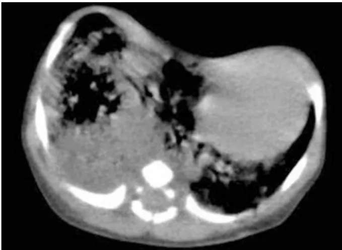
diagnosed according to the chest radiographs and computed tomography scan. The first chest radio-graphs and computed tomography scan revealed multiple small cysts filled with air in the middle lobes of the lungs and the heart was pushed to the left of the chest (Figure 1, Figure 2).
Figure 1. First days chest radiograph; multiple small cys-tic filled with air in the middle lobe of the lung and the heart was pushed to left of chest
Figure 2. First computed tomography scan; multiple small cystic filled with air in the middle lobe of the lung and heart was pushed to left of chest
In addition, the chest radiographs and comput-ed tomography scan designatcomput-ed a better sign, which is the normal position of the heart (Figure 3,
Fig-ure 4). Although the surgeons suggested lobectomy considering the patient is not completely asymp-tomatic, family did not accept this operation due to the risk of death. The patient was discharged from the hospital until the next control.
Figure 3. The last days chest radiograph; multiple small cystic filled with air in the middle lobe of the lung and posi-tion of the heart was normal
Figure 4. Second contrast-enhanced computed tomogra-phy scan; multiple small cystic filled with air in the middle lobe of the lung and position of the heart was normal
DISCUSSION
This case may be the first in the literature that the patient was ventilated for 50 days and had weaned from the mechanical ventilator at 50th day of his life.
Chen et al. reported seven patients with respi-ratory distress begins immediately after the birth were subsequently diagnosed with CCAM (%50 cases), 62% of patients had type II CCAM and 56% of the lesions were on the right lung [7]. Parikh et al. found all of their 22 patients were asymptomatic
İ. Yolbaş et al. Congenital cystic adenomatoid malformation of the lung 667
Dicle Tıp Derg / Dicle Med J www.diclemedj.org Cilt / Vol 40, No 4, 345-349
[8]. Some studies reported that 50-72% of the pa-tients with CCAM were asymptomatic at birth and 5-60% of radiographs taken after the birth were nor-mal [9]. Our case was symptomatic type II CCAM with abnormal lung radiographs at birth and then symptoms of the patient regressed slowly.
18-20% of CCAM patients can born with Prune Belly Syndrome, pulmonary sequestration, dia-phragmatic hernia, agenesis of a lung, jejunal atre-sia, cardiac and renal anomalies [10]. Our case had only pectus excavatum abnormality.
CCAM is usually diagnosed with lung X-ray, antenatal ultrasonography and computed tomog-raphy or lung biopsy [5]. Our case was diagnosed with images of the chest radiographs and computed tomography scan.
Polyhydramnios and microcystic type of le-sions in patients with CCAM are associated with a poor prognosis [11]. The major cause of morbidity is respiratory distress caused by compromised pul-monary function or pulpul-monary hypoplasia. Surgery should be considered in patients with large CCAM and in the case of poor prognosis11. Dommergues at al. treated 17 patients with CCAM without acute polyhydramnios or hydrops with conservative treat-ment [12]. Dommergues at al. also treated 17 pa-tients with CCAM without acute polyhydramnios or hydrops by conservative treatment [12]. In our case, although the surgeons suggested lobectomy considering the patient had polyhydramnios so was not completely asymptomatic, family did not accept this operation due to the risk of death. Therefore, we treat the patient with conservative treatment, then the patient’s symptoms regressed.
In summary, CCAM should be considered among the causes of the respiratory distress in neo-nates, and it may mix wit with congenital pneumo-nia, RDS.
REFERENCES
1. Ch’in KY, Tang MY. Congenital asdenomatous malformation of one lobe of a lung with general anasarca. Arch Pathol 1949;48:221-229.
2. Revillon Y, Jan D, Plattner V, et al. Congenital cystic asdeno-matous malformation of the lung: prenatal management and prognosis. J Pediatr Surg 1993;28:1009-1011.
3. Bagalan P, Nahom A, Giorlandino C, et al. Cystic asdeno-matous malformation of the lung: Clinical evolution and management. Eur J Pediatr 1999;158:879-882.
4. Ioachimescu OC, Mehta AC. From cystic pulmonary airway malformation, to bronchioloalveolar carcinoma and adeno-carcinoma of the lung. Eur Respir J 2005;26:1181. 5. Sauvat F, Michel J, Benachi A, Edmond A, Revillon Y.
Man-agement of Asymptomatic Neonatal Cystic Asdenomatous Malformations. J Pediatr Surg 2003;38:548-552.
6. Stocker JT, Madewell JE, Drake RM. Congenital cystic as-denomatous malformation of the lung: Clussification and morphologic spectrum.. Hum Pathol 1977;8:155-171. 7. 9. Chen HW, Hsu WM, Lu FL, et al. Management of
Con-genital Cystic Asdenomatous Malformation and Broncho-pulmonary Sequestration in Newborns. Pediatr Neonatol 2010;51:172−177.
8. 10. Parikh D, Samuel M. Congenital cystic lung lesions: Is surgical resection essential? Pediatr Pulmonol 2005;40:533-537.
9. Van Leeuwen K, Teitelbaum DH, Hirschl RB, et al. Prenatal diagnosis of congenital cystic asdenomatous malformation and its postnatal presentation, surgical indications, and nat-ural history. J Pediatr Surgery 1999;34:94-798.
10. Hansen TN, Corbet A. Anomalies of the airways, medias-tinum and lung parenchyma. In: Taeusch HW, Ballard RA, Gleason CA (Eds). Avery’s Diseases of the newborn. Phila-delphia: Elsevier Saunders 2005: p. 747-749.
11. Adzick NS, Harrison MR, Crombleholme TM, et al. Fetal lung lesions: management and outcome. Am J Obstet Gy-necol 1998;179:884-889.
12. Dommergues M, Louis-Sylvestre C, Mandelbrot L, et al. Congenital asdenomatous malformation of the lung: When is active fetal therapy indicated? Am J Ob Gynecol 1997;177:953-958.