Ankara University
Institute of Nuclear Sciences
Owner on behalf of Institute of Nuclear Sciences,
Ankara University,
Director
Niyazi MERİÇ, Ph.D.
http://jns.ankara.edu.tr
Editor-in-Chief
Haluk YÜCEL, Ph.D.
Assistant Editor-in-Chief
George S. POLYMERIS, Ph.D.
Editorial Board
Birol ENGİN, Ph.D.
Erkan İBİŞ, M.D.
Gaye Ö. ÇAKAL, Ph.D.
Güneş TANIR, Ph.D.
Hamit HANCI, M.D.
Ioannis LIRITZIS, Ph.D.
İsmail BOZTOSUN, Ph.D.
M.Salem BADAWI, Ph.D.
Mustafa KARADAĞ, Ph.D.
Niyazi MERİÇ, Ph.D.
Osman YILMAZ, Ph.D.
Özlem BİRGÜL, Ph.D.
Özlem KÜÇÜK, M.D.
Slobodan JOVANOVIC, Ph.D.
Turan OLĞAR, Ph.D.
Volume 2, No. 2
May 2015
ISSN: 2148-3981
Journal of Nuclear Sciences
Not for reproduction, distribution or commercial use.
Provided for non-commercial research and education use.
*Corresponding author.
E-mail address: [email protected] (A. Nourreddine) Journal of Nuclear Sciences, Vol. 2, No.2 , May 2015, 53-58. Copyright © Ankara University Institute of Nuclear Sciences
Journal of Nuclear Sciences
ISSN: 2147-7736
J o ur na l h om e page: h t tp :/ / j n s . a n k a r a . e d u . t r /
DOI: 10.1501/nuclear_0000000013
Present state of the art of a fast neutron dosimeter incorporating RPL
detectors
Y.O. Salem, A. Nourreddine*, A. Nachab, C. Roy, A. Pape
Groupe RaMsEs, Institut Pluridisciplinaire Hubert Curien (IPHC), UMR 7178 CNRS/IN2P3, 23 rue du Loess, BP 28, F-67037 Strasbourg Cedex 2, France
Received 23.09.2014 received in revised form 03.02.2015; accepted 04.05.2015
ABSTRACT
The recently introduced radiophotoluminescent (RPL) detectors offer a unique combination of advantages for radiation monitoring that include rapid exploitation, stability to fading, reusability, and insensitivity to light, temperature and humidity. We look at the behavior of an RPL-based fast neutron dosimeter capable of measuring neutrons in an n- field. The tested dosimeter consists of an ordered assembly of Al foil, RPL detector (I), Al foil, polyethylene converter, RPL detector (II) and Al foil encased in a polyethylene container. The difference between the two RPL configurations represents the (n,p) protons and is related to the fast neutron dose. The dosimeter response is linear and shows an acceptable angular dependence. However the measured detection threshold for this dosimeter is too high for routine monitoring. This threshold could be lowered at to a more practicable value if next generation improvements in RPL detectors and the reader are applied. The main shortcomings we encountered are (i) a 1.7 µm thick dead layer at the front surface of the detectors that render them insensitive to a large fraction of the recoil protons and (ii) an intrinsic detector background that could be reduced if the reader were able to separate individual densely ionized zones created by the recoil protons from the -ray signal.
Keywords: Ambient dosimetry; fast neutrons; Ag-doped phosphate glass; radiophotoluminescence; MCNPX 1. Introduction
Evaluation of the dose received by an individual exposed to neutrons constitutes a current problem of radiation protection. The objective of the present study is to realize a neutron dosimeter using the radiophotoluminescence (RPL) of Ag-activated phosphate glass. Since discrimination against
radiation has always been a concern in the development of neutron dosimeters, any technique must take a neutron dosimeter’s sensitivity into account [1]. An RPL dosimeter has been used for the detection of , X and radiation [2], but it can be made indirectly sensitive to neutrons by incorporating a neutron converter appropriate for thermal or fast neutrons.
RPL is a phenomenon where a Ag-activated phosphate glass emits a fluorescence when excited with ultraviolet light after exposure to ionizing radiation. Taking advantage of the relatively low sensitivity of RPL glass to fast neutrons [3], Miljanić et al. [4] used it for photon dosimetry in a
mixed n- field. In our case, we want to make a
neutron dosimeter for use in n- fields. In an article
by Girod et al. [5], RPL dosimeters were studied for area and criticality monitoring. In the same paper, a voluminous (n,) converter was designed to detect fast neutrons by thermalization and radiative capture in cadmium. Then simultaneous measurements with a neutron-sensitive and a neutron-insensitive detector enabled each component of a mixed n- field to be found, with differences between measured and reference values in the criticality situation not exceeding 30%. Here we study the feasibility of another technique for measuring neutrons, namely RPL glass associated with an (n,p) converter for ambient dosimetry. Our dosimeter has been characterized with the neutrons from an 241Am-Be source. The angular dependence of the dosimeter response was measured for five angles from 0 to 60 to the normal as stipulated by ISO 8529-3 [6].
Salem et. al/ Journal of Nuclear Sciences Vol 2(2) (2015) 53‐58
54
2. RPL glass
The RPL glass employed is a rigid detector [2] consisting of a plate of silver-doped phosphate glass (35 x 7 x 1.5 mm3) containing by weight: 48.33% O, 13.24% Na, 6.18% Al, 31.53% P and 0.72% Ag with a 2.6 g cm-3 density. This elemental analysis was made by Scanning Electron Microscopy (SEM) at the Institute of Physics and Chemistry of Materials of Strasbourg. During irradiation, luminescence centers in the glass are activated. Before reading a detector, the centers must undergo a stabilizing heat treatment (“preheating’’) at 100°C for 1 h [2]. When illuminated under the UV laser beam of the FGD-660 readout module, the de-excitation of the centers is accompanied by an orange luminescence with an intensity proportional to the number of trapped electrons and thus proportional to the dose. RPL centers do not disappear after a readout operation [7] which allows rereading a detector. Upon heating to 400°C for 1 h, a detector is reset and can be reused. The highly sensitive reading technique of the reader based on pulsed UV laser excitation enables measuring , X and radiation from low to high levels of about 10 Gy.
3. Experimental
3.1 Preliminary study
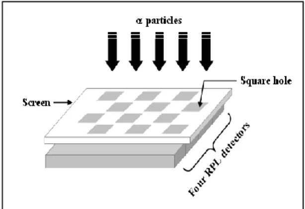
RPL glass is transparent with front and rear faces that are distinguished by a minuscule identification code. We designate the coded side as the rear face. As already alluded to, and as described in the next subsection, the dosimeter signal for neutrons is a number of (n,p) protons. However in some of early trials we did not register any protons, so to investigate the reason for this non-registration we performed the following tests with α-particles. A paper screen with square holes covering four RPL detectors was implemented as shown in Fig. 1. The side of a square hole was the 7 mm width of an RPL detector. Irradiations were performed with a 5 cm diameter, 25 µm thick α-particle source (153 α s-1 in 2) [8] in contact with the screen for about 24 h. The first experiment consisted of irradiating four detectors by their front face; the second consisted of irradiating the four detectors, this time two by the front face and two by the rear face. The -particle energies incident on the detectors varied between 0 and 5.48 MeV.
The response of the detectors, shown in Fig. 2, shows that the four detectors irradiated by the front face registered α-particles passing through all the holes of the paper screen (Fig. 2a). The same was true for detectors 1 and 3 (Fig. 2b) irradiated by their front face, but detectors 2 and 4 irradiated by the rear face were particle-insensitive. This experiment demonstrates that to detect α-particles of 5.48 MeV, and presumably other charged
particles in the energy range encountered here, a sense of orientation of an RPL detector must be respected. With this information, proton signals could be detected consistently. γ-rays are detected equally well by both faces.
Fig. 1. Diagram of the experiment with α-particles to determine the charged-particle-sensitive face of an RPL detector.
3.2 Dosimeter configuration
Fig. 3 shows the configuration adopted for the fast neutron dosimeter. It consists of an assembly of two RPL detectors (each 1.5 mm thick), three aluminium foils (each 0.9 mm thick) and a polyethylene converter (1 mm thick in which proton equilibrium is reached for (n,p) reactions with
241
Am-Be neutrons [9] placed in a closed polyethylene case. Neutrons are detected by their (n,p) collisions in the hydrogen-rich converter. The aluminium sheets at the left and the right of the stack are used to stop any recoil protons ejected from the polyethylene of the case. The maximum neutron energy from the 241Am-Be source is 10 MeV and it can produce recoil protons with the same energy. A 0.9 mm aluminium foil will stop protons of 12 MeV [10]. RPL() registers -ray exposure most of which comes from the polyethylene case; RPL() detects -rays plus protons from the converter. Both detectors are assumed to receive the same quantity of n and radiation. The fast neutron dose is measured by recoiling protons, taken to be the difference between RPL() and RPL() readings Rn,p in
arbitrary units of luminescence intensity :
Rn,p = RRPL() – RRPL() (1)
SEM measurements did not detect silver in the first 0-1.7 µm in a profile of the front face of an RPL detector which implies the absence of Ag-dependent luminescence centers over this thickness. The consequence from the proton range-energy curve is that (n,p) protons, and also the impinging neutrons, must have E 115 keV to be detected.
We mention here the possibility of lowering the low energy threshold for protons detected to less than 115 keV by chemically removing a layer from the RPL glass detector. This can be done by etching with NaOH [11]. Readings after successive removal
of RPL glass layers could constitute a crude neutron spectroscopy.
Fig. 2. Responses of RPL detectors to α-particles. (a) irradiation by the front face of four detectors; (b) irradiation of four detectors, two by the front face [RPL(1) et RPL(3)] and two by the rear face [RPL(2) et RPL(4)], showing that the front face is the one sensitive to α-particles.
Fig. 3. Exploded diagram (not to scale) of the RPL fast neutron dosimeter. Al : aluminium; RPL :
Radiophotoluminescent detector; (CH2)n :
polyethylene converter. 3.3 Irradiation conditions
The neutron irradiations were performed with the laboratory’s 241
Am-Be calibrator. The neutron source intensity is estimated to be (2.24 0.12) 106 s-1, determined by Bonner sphere spectrometry [12] for an ambient neutron dose equivalent rate H*(10) of 48.8 2.4 µSv h-1 at 75.0 0.1 cm. The neutron fluence rate and H*(10) calculated using the MCNPX code [13] through a point detector (F5 tally) for the same distance are 43.5 0.5 cm-2 s-1 and 50.6 0.5 µSv h-1, respectively. These values are to be compared with the values of 41.0 1.9 cm-2 s-1 and 48.8 2.4 µSv h-1 obtained by Bonner sphere spectrometry [12]. For our simulation, the
241Am-Be source structure was taken as reported by
the supplier [14] and the neutron energy distribution (4·10-7 E
n 10 MeV) with an average neutron
energy of 4.4 MeV was provided by ISO 8529-1 [15]. H*(10) was obtained at the point detector using the fluence-to-ambient dose equivalent conversion coefficients recommended by ICRP 74 [16]. In our case, the dosimeter is used to evaluate the neutron dose only, although -rays are present. During irradiation, the dosimeter was placed on a holder rotating around the 241Am-Be source at a distance of 19.0 0.1 cm. The source is shielded with 1 mm of lead that absorbs over 99.6% of the 59 keV 241Am γ-rays. To reduce the possibility of detecting scattered neutrons, the irradiations were performed in a large hall with a minimum of material in proximity. The ambient dose equivalents
H*(10) of incident neutrons on the dosimeter were
in the range 2-433 mSv.
4. Results and discussion
4.1 Dosimeter response
Fig. 4 shows that the dosimeter response to 241 Am-Be neutrons varies linearly as a function of H*(10) with a correlation coefficient r² = 0.99. From 2 to 433 mSv, the signal from recoil protons detected follows the linear form:
Rn,p = (4.17 ± 0.08) H*(10) (2)
where Rn,p is obtained from relation (1). All data
points of Fig. 4 were obtained with the prescribed single preheating before a reading of the RPL detector. Between measurements the detectors were reset by heating at 400°C for 1 h. The signal in
Salem et. al/ Journal of Nuclear Sciences Vol 2(2) (2015) 53‐58
56 RPL() is 9.5 times the proton signal in RPL(), so at low doses the statistical uncertainty on the (n,p) proton signal limits a determination of H*(10) to values above about 2 mSv.
Fig. 4. Dosimeter response to 241Am-Be neutrons as
a function of ambient neutron dose equivalent H*(10) at normal incidence. Errors are statistical.
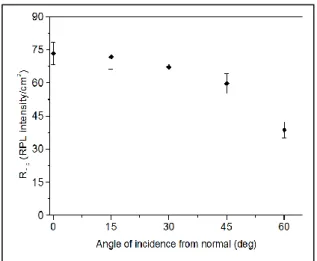
4.2 Angular dependence
Fig. 5 presents the angular dependence of the dosimeter up to 60° to the normal of the detector for a distance of 19.0 0.1 cm. The arithmetical mean of the decreasing dosimeter response at the five angles is 15% lower than the response at normal incidence and satisfies the ISO 21909 [17] requirement stipulating that this difference should not be greater than 30%.
Fig. 5. Dosimeter response as a function of incident
angle to 241Am-Be neutrons. Errors are statistical.
4.3 Preheating and loss of signal
RPL luminescence centers are quasi-stable and are re-readable [7]. Protons from the (n,p) scattering have energies ranging from zero to several MeV
and we would like to know if the prescribed preheating at 100°C for 1 h before a reading has caused any loss of information. For this purpose we have irradiated three dosimeters for a total period of 567 h (corresponding to 433 mSv) according to the following protocols:
Dosimeter (D1): Reading at a period of 567 h.
Dosimeter (D2): Reading after 141.27 h followed by resetting; reading after 235.81 h followed by resetting; reading after 189.73 h (total irradiation time 567 h).
Dosimeter (D3): Readings at 141.27, 377.08 and 567 h, each with a preheating but no resetting.
Readings from Table 1, i.e., amount of RPL signal per hour of irradiation, after one preheating from dosimeters D1, D2, D3 are: D1: 908/567 h = 1.60 D2: 213/141.3 h =1.51 338/235.8 h = 1.43 279/189.7 h = 1.47 D3: 217/141.3 h = 1.54
Average: 1.51 RPL signal per hour measured by the reader after one preheating.
Let x be the amount signal lost in a second preheating and y the signal lost in a third preheating. Then, using readings and irradiation time differences from Table 1, one can write Eq. 1: 493 = 217x + 356
Eq. 2: 690 = 217xy +493x + (1.51)(567 – 377) In Eq. 1, the 356 = (1.51)(377 – 141) represents the new counts added after the detector was read at 141 h.
In Eq. 2, 217xy represents the counts remaining after the second preheating which will now undergo a loss y in a third preheating. The (1.51)(567 – 377) term represents the new counts added after the resetting at 377 h.
Solving Eqs. 1 and 2 for x and y with the exact time values and the uncertainties gives :
x = 0.63 ± 0.10, fraction of RPL signal lost in a second preheating
y = 0.68 ± 0.38, fraction of RPL signal lost in a third preheating
The relatively large loss of signal in the preheatings at 100 °C for 1 h is not surprising because heating at 400 °C for 1 h totally resets the detector. The amount of signal lost in a 1st preheating was not measured, but the fractions x and y of new signal lost in the 2nd and 3rd preheatings indicate that about 60% is also lost in the 1st preheating.
Table 1. Recoil proton signals from the (n,p) converter detected with different irradiation, preheating, reading and detector reset protocols for a total irradiation time of 567 h. The numbers are the values of luminescence intensity furnished by the reader in arbitrary units, with uncertainties at 1σ. The uncertainties for detectors D1 and D3, which were never erased but which underwent respectively a preheating at 567 h and preheatings at 141.27, 377.08 and 567 h are the statistical uncertainties of the readings at 567 h. The uncertainty for detector D2, which was erased after each reading, is the quadratic sum of the statistical uncertainties of the readings at 141.27, 235.81 and 189.73 h.
D1
D2
D3
908
21.2 at 567 h
213
5.8 at 141.27 h
217
5.5 at 141.27 h
338
10.4 at 235.81 h
493
16.4 at 377.08 h
279
9.9 at 189.73 h
690
21.1 at 567 h
Total
908
21.2 at 567 h
Total
830
15.5 for 567 h
Total
690
21.1 at 567 h
5. Conclusion
This study has demonstrated that fast neutrons can be detected in a mixed n- field by a compact dosimeter incorporating RPL Ag-doped glass detectors associated with a neutron-proton converter. The dosimeter response is closely linear in H*(10) over the range 2-433 mSv of neutrons with an acceptable angular dependence up to 60 deg to the normal, which makes it applicable for ambient dosimetry. Applications should be possible in the nuclear power industry, as in fuel fabrication or retreatment centers, certain zones of nuclear power plants, and around some industrial or research accelerators. The charged-particle-sensitive face of the RPL detectors registers (n,p) protons with E ≥ 115 keV.
References
[1]
“
Operational dose equivalent quantities for neutrons”
, ICRU report 66 (2001).[2] Chiyoda Techno Glass Corporation, 2008,
“
User’s manual for reading module FGD-660”,
Ref : AS-04-21-0010-R0.[3] S. Croft, D. Weaver,
“
The application of radiophotoluminescent glass to dosimetry in mixed n- fields”
, Radiat. Prot. Dosim. 17, 67-70 (1986).[4] S. Miljanić, M. Ranogajec-Komor, S. Blagus, J.K. Pàlfalvi, T. Pàzmàndi, S. Deme, P. Szàntó,
“
Response of radiophotoluminescent dosimeters to neutrons”
, Radiat. Meas. 43, 1068-1071 (2008). [5] M. Girod, L. Bourgois, G. Cornillaux, L. Andre,“
Study and presentation of a fast neutron and photon dosimeter for area and criticality monitoringusing radiophotoluminescent glass
”
, Radiat. Prot. Dosim. 112, 359-370 (2004).[6] International Organisation for Standardisation. Neutron reference radiations – Part 3:
“
Calibration of area and personal dosimeters and determination of their response as a function of neutron energy and angle of incidence”
, ISO 8529-3 (2000). [7] Y. Miyamoto, K. Kinoshita, S. Koyama, Y. Takei, H. Nanto, T. Yamamoto, M. Sakakura, Y. Shimotsuma, K. Miuta, K. Hira,“
Emission and excitation mechanism of radiophotoluminescence in Ag+-activated phosphate glass”
, Nucl. Instr. Meth. Phys. Res. A 619, 71-74 (2010).[8]
“
Source pour l’étalonnage des moniteurs de contamination surfacique”
, type AM241 ESAL20, N° de série : 0017. Certificat d’étalonnage N°CT/020169/02/0421. LEA Laboratoire Etalons d’Activité, 26701 Pierrelatte Cedex, France. [9] M. Trocmé, S. Higueret, D. Husson, A. Nourreddinne, T.D. Lê,“
A new compact device for efficient neutron counting using a CMOS active pixel sensor”
, Radiat. Meas. 43, 1100-1103 (2008). [10] J. Biersack, J. Ziegler,http://www.srim.org/SRIM/SRIM2003.htm. [11] K. Becker,
“
Range and depth dose distribution of low energy charged particles in dosimeter glasses”,
1st International Congress of the International Radiation Protection Assn., Rome, inRadiation Protection, Part 1, W. S. Snyder, H. H.
Abee, L. K. Burton, R. Maushart, A. Benco, F. Duhamel, B. M. Wheatley, eds., 1968, pp. 135-140.
Salem et. al/ Journal of Nuclear Sciences Vol 2(2) (2015) 53‐58
58 [12] K. Amgarou, M. Trocmé, J.M. Garcìa-Fusté, M. Vanstalle, E. Baussan, A. Nourreddine, C. Domingo,
“
Characterization of the neutron field from the 241Am-Be isotopic source of the IPHC irradiator”,
Radiat. Meas. 50, 61-66 (2013).[13] D.B. Pelowitz,
“
MCNPX Version 2.6.0 (Los Alamos National Laboratory)”
, LA-CP-07-1473 (November 2007).[14] High technology sources LTD. Americium-241/Beryllium.
http://www.hightechsource.co.uk/Americium_Beryl lium.pdf
http://osrp.lanl.gov/Documents/SFCertificates/USA -0631-S.pdf
[15] International Organisation for Standardisation. Neutron reference radiations – Part 1:
“
Characteristics and methods of production”,
ISO 8529-1 (2001).[16] International Commission on Radiological Protection,
“
Conversion coefficients for use in radiological protection against external radiation”,
ICRP Publication 74.
[17] International Organisation for Standardisation,
![Fig. 2. Responses of RPL detectors to α-particles. (a) irradiation by the front face of four detectors; (b) irradiation of four detectors, two by the front face [RPL(1) et RPL(3)] and two by the rear face [RPL(2) et](https://thumb-eu.123doks.com/thumbv2/9libnet/4101296.60059/4.892.111.780.203.467/responses-detectors-particles-irradiation-detectors-irradiation-detectors-rpl.webp)