Relationship between endothelial dysfunction and
cardiovascular risk factors and the extent and
severity of coronary artery disease
Endotel fonksiyonu bozukluğu ile kalp damar hastalığı risk faktörleri ve
koroner arter hastalığı yaygınlığı ve ciddiyeti arasındaki ilişki
Department of Cardiology, Istanbul Bilim University Faculty of Medicine, Istanbul;
#Department of Cardiology, Florence Nightingale Hospital, Istanbul
Hilal Kurtoğlu Gümüşel, M.D., Alp Burak Çatakoğlu, M.D., Özlem Yıldırımtürk, M.D.,#
Selen Yurdakul, M.D.,# Funda Helvacıoğlu, M.D., Murat Ziyrek, M.D.,
Ruken Hanavdeloğulları, M.D., Vedat Aytekin, M.D.,# Saide Aytekin, M.D.#
Objectives: Endothelial dysfunction (ED) is associated with coronary artery disease (CAD) and cardiovascular risk fac-tors. The relationship between cardiovascular risk factors, ED and the presence, extent and severity of CAD, was evaluated in patients with and without angiographically defined CAD in our study.
Study design: Eighty patients with CAD and 20 subjects with normal coronary arteries were included. Endothelial function was evaluated by endothelium-dependent, flow-mediated dil-atation (FMD) and nitroglycerine-mediated dildil-atation (NMD) measurements, using brachial artery Doppler ultrasonogra-phy (USG). Cardiovascular risk factors were identified. The extent and severity of CAD was determined via vessel and modified Gensini scores.
Results: FMD% and NMD% were significantly decreased in the CAD(+) group compared with the CAD(–) group (p=0.0001). In the CAD(+) group, the cut-off values of FMD% and NMD% in distinguishing between single-vessel and multi-vessel diseases were 8.5% (sensitivity: 95%, specificity: 62%) and 13.6% (sensitivity: 91%, specificity: 62%), respectively. Additionally, a modified Gensini score was significantly cor-related with both FMD and NMD (r=-0.825, r=-0.778, respec-tively; p=0.0001) in the CAD(+) group.
Conclusion: ED was more prevalent in the CAD(+) group, and the degree of ED correlated well with the extent and se-verity of CAD.
Amaç: Endotel fonksiyonu bozukluğu (EFB), kalp damar has-talığı risk faktörleri ve koroner arter hashas-talığı (KAH) ile ilişkilidir. Çalışmamızın amacı, anjiyografik olarak saptanmış KAH’sı olan ve olmayan hastalarda, KAH risk faktörleri ile EFB ilişkisi-nin değerlendirilmesi ve KAH varlığı, yaygınlığı ve ciddiyeti ile EFB arasındaki ilişkinin gösterilmesidir.
Çalışma planı: Koroner arter hastalığı saptanan 80 has-ta çalışma grubuna, normal koroner arterleri olan 20 hashas-ta kontrol grubuna alındı. EFB, brakiyal arter ultrasonografisi ile “endotel bağımlı vazodilatatör yanıt” (EBVY) ve “nitrat bağımlı vazodilatatör yanıt” (NBVY) ölçümleri yapılarak değerlendiril-di. KAH risk faktörleri kaydedildeğerlendiril-di. KAH yaygınlığı ve ciddiyeti, hasta damar sayısı ve Gensini skoru ile belirlendi.
Bulgular: Koroner arter hastalığı (+) grupta KAH (–) gruba göre EBVY ve NBVY anlamlı olarak daha düşük bulundu (p=0.0001). KAH (+) grupta, EBVY için; %8.5 cut-off değerinin %95 duyarlılık ve %62 özgüllükle, NBVY için ise; %13.6 cut-off değerinin %91 duyarlılık ve %62 özgüllükle tek damar ve çok damar hastalığı ayrımını sağlayabileceği bulundu. KAH (+) grupta Gensini skor ile EBVY ve NBVY arasında çok güçlü düzeyde korelasyon saptandı (sırasıyla, r=-0.825, r=-0.778; p=0.0001).
Sonuç: Çalışmamızda KAH (+) grupta EFB’nin daha fazla olduğunu ve KAH yaygınlığı ve ciddiyeti ile EFB derecesinin arttığını tespit ettik.
Received:October 20, 2012 Accepted:January 17, 2014
Correspondence: Dr. Saide Aytekin. Florence Nightingale Hastanesi, Abidei Hürriyet Cad., No: 290, Çağlayan, Şişli, İstanbul.
Tel: +90 212 - 375 65 65 e-mail: [email protected]
© 2014 Turkish Society of Cardiology
T
he endothelium is an autocrine, paracrine, and en-docrine organ, which plays a key role in the regu-lation of vascular tone, thrombogenesis, lipid metabo-lism, inflammation, and vessel growth.[1-3] Endothelial dysfunction (ED), a hallmark of vascular diseases, often leads to atherosclerosis. Traditional risk factors such as hypertension (HT),[4-6] cigarette smoking,[7] diabetes mellitus (DM),[8] age,[9-12] and hypercholes-terolemia[13] may induce ED in the coronary arteries, due to decreased bioavailability of nitric oxide (NO). A dysfunctional endothelium loses its ability to pro-tect the vascular system as a result of the reduction of anti-atherosclerotic and anti-thrombotic properties. ED promotes the development and progression of ath-erosclerosis.The aim of the present study was to evaluate the relationship between ED and the presence, extent and severity of coronary artery disease (CAD) in patients with and without angiographically defined CAD. The potential risk factors that might affect ED in the CAD(+) group were also analyzed. Among the various methods used to assess endothelial function was the brachial artery Doppler ultrasonography (USG), which measured endothelium-dependent, flow-mediated dil-atation (FMD) and nitroglycerin-mediated dildil-atation (NMD) in the brachial artery. This is a noninvasive, highly reproducible, and simple method to use, which is based on high-sensitivity ultrasound waves.[14]
PATIENTS AND METHODS
Study Population
This study included 100 patients seen between Febru-ary and May 2008 with suspected CAD due to typical chest pain or ischemic findings on a treadmill exercise test or myocardial scintigraphy who underwent coro-nary angiography. Eighty patients (62.4±9.9 years, 65% male) who had angiographically proven CAD were defined as the study group. Twenty subjects who had normal coronary arteries (57.7±10.5 years; 30% male) served as the control group. Informed consents were obtained from all the patients in each group. The study was approved by the local ethics committee. Pa-tients with acute coronary syndrome, severe left ven-tricular dysfunction (left venven-tricular ejection fraction <35%) and a history of coronary angioplasty or coro-nary artery bypass surgery were excluded from the study. CAD was defined as a visually estimated per-cent diameter stenosis of greater than 25% in any of
the major epicardial coro-nary arteries. Clinically important CAD was de-fined as greater than 50% diameter stenosis in any of the major epicardial coro-nary arteries by consensus of two observers who were blinded to the results of the ultrasound studies. All study subjects underwent a complete physical exam-ination. Biochemical and electrocardiographic
mea-surements were also performed. The following risk factors for atherosclerosis were assessed: smoking, HT, hyperlipidemia (HL), DM, obesity, and a positive family history of CAD. Obesity was defined as a body mass index (BMI) above 30 kg/m². HT was defined as blood pressure ≥140/90 mmHg or use of anti-hyper-tensive drugs. DM was defined as fasting blood glu-cose level ≥126 mg/dl, in two separate measurements, or the use of anti-diabetic agents. Endothelial function was evaluated by brachial artery Doppler USG. The extent and severity of CAD was determined accord-ing to the number of diseased vessels and a modified Gensini score.[15]
Assessment of vascular function
The noninvasive determination of ED was performed according to the method described by Corretti et al.[16] A high-resolution echocardiography Doppler ultra-sound (General Electric Vivid I) with an 8.0 MHz linear array transducer was used to measure the flow velocity and diameter of the left brachial arteries.
Each subject was studied in the morning, after abstaining from food, alcohol, caffeine, and tobacco for 8h-12h before the examination.[17] All ultrasound images were recorded for subsequent blinded analy-sis. Recordings of both B-mode and pulsed Doppler spectral curve were taken at rest, during reactive hy-peremia and following the sublingual application of isosorbide dinitrate. After baseline measurements, a sphygmomanometer cuff placed around the left up-per arm proximal to the imaged artery segment was inflated to the pressure of 240 mmHg for 5 minutes to occlude arterial flow. To verify that suprasystolic compression of the brachial artery caused adequate increase in blood flow, flow velocity was measured at
Abbreviations:
BMI Body mass index CAD Coronary artery disease DM Diabetes mellitus ED Endothelial dysfunction FMD Flow-mediated dilatation HDL-C High-density lipoprotein cholesterol HL Hyperlipidemia HT Hypertension NMD Nitroglycerin mediated dilatation NTG Nitroglycerin ROC Receiver operator characteristic USG Ultrasonography
rest and again within 15 seconds after cuff deflation. Blood flow, pressure and end-diastolic diameter were recorded at 30-second intervals for 300 seconds after cuff release and at six, eight and ten minutes until re-covery to baseline values. After re-establishing base-line conditions 15-20 minutes later, measurements of the arterial diameter and flow velocity were repeated, followed by sublingual isosorbide dinitrate adminis-tration at a dose of 5 mg in order to assess endothe-lium-independent vasodilatation. Four minutes later, measurements of arterial diameter and flow velocity were repeated. The arterial diameter was measured in millimeters as the distance between the anterior wall media-adventitial interface (“m” line) and the poste-rior wall intima-lumen interface at end-diastole, coin-cident with the R wave on the continuously recorded electrocardiogram at two sites along the artery. The maximum FMD and NMD diameters were calculat-ed as the average of the three consecutive maximum diameter measurements after hyperemia and nitro-glycerin, respectively. FMD% was calculated as the percent change from the basal diameter value during reactive hyperemia. NMD% was calculated as the percent change in diameter from the resting value to the value after nitroglycerin administration.
Assessment of coronary atherosclerosis
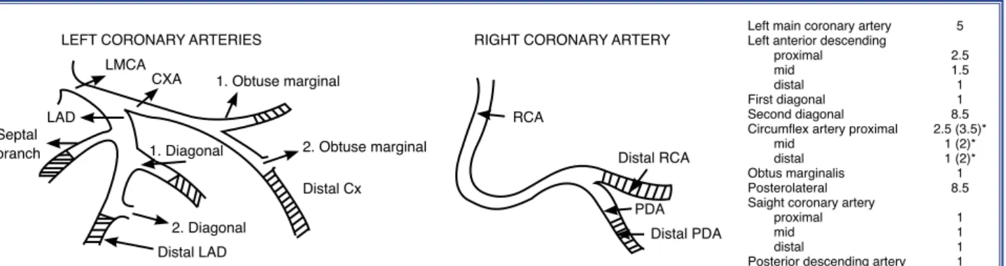
All patients underwent selective coronary angiogra-phy after brachial artery Doppler USG. After femoral artery cannulation, the Judkins technique was used for the cannulation of the left and right coronary arteries. All angiograms were evaluated by two experienced in-vasive cardiologists blinded to the study. The severity of CAD in major epicardial coronary arteries was evaluated according to the modified Gensini scoring system.15 Modified Gensini scores grade the degree
of narrowing of the coronary arteries as: 1: 1-25% narrowing, 2: 26-50% narrowing, 4: 51-75% narrow-ing, 8: 76-90% narrownarrow-ing, 16: 91- 99% narrownarrow-ing, and 32: total occlusion. The score is multiplied by a factor that reflects the importance of the position of the lesion in the coronary arterial tree (Figure 1).
The extent of CAD was assessed by the vessel score. The number of vessels with significant stenosis (presence of at least one lesion causing at least ≥50% diameter stenosis) was determined to be the vessel score. Scores ranged from 0 to 4, depending on the number of vessels involved. Left main artery stenosis was scored as a one-vessel disease. CAD involving only one vessel was defined as ‘single-vessel disease’ while, when two or more coronary arteries were in-volved, it was defined as ‘multi-vessel disease’.
Statistical analysis
Statistical analyses were performed using the Statisti-cal Package for the Social Sciences (SPSS-15) soft-ware package. Descriptive statistics were calculated, including means and standard deviations for continu-ous variables, frequencies for qualitative variables, and percentages for categorical variables. To test the normal distribution of the samples, a Kolmogorov-Smirnov test was performed. Parameters normally dis-tributed were compared by Student’s t-test, whereas uneven distributions were compared by Mann-Whit-ney U test. Chi-square or Fisher’s test was used to compare qualitative variables, and the Student’s t-test was used to compare quantitative variables. Pearson’s correlation test was used to test the correlations be-tween variables. Multivariate analysis was performed using multiple linear regression models. Multiple lin-ear regression analysis was used to assess the
relation-LEFT CORONARY ARTERIES LMCA
CXA 1. Obtuse marginal
2. Obtuse marginal
RIGHT CORONARY ARTERY
RCA Distal RCA Distal PDA PDA 2. Diagonal 1. Diagonal Distal LAD Septal branch LAD Distal Cx
Left main coronary artery 5 Left anterior descending
proximal 2.5
mid 1.5
distal 1
First diagonal 1 Second diagonal 8.5 Circumflex artery proximal 2.5 (3.5)*
mid 1 (2)*
distal 1 (2)* Obtus marginalis 1 Posterolateral 8.5 Saight coronary artery
proximal 1
mid 1
distal 1
Posterior descending artery 1
Figure 1. Position of the lesion in coronary arterial tree. LMCA: Left main coronary artery; LAD: Left anterior descending artery; Cx: Circumflex artery; RCA: Right coronary artery; PDA: Posterior descending artery.
ol (TC) and LDL-C levels. Other cardiovascular risk factors were similar between the groups. There were no significant differences between the two groups with regard to medical therapy.
Brachial artery Doppler ultrasound measurements
In the study population (n=100), mean FMD% was 9.5±4.5% and mean NMD% was 14.8±5.6%. There was a significant correlation between FMD% and NMD% (r=0.950, p<0.001). In the CAD(+) group, mean FMD% was 7.6±2.8% and mean NMD% was 12.4±3.4%. In the CAD(–) group, mean FMD% was 16.8±1.8% and mean NMD% was 24.1±2%. FMD% and NMD% values were markedly reduced in the CAD(+) group compared to the CAD(–) group (p<0.001 and p<0.001, respectively) (Figure 2).
Cardiovascular risk factors and brachial artery Doppler ultrasonography measurements
In the univariate analysis, it was observed that diabetic patients had significantly lower FMD% and NMD% values than non-diabetic patients in the CAD(+) group (p=0.037 and p=0.035, respectively) (Table 2). However, when we used the Bonferroni adjustment, there was no significant difference between diabetic patients and non-diabetic patients. No significant re-lationship was observed between the other risk factors and FMD% and/or NMD% in the CAD(+) group. ship between coronary risk factors and FMD% and
NMD%. Variables with a p<0.1 value in the univari-ate analysis were included in the multivariunivari-ate analy-sis. Smoking status, age, gender, DM, HL, triglyceride (TG), and high-density lipoprotein cholesterol (HDL-C) levels and the presence of CAD were analyzed in the multivariate model. A receiver operator character-istics (ROC) curve analysis was performed to identify the predictive index value of FMD and NMD for sin-gle-vessel disease or multi-vessel disease. Statistical significance was defined as p<0.05.
RESULTS
Patient population
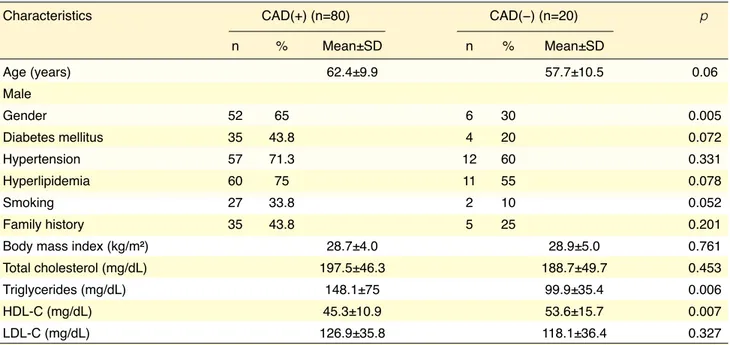
Clinical and demographic characteristics of the study population are presented in Table 1. The num-ber of male patients were significantly higher in the CAD(+) group (p=0.005). The number of smokers were lower in the CAD(–) group compared with the CAD(+) group, which was not statistically signifi-cant (p=0.052). Serum HDL-C level was decreased in the CAD(+) group compared with the other group (p=0.007); however, TG level was found to be high-er in the CAD(+) group compared with the CAD(–) group (p=0.006). There was no significant difference between the groups in terms of serum total
cholester-Table 1. Clinical and demographic characteristics
Characteristics CAD(+) (n=80) CAD(−) (n=20) p
n % Mean±SD n % Mean±SD Age (years) 62.4±9.9 57.7±10.5 0.06 Male Gender 52 65 6 30 0.005 Diabetes mellitus 35 43.8 4 20 0.072 Hypertension 57 71.3 12 60 0.331 Hyperlipidemia 60 75 11 55 0.078 Smoking 27 33.8 2 10 0.052 Family history 35 43.8 5 25 0.201
Body mass index (kg/m²) 28.7±4.0 28.9±5.0 0.761
Total cholesterol (mg/dL) 197.5±46.3 188.7±49.7 0.453
Triglycerides (mg/dL) 148.1±75 99.9±35.4 0.006
HDL-C (mg/dL) 45.3±10.9 53.6±15.7 0.007
LDL-C (mg/dL) 126.9±35.8 118.1±36.4 0.327
In the multiple linear regression analysis, lower HDL-C (B=0.001, 95% confidence interval (CI) (0.000, 0.001); p=0.016) and the presence of CAD (B= -0.082, 95% CI (-0.099, -0.066); p<0.001) were determined as independent risk factors for FMD
(R²=0.76). For NMD, lower HDL-C (B=-0.001, 95% CI (0.000, 0.001); p<0.003), and the pres-ence of CAD (B=-0.104, 95% CI (-0.124, -0.084); p<0.001) were established as independent risk fac-tors (R²=0.79).
Figure 2. Comparison of the FMD% and NMD% in CAD(+) and CAD(−) patients. (p<0.001; p<0.001). CAD: Coronary artery disease; FMD: Flow-mediated dilatation; NMD: Nitroglycerine mediated dilatation.
18.0 16.0 14.0 12.0 10.0 8.0 6.0 4.0 2.0 0.0 CAD+ CAD– 7.6% p<0.001 FMD % 16.8% 25.0 15.0 20.0 10.0 5.0 0.0 CAD+ CAD– 12.4% p<0.001 NMD % 24.1%
Table 2. Brachial artery Doppler ultrasound measurements, vessel scores and modified Gensini scores in diabetic and non-diabetic patients
CAD(+) Group (n=80) p
DM(+) (n=35) DM(−) (n=45)
Brachial artery diameter (mm) 4.2±0.7 4.5±0.6 0.062
Flow-mediated dilatation (%) 6.9±2.3 8.2±3 0.037
Nitroglycerine mediated dilatation (%) 11.5±2.7 13.1±3.8 0.035
Vessel score 2.4±0.9 1.9±1.1 0.026
Gensini score 76.4±45 51.5±33.3 0.010
CAD: Coronary artery disease; DM: Diabetes mellitus.
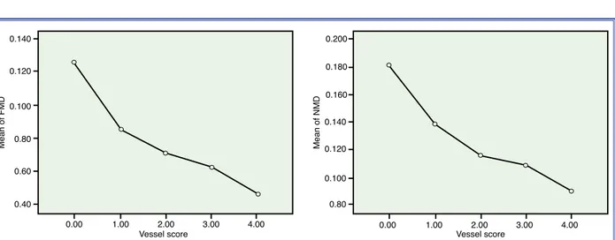
Figure 3. Relationship between FMD%, NMD% and vessel score in the CAD(+) group. (r=-0.560, p=0.0001; r=-0.522, p=0.0001). CAD: Coronary artery disease, FMD: Flow-mediated dilatation, NMD: Nitroglycerine mediated dilatation.
Mean of FMD 0.140 0.120 0.100 0.80 0.60 0.40 0.00 1.00 2.00 3.00 4.00 Vessel score Mean of NMD 0.200 0.180 0.160 0.140 0.120 0.100 0.80 Vessel score 0.00 1.00 2.00 3.00 4.00
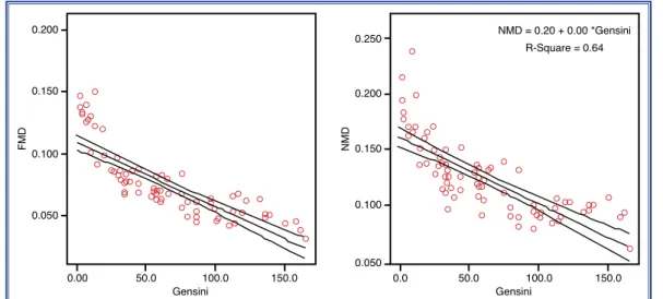
modified Gensini scores and both FMD and NMD in the CAD(+) group (r=-0.825, p<0.001; r=-0.778, p<0.001, respectively) (Figure 4).
DISCUSSION
Atherosclerosis is a progressive, inflammatory pro-cess that initially involves ED. Growing evidence indicates that ED may also influence the progres-sion of atherosclerotic leprogres-sions.[18] Many studies have investigated the relationship between coronary risk factors and endothelial function. Benjamin et al.[19] studied the relationship between coronary risk fac-tors and endothelial function in 2,883 participants in the Framingham study. They revealed that in stepwise multivariable linear regression models, FMD% was inversely correlated to age, systolic blood pressure, BMI, lipid-lowering medication, and smoking and positively related to female gender, heart rate, and prior walk test. Kirma et al.[20] investigated the rela-tionship between endothelial function and coronary risk factors in patients with angiographically proven CAD. In univariate analysis, they found a negative correlation between FMD% and age, BMI, and DM. In multiple stepwise regression analysis, FMD% was found to be correlated with DM and advanced age, but not with BMI. However, FMD% was not associated with HL, family history of premature atherosclerosis, HT, or smoking.
We investigated the relationship between coronary
Endothelial dysfunction and the extent and severity of coronary artery disease
In the CAD(+) group, 18 patients had single-vessel disease, 56 patients had multi-vessel disease, and 6 patients had noncritical lesions. The mean number of diseased vessels was 2.1±1.2 and the mean modified Gensini score was 62.4±43.4. We obtained a strong, negative correlation between the number of diseased vessels and both FMD and NMD in the CAD(+) group (r=-0.560, p<0.001; r=-0.522, p<0.001, respectively) (Figure 3). Moreover, a strong, positive correlation was observed between a modified Gensini score and the mean number of diseased vessels (r=0.601, p<0.001).
Predictive Value of Percent Change in FMD and NMD in Distinguishing Single-Vessel from Multi-Vessel Disease
The cut-off value for FMD% for predicting multi-vessel disease was <8.5% (area under ROC curve = 0.838; CI 0.71-0.96%) with 95% sensitivity and 62% specificity. The positive predictive value and negative predictive value for FMD were 89% and 84%, re-spectively. For NMD%, the cut-off value was <13.6% (area under ROC curve = 0.84; CI 0.70-0.97%) with 91% sensitivity and 62% specificity. The positive pre-dictive value and negative prepre-dictive value for NMD were 89% and 89%, respectively.
Relationship between Modified Gensini Score and Percent Change in FMD and NMD
We obtained a strong, negative correlation between
Figure 4. Relationship between FMD%, NMD% and Gensini score in the CAD(+) group. (r=-0.825, p=0.0001; r=-0.778, p=0.0001). CAD: Coronary artery disease; FMD: Flow-mediated dilatation; NMD: Nitroglycerine mediated dilatation.
FMD 0.200 0.150 0.100 0.050 0.00 50.0 100.0 150.0 Gensini NMD 0.250 0.150 0.200 0.100 0.050 0.0 50.0 100.0 150.0 Gensini NMD = 0.20 + 0.00 *Gensini R-Square = 0.64
Cigarette smoking is strongly associated with CAD and atherosclerosis. While smoking causes ED[29] as a result of impairment in endothelium-dependent va-sorelaxation, the exact mechanisms involved are not completely understood.[30] In our study, the number of smokers was lower in the CAD(–) group compared with the CAD(+) group, which was not statistically significant (p=0.052). In contrast to previous studies, we found no relationship between smoking and ED in the CAD(+) group. Benjamin et al.[19] found that FMD% was increased in smokers who did not smoke within six hours prior to testing than in those who did. In our study, all participants abstained from tobacco from 8h-12h before the study..
The extent and severity of CAD is of particular importance in prognosis and treatment. Neunteufl et al.[31] observed a strong correlation between FMD and the number of diseased vessels and maximum percent diameter stenosis. In our study, we found a strong cor-relation between vessel score, modified Gensini score and FMD% and NMD%.
Consequently, cardiovascular risk factors may cause ED in both healthy individuals and patients with CAD. Noninvasive techniques such as exercise electrocardiogram (ECG), stress echocardiography, myocardial perfusion scintigraphy, positron emission tomography, and cardiac magnetic resonance imag-ing allow earlier diagnosis of CAD in order to prevent cardiovascular diseases. However, new diagnostic tests for the early detection of disease that are lower in cost and higher in sensitivity and specificity are re-quired. Brachial artery Doppler USG is a non-inva-sive and reproducible method that elicits diagnosis of premature atherosclerosis in a subclinical stage.[32-34] Additionally, impairment in endothelial function, as-sessed by the brachial artery Doppler USG method, is closely related to the angiographic extent and severity of CAD. It is important to detect subclinical athero-sclerosis and prevent cardiovascular diseases.
Study limitations
The major limitation of our study is the small num-ber of patients in the study population. Brachial artery Doppler USG is a widely used, reproducible and reli-able method, based on the current literature. Neverthe-less, long-term studies involving larger study popula-tions are warranted to establish the beneficial effect of brachial artery Doppler USG technique in predicting risk factors and brachial artery Doppler USG
mea-surements in 80 patients with angiographically proven CAD. We also investigated the relationship between ED and modified Gensini and vessel scores. In our study, in the multiple linear regression analysis for FMD%, lower HDL-C and the presence of CAD were established as independent risk factors. For NMD%, lower HDL-C and the presence of CAD were deter-mined as independent risk factors.
Gender differences in endothelial function have been the focus of considerable research interest. In a previous study, Uehata et al.[21] observed increased FMD% in young healthy females compared with males. Urstad et al.[22] revealed that FMD% was similar in men and women at about 55 years, while FMD% and NMD% values were similar in men of both age groups. However, women at 55 years had lower FMD% than women at 35 years. Taddei et al.[23] demonstrated an age-related decline in men, but en-dothelial function was preserved until the onset of menopause in women. In our study, the number of males was significantly higher than the number of fe-males in the CAD(+) group. However, no significant gender difference was obtained regarding FMD% and NMD%. The possible reason may be attributed to the mean ages of female patients being high in both groups in our study.
Diabetes mellitus (DM) is associated with accel-erated atherosclerosis and an increased prevalence of cardiovascular disease. Simova II et al.[24] revealed in their study that diabetic patients had a significantly lower FMD compared with non-diabetic patients. In our study, modified Gensini scores and vessel scores were significantly higher in diabetic CAD(+) patients. Also, diabetic patients had lower FMD% and NMD% values in the CAD(+) group. However, due to the lim-ited number of patients, when the Bonferroni adjust-ment was used, there was no significant difference between diabetic patients and non-diabetic patients.
Elevated serum levels of TC and blood pressure has been shown to impair endothelial function.[25,26] Anti-hypertensive and lipid-lowering therapy improve endothelial function and decrease cardiac event rates. [27,28] In our study, we did not observe any significant relationship between HT, HL and FMD% or NMD%. This may have resulted from the fact that nearly all of the patients were under optimal anti-hypertensive and lipid-lowering therapy.
cardiovascular events at a subclinical stage.
In conclusion, we observed that ED was more prevalent in the CAD(+) group and that the degree of ED correlated well with the extent and severity of CAD. Based on these results, we suggest that evalu-ation of ED with a non-invasive ultrasonographic method may be beneficial for the diagnosis and pre-diction of the severity of CAD.
Conflict-of-interest issues regarding the authorship or article: None declared
REFERENCES
1. Furchgott RF, Zawadzki JV. The obligatory role of endothelial cells in the relaxation of arterial smooth muscle by acetylcho-line. Nature 1980;288:373-6. CrossRef
2. Vane JR, Anggård EE, Botting RM. Regulatory functions of the vascular endothelium. N Engl J Med 1990;323:27-36. 3. Lüscher TF, Vanhoutte PM. The endothelium: modulator of
cardiovascular function. Boca Raton, Fla: CRC Press; 1990. 4. Panza JA, Quyyumi AA, Brush JE Jr, Epstein SE. Abnormal
endothelium-dependent vascular relaxation in patients with essential hypertension. N Engl J Med 1990;323:22-7. CrossRef
5. Taddei S, Virdis A, Mattei P, Salvetti A. Vasodilation to ace-tylcholine in primary and secondary forms of human hyper-tension. Hypertension 1993;21:929-33. CrossRef
6. Perticone F, Ceravolo R, Maio R, Ventura G, Zingone A, Perrotti N, et al. Angiotensin-converting enzyme gene poly-morphism is associated with endothelium-dependent vaso-dilation in never treated hypertensive patients. Hypertension 1998;31:900-5. CrossRef
7. Zeiher AM, Schächinger V, Minners J. Long-term cigarette smoking impairs endothelium-dependent coronary arterial va-sodilator function. Circulation 1995;92:1094-100. CrossRef
8. Johnstone MT, Creager SJ, Scales KM, Cusco JA, Lee BK, Creager MA. Impaired endothelium-dependent vasodilation in patients with insulin-dependent diabetes mellitus. Circula-tion 1993;88:2510-6. CrossRef
9. Taddei S, Virdis A, Mattei P, Ghiadoni L, Gennari A, Fasolo CB, et al. Aging and endothelial function in normotensive subjects and patients with essential hypertension. Circulation 1995;91:1981-7. CrossRef
10. Herrington DM, Fan L, Drum M, Riley WA, Pusser BE, Crouse JR, et al. Brachial flow-mediated vasodilator respons-es in population-based rrespons-esearch: methods, reproducibility and effects of age, gender and baseline diameter. J Cardiovasc Risk 2001;8:319-28. CrossRef
11. Celermajer DS, Sorensen KE, Spiegelhalter DJ, Georgako-poulos D, Robinson J, Deanfield JE. Aging is associated with endothelial dysfunction in healthy men years before the age-related decline in women. J Am Coll Cardiol 1994;24:471-6.
12. Yavuz BB, Yavuz B, Sener DD, Cankurtaran M, Halil M, Ulger Z, et al. Advanced age is associated with endothe-lial dysfunction in healthy elderly subjects. Gerontology 2008;54:153-6. CrossRef
13. Creager MA, Cooke JP, Mendelsohn ME, Gallagher SJ, Cole-man SM, Loscalzo J, et al. Impaired vasodilation of forearm resistance vessels in hypercholesterolemic humans. J Clin In-vest 1990;86:228-34. CrossRef
14. Celermajer DS, Sorensen KE, Gooch VM, Spiegelhalter DJ, Miller OI, Sullivan ID, et al. Non-invasive detection of endo-thelial dysfunction in children and adults at risk of atheroscle-rosis. Lancet 1992;340:1111-5. CrossRef
15. Gensini GG. Coronary angiography. Mount Kisco (NY): Fu-tura Publishing Co; 1975.
16. Corretti MC, Anderson TJ, Benjamin EJ, Celermajer D, Charbonneau F, Creager MA, Deanfield J, et al. Guidelines for the ultrasound assessment of endothelial-dependent flow-mediated vasodilation of the brachial artery: a report of the In-ternational Brachial Artery Reactivity Task Force. J Am Coll Cardiol 2002;39:257-65. CrossRef
17. Williams SB, Goldfine AB, Timimi FK, Ting HH, Roddy MA, Simonson DC, et al. Acute hyperglycemia attenuates endothe-lium-dependent vasodilation in humans in vivo. Circulation 1998;97:1695-701. CrossRef
18. Vogel RA. Cholesterol lowering and endothelial function. Am J Med 1999;107:479-87. CrossRef
19. Benjamin EJ, Larson MG, Keyes MJ, Mitchell GF, Vasan RS, Keaney JF Jr, et al. Clinical correlates and heritability of flow-mediated dilation in the community: the Framingham Heart Study. Circulation 2004;109:613-9. CrossRef
20. Kirma C, Akcakoyun M, Esen AM, Barutcu I, Karakaya O, Saglam M, et al. Relationship between endothelial function and coronary risk factors in patients with stable coronary ar-tery disease. Circ J 2007;71:698-702. CrossRef
21. Uehata A, Lieberman EH, Meredith IT, Anderson TJ, Polak J, Selwyn AP, et al. Non-invasive assessment of flow-medi-ated vasodilation in brachial arteries: diminished response in young males compared to females. Circulation 1992;86:1-620.
22. Jensen-Urstad K, Johansson J. Gender difference in age-relat-ed changes in vascular function. J Intern Mage-relat-ed 2001;250:29-36. CrossRef
23. Taddei S, Virdis A, Ghiadoni L, Mattei P, Sudano I, Bernini G, et al. Menopause is associated with endothelial dysfunction in women. Hypertension 1996;28:576-82. CrossRef
24. Simova II, Denchev SV, Dimitrov SI, Ivanova R. Endothelial function in patients with and without diabetes mellitus with different degrees of coronary artery stenosis. J Clin Ultra-sound 2009;37:35-9. CrossRef
25. Ward NC, Croft KD, Hodgson J, Rich L, Beilin LJ, Pud-dey IB. Brachial artery vasomotor function is inversely as-sociated with 24-h ambulatory blood pressure. J Hypertens 2004;22:967-72. CrossRef
Key words: Coronary artery disease; cardiovascular risk factors;
coronary angiography; coronary disease; endothelial dysfunction.
Anahtar sözcükler: Koroner arter hastalığı; kardiyovasküler risk
fak-törleri; koroner anjiyografi; koroner hastalık; endotel disfonksiyonu.
26. Sorensen KE, Celermajer DS, Georgakopoulos D, Hatcher G, Betteridge DJ, Deanfield JE. Impairment of endothelium-dependent dilation is an early event in children with familial hypercholesterolemia and is related to the lipoprotein(a) level. J Clin Invest 1994;93:50-5. CrossRef
27. Taddei S, Virdis A, Ghiadoni L, Sudano I, Salvetti A. Effects of antihypertensive drugs on endothelial dysfunction: clinical implications. Drugs 2002;62:265-84. CrossRef
28. Koh KK, Son JW, Ahn JY, Kim DS, Jin DK, Kim HS, et al. Simvastatin combined with ramipril treatment in hypercho-lesterolemic patients. Hypertension 2004;44:180-5. CrossRef
29. Esen AM, Barutcu I, Acar M, Degirmenci B, Kaya D, Turk-men M, et al. Effect of smoking on endothelial function and wall thickness of brachial artery. Circ J 2004;68:1123-6. CrossRef
30. Raij L, DeMaster EG, Jaimes EA. Cigarette smoke-induced endothelium dysfunction: role of superoxide anion. J Hyper-tens 2001;19:891-7. CrossRef
31. Neunteufl T, Katzenschlager R, Hassan A, Klaar U, Schwar-zacher S, Glogar D, et al. Systemic endothelial dysfunction is
related to the extent and severity of coronary artery disease. Atherosclerosis 1997;129:111-8. CrossRef
32. Jambrik Z, Venneri L, Varga A, Rigo F, Borges A, Picano E. Peripheral vascular endothelial function testing for the diag-nosis of coronary artery disease. Am Heart J 2004;148:684-9. 33. Kaku B, Mizuno S, Ohsato K, Murakami T, Moriuchi I, Arai
Y, et al. The correlation between coronary stenosis index and flow-mediated dilation of the brachial artery. Jpn Circ J 1998;62:425-30. CrossRef
34. Ghaffari S, Toufan M. The value of endothelium dependent vasodilatation in diagnosing coronary artery disease and its comparison with the results of routine diagnostic tests. Saudi Med J 2007;28:1344-9.