375
http://journals.tubitak.gov.tr/medical/ © TÜBİTAK
doi:10.3906/sag-1709-143
Anticandidal activities of lactic acid bacteria isolated from the vagina
Sevda ER1,*, Ayşe İSTANBULLU TOSUN2, Gizem ARIK3, Merih KIVANÇ31Department of Medical Services and Techniques, Yunus Emre Vocational School of Health Services,
Anadolu University, Eskişehir, Turkey
2Department of Medical Microbiology, Faculty of Medicine, İstanbul Medipol University, İstanbul, Turkey
3Department of Biology, Faculty of Sciences, Anadolu University, Eskişehir, Turkey
* Correspondence: [email protected] 1. Introduction
Human microbiota colonized in human body consists of trillions of microorganisms. Different microbial communities were located in the vagina, mouth, skin, gastrointestinal tract, nose, urethra, and other parts of the body (1).
Lactobacillus species are dominant in the vaginal microbiota of healthy women (2). Premenopausal healthy women have 107–108 colony forming unit/gram lactobacilli in their vaginal fluid (3). Furthermore, Staphylococcus spp., Ureaplasma spp., Corynebacterium spp., Streptococcus spp., Peptostreptococcus spp., Gardnerella vaginalis, Bacteroides spp., Mycoplasma spp., Enterococcus spp., Escherichia coli, Veillonella spp., Bifidobacterium spp., and Candida spp. are also found in the vagina (4).
The lactobacilli in the vaginal microbiota protect the flora against the colonization of other sexually transmitted infectious agents such as bacterial vaginosis, urinary tract infections, vulvovaginal candidiasis, and AIDS. It was suggested that this is carried out through adhesion to the
vaginal epithelium cells by competing with the pathogens and by producing antimicrobials such as bacteriocin, hydrogen peroxide, and lactic acid (5).
Vulvovaginal candidiasis is a common infection seen in women throughout their lives. In some in vitro studies, some lactic acid bacterial strains were shown to inhibit the adhesion and development of Candida albicans (6). Drutz reported that oral administration of Lactobacillus acidophilus has a protective effect against Candida vaginitis (7).
In the present study, it was aimed to investigate the anticandidal activities of lactic acid bacteria isolated from the vagina of the healthy women on the vaginal Candida isolates. 2. Material and methods
2.1. Isolation of microorganisms from the samples taken from the vagina
Lactic acid bacteria and yeast isolates were obtained through a physician from the samples taken from 30 healthy volunteer women aged between 20 and 40 Background/aim: Lactic acid bacteria prevent the overgrowth of pathogenic agents and opportunistic pathogens in the vagina. Moreover, lactic acid bacteria contribute to the preservation of vaginal microbiota by producing antimicrobial agents. Previous studies showed that some lactic acid bacteria exhibited antimicrobial activity against Candida species causing yeast vaginosis as well as many bacterial pathogens.
Materials and methods: The antifungal activities of various lactic acid bacteria isolated from the vagina of healthy women on some Candida species isolated from the vagina were investigated by agar diffusion technique.
Results: Most of the lactic acid bacteria that belong to the species of Lactobacillus crispatus, L. fermentum, L. acidophilus, L. paracesei subsp. paracesei, L. pentosus, and L. plantarum exhibited antifungal activity in varying ratios against C. albicans, C. glabrata, and C. tropicalis strains isolated from the vagina.
Conclusion: The lactic acid bacteria are useful microorganisms associated with a variety of probiotic properties. In this sense, our lactic acid bacteria isolates with high antifungal activity may be promising candidates as probiotic microorganisms in the inhibition of vaginal candidiasis, which is one of the most prevalent problems, or in the protection against candidiasis. We will continue our studies in this area.
Key words: Agar diffusion technique, anticandidal activity, Candida, lactic acid bacteria
Received: 28.09.2017 Accepted/Published Online: 21.10.2018 Final Version: 11.02.2019 Research Article
years at the Gynecology Clinic of Medipol University Hospital, İstanbul. All were premenopausal and were not menstruating at the time of collection. All the studied women were clinically normal. They had not received antibiotics in the last 3 months. Vaginal samples were planted on the PDA (potato dextrose agar), chocolate agar, MRS (de Man, Rogosa, and Sharpe) agar, and M17 agar in the laboratory. For lactic acid bacteria isolation; MRS agar and M17 agar petri dishes were inoculated. The dishes were then incubated at 37 °C in 5% CO2 for 48–72 h. For yeast isolation, the PDA was inoculated and then the petri dishes were incubated at 30 °C for 5 days. Isolation of colonies developed in the petri dishes after incubation was performed. Primarily, Gram stains and catalase tests of the isolated microorganisms were performed. Gram-positive, catalase-negative bacterial isolates were separated as lactic acid bacteria and oxidase; mobility tests, development at different temperatures (4, 15, and 45 °C), development at different salt concentrations (6%, 7.5%, and 10% NaCl), development at different pH levels (pH 3.9 and 9.6), H2S formation, and ammonia formation from arginine were performed (8–9).
The microorganisms isolated from MRS agar, M17 agar, and PDA were stored at −85 °C. The permission of the ethical committee of our study was taken from the Ethics Committee of Non-Interventional Clinical Researches at the Istanbul Medipol University on Dec. 11, 04. All applicable international, national, and/or institutional guidelines for the care and use of human were followed. 2.1.1. Determination of the use of carbohydrates with API CHL 50 by lactic acid bacteria isolates
The test was carried out by sowing API CHL 50 kits in accordance with the administrator’s instructions and the microorganisms were defined according to the carbohydrate sources they used. As a result of the test, the color change results of the isolates were entered into the database optimized by the management company and species identification was obtained as the % rates.
2.1.2. Definition of the yeast isolates by MALDI-TOF mass spectrometry
The colony sample was taken from the yeast isolate which was activated in PDA at 37 °C for 48 h and was planted on a 48-well plate. 0.3 μL of matrix solution was added onto the plate surface. In the device, the result obtained by performing the robust protein ionization and molecular weight measurement of the microorganism was based on the definition of microorganism by comparing it with the database. This part of the study was performed by BioMérieux Diagnostics Incorporated Company (Marcy-l’Étoile, France).
2.1.3. 16srRNA sequence analysis
16s rRNA sequence analysis was performed for genotypic identification of the bacteria isolate. The genomic DNA
of the isolate was purified using GeneJET genomic DNA purification kit (Thermo Fisher Scientific, Waltham, MA, USA). The obtained genomic DNA was used as template DNA and PCR reaction was performed for 16s rRNA gene locus. 27F 5’ AGAGTTTGATCMTGGCTCAG-3’ and 1492R 5’TACGGYTACCTTGTTACGACTT-3’ universal primers were used. PCR reaction components include 2.5 µL of 10X Taq buffer (+ KCl–MgCl2), 2.5 µL of 25 mM MgCl2, 2.5 µL of 2.5 mM dNTP mix, 2.5 µL of 2.5 mM 27F primer, 2.5 µL of 2.5 mM 1492R primer, 0.25 µL of Taq polymerase (5 U/µL), 11.75 µL of nuclease-free ddH2O, and 1 µL of template DNA. PCR products obtained from the reaction were screened in 1% agarose gel. 1492R and 907R (5’-CCGTCAATTCMTTTRAGTTT - 3’) primers were used for the sequence analysis of nearly 1400 base pair region (10). The sequence analysis of isolate was performed by MedSanTek Laboratory Supplies Trade & Industry Ltd (İstanbul, Turkey).
2.1.4. Determination of lactic acid production of the lac-tic acid bacteria isolates
The lactic acid bacteria isolate was incubated in the MRS broth medium at 37 °C and 5% CO2 for 48 h. Following the incubation, 1 mL of fresh lactic acid bacteria culture was transferred to a clean flask and filled up to 100 mL by sterile dH2O. Two or three drops of phenolphthalein indicator were added and titrated with 0.1 M NaOH solution. The amount of NaOH was recorded. Acid produced by the culture was calculated as percent titrable acidity. Lactic acid amount produced by the bacteria was calculated by the formula below. The study was conducted in duplicate (11).
Acidity %: 0.1 N NaOH (mL) amount used × 0.9/mL 2.1.5. Determination of hydrogen peroxide production of the lactic acid bacteria isolates
5 mL of distilled water was added to the lactic acid bacteria cultures and centrifuged at 5000 rpm for 15 min. After centrifugation, the clear liquid formed on top was removed and filtered through Whatman No. 42 filter paper (Buckinghamshire, UK). After filtration, 4 mL of the obtained filtrate was taken into a separate tube. On top of this filtrate, 0.5 mL of sulfuric acid, 0.5 mL of ammonium molybdenum, and 0.5 mL of potassium iodide solution were added, and after each chemical addition, the samples were thoroughly vortexed. After all these processes were carried out, the optical densities of the obtained liquid were determined at 350 nm wavelength using a Shimadzu UV-1800 spectrophotometer (Kyoto, Japan). The obtained optical density (OD) values were calculated in terms of μg/ mL according to the previously prepared standard curve (12).
2.2. Determination of anticandidal activity
Anticandidal activity was investigated by agar spot technique. For this purpose, the lactic acid bacteria
were incubated in MRS and M17 broth for 48 h. After incubation, the lactic acid bacteria were adjusted to 0.5 McFarland turbidity in 0.85% physiological saline. 5 μL of the turbidity-adjusted lactic acid bacteria sample was instilled on the Mueller–Hinton soft agar containing glucose. After the petri dish was allowed to stand at room temperature for 30 min, it was allowed to incubate at 37 °C for 48 h.
After the incubation, the Candida isolates developed in potato dextrose broth at 37 °C for 48 h were adjusted to 0.5 McFarland turbidity in a 0.85% physiological saline solution. Potato dextrose soft agar inoculation was performed on the Candida isolates with turbidity adjustment. After the soft agar was thoroughly mixed, 7 mL of the mixture was slowly poured onto the surface of the petri dishes in which lactic acid bacteria were located. After the agar was frozen, the petri dishes were allowed to incubate at 37 °C for 48 h. After the incubation, zones around the lactic acid bacteria were observed and zone diameters were measured and recorded (13). The study was repeated twice.
3. Results
3.1. Isolation of microorganisms from the vaginal sam-ples
Lactic acid bacteria and yeast isolates were obtained from samples taken from 30 healthy females aged between 20 and 40 years at the Gynecology Clinic of Medipol University Hospital, İstanbul. It was made sure that the healthy women were not pregnant and that they had not used antibiotics in the last 3 months. It was demonstrated in the study that the 49 isolates isolated from MRS and M17 agars were catalase-negative (−), gram-positive (+). 14 isolates (8MR11, 13P1, 18P1, 19P3, 21P2, 24P1, 30P1, 14P1, 27P2, 13P2, 17P2, 16P1, 1C3, 5MR2 isolates), on the other hand, were found to be yeast. The isolates isolated from MRS, M17, potato dextrose, and chocolate agars were named to include “MR”, “M”,“P”, and “ C ” codes, respectively.
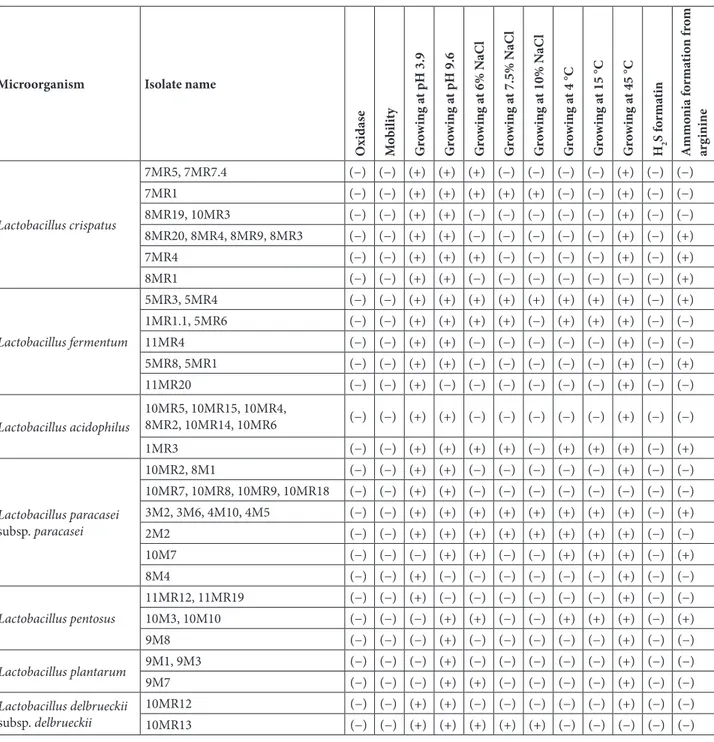
In Table 1, the test results of 49 gram-positive (+) bacilli and catalase-negative (−) isolates for oxidase activity, growth at different temperatures, development at different salt concentrations, formation of hydrogen sulphide, and formation of ammonia from arginine are illustrated. According to these results, it was determined that all of the 49 isolates were oxidase-negative and immobilized. If we look at the development at different pH levels, 42 isolates developed at pH 3.9, while 7 isolates showed no improvement. Furthermore, 45 isolates showed a pH of 9.6 and 4 of them did not. Regarding the developments in different salt concentrations, 19 isolates were produced in the medium containing 6% NaCl and 30 isolates were not produced. 12 isolates were produced in the medium
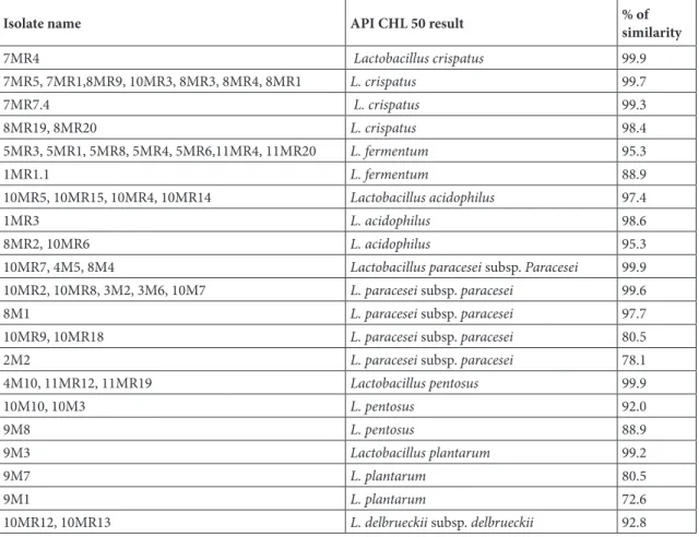
containing 7.5% NaCl, and 37 isolates were isolated. In the medium containing 10% NaCl, 9 isolates were produced and 40 isolates were not produced. If we look at the developments at different temperatures, 13 isolates grew at 4 and 15 °C and 36 did not show any growth. At 45° C, 43 isolates showed growth, 6 did not show any growth. As for the formation of hydrogen sulphide, no hydrogen sulphide or gas formation was observed in any of the 49 isolates. Ammonia was also formed from 18 isolate arginine and no ammonia was formed from 31 isolate arginine (Table 1). 3.1.1. Determination of carbohydrate utilization status of the lactic acid bacteria isolates with API CHL 50 system Identification results of 49 gram-positive (+) bacilli and catalase-negative (−) bacterial isolates isolated with the API CHL50 system are illustrated in Table 2. According to these results; of the 49 isolates, 8 were identified as Lactobacillus acidophilus, 3 as Lactobacillus plantarum, 9 as Lactobacillus pentosus, 8 as Lactobacillus fermentum, 17 as Lactobacillus paracesei subsp. paracesei, 13 as Lactobacillus crispatus, and 2 as Lactobacillus delbrueckii subsp. delbrueckii.
3.1.2. Definition of yeast isolates by the MALDI-TOF mass spectrometry
By the MALDI-TOF Mass Spectrometry, 10 of 14 yeast isolates were Candida albicans, 3 was C. glabrata, and 1 was C. tropicalis.
3.1.3. Results of the 16s rRNA sequence analysis
The 5MR1, 5MR6, and 10MR5 isolates with high anticandidal activity were identified as Lactobacillus jensenii (GenBank accession no: MH327499), Enterococcus faecalis (GenBank accession no: MH327502) with 99% similarity according to genotypic characterization results by 16S rRNA sequence analysis, and L. jensenii (GenBank accession no: MH327501), respectively.
3.1.4. Lactic acid and hydrogen peroxide production of the lactic acid bacteria isolates
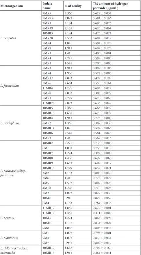
Table 3 illustrates the amounts of lactic acid and hydrogen peroxide in the lactic acid bacteria isolated from the vagina. While the lactic acid production of 49 isolates was between %0.91 and %2.684, it was found that the 10M7 isolate produced the lowest and the 5MR6 isolate produced the highest amount of lactic acid. As for the production of hydrogen peroxide, it was established that the production of hydrogen peroxide of 72 isolates was between 0.308 and 0.863 μg/mL. Furthermore, it was found that the 5MR8 isolate produced the lowest and 10M3 isolate produced the highest amount of hydrogen peroxide.
3.2. Anticandidal activities of the lactic acid bacteria iso-lates
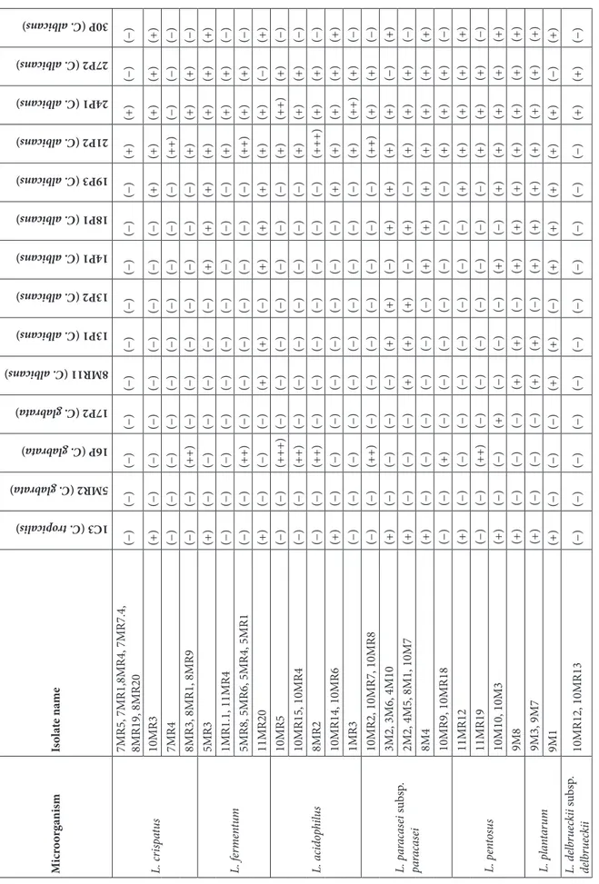
Table 4 illustrates the anticandidal activity results of 49 lactic acid bacteria isolated and identified from the vagina against the 14 Candida species (C. tropicalis 1C3, C. glabrata
5MR2, C. glabrata 16P, C. glabrata 17P2, C. albicans 8MR11, C. albicans 13P1, C. albicans 13P2, C. albicans 14P1, C. albicans 18P1, C. albicans 19P3, C. albicans 21P2, C. albicans 24P1, C. albicans 27P2, C. albicans 30P) isolated from the vagina again. As far as the results are concerned, most of the lactic acid bacteria had variation of 8–44 mm
of zone formation against the 14 Candida isolates. It was found that the lowest zone formation (8 mm) and the L. acidophilus 10MR14 isolate took place on the C. albicans 30P, and the highest zone formation (44 mm), on the other hand, L. acidophilus 10MR5 isolate took place on the C. glabrata 16P isolate.
Table 1. Biochemical activity tests of gram-positive (+) bacilli and catalase-negative (−) bacteria. (+): positive, (−): negative.
Microorganism Isolate name
O xi das e Mo bi lit y G ro w in g a t pH 3.9 G ro w in g a t pH 9.6 G ro w in g a t 6% N aC l G ro w in g a t 7.5% N aC l G ro w in g a t 10% N aC l G ro w in g a t 4 °C G ro w in g a t 15 °C G ro w in g a t 45 °C H2 S f orma tin A mmo ni a f orma tio n f ro m arg inine Lactobacillus crispatus 7MR5, 7MR7.4 (−) (−) (+) (+) (+) (−) (−) (−) (−) (+) (−) (−) 7MR1 (−) (−) (+) (+) (+) (+) (+) (−) (−) (+) (−) (−) 8MR19, 10MR3 (−) (−) (+) (+) (−) (−) (−) (−) (−) (+) (−) (−) 8MR20, 8MR4, 8MR9, 8MR3 (−) (−) (+) (+) (−) (−) (−) (−) (−) (+) (−) (+) 7MR4 (−) (−) (+) (+) (+) (−) (−) (−) (−) (+) (−) (+) 8MR1 (−) (−) (+) (+) (−) (−) (−) (−) (−) (−) (−) (+) Lactobacillus fermentum 5MR3, 5MR4 (−) (−) (+) (+) (+) (+) (+) (+) (+) (+) (−) (+) 1MR1.1, 5MR6 (−) (−) (+) (+) (+) (+) (−) (+) (+) (+) (−) (−) 11MR4 (−) (−) (+) (+) (−) (−) (−) (−) (−) (+) (−) (−) 5MR8, 5MR1 (−) (−) (+) (+) (−) (−) (−) (−) (−) (+) (−) (+) 11MR20 (−) (−) (+) (−) (−) (−) (−) (−) (−) (+) (−) (−) Lactobacillus acidophilus 10MR5, 10MR15, 10MR4, 8MR2, 10MR14, 10MR6 (−) (−) (+) (+) (−) (−) (−) (−) (−) (+) (−) (−) 1MR3 (−) (−) (+) (+) (+) (+) (−) (+) (+) (+) (−) (+) Lactobacillus paracasei subsp. paracasei 10MR2, 8M1 (−) (−) (+) (+) (−) (−) (−) (−) (−) (+) (−) (−) 10MR7, 10MR8, 10MR9, 10MR18 (−) (−) (+) (+) (−) (−) (−) (−) (−) (−) (−) (−) 3M2, 3M6, 4M10, 4M5 (−) (−) (+) (+) (+) (+) (+) (+) (+) (+) (−) (+) 2M2 (−) (−) (+) (+) (+) (+) (+) (+) (+) (+) (−) (−) 10M7 (−) (−) (−) (+) (+) (−) (−) (+) (+) (+) (−) (+) 8M4 (−) (−) (+) (−) (−) (−) (−) (−) (−) (+) (−) (−) Lactobacillus pentosus 11MR12, 11MR19 (−) (−) (+) (−) (−) (−) (−) (−) (−) (+) (−) (−) 10M3, 10M10 (−) (−) (−) (+) (+) (−) (−) (+) (+) (+) (−) (+) 9M8 (−) (−) (−) (+) (−) (−) (−) (−) (−) (+) (−) (−) Lactobacillus plantarum 9M1, 9M3 (−) (−) (−) (+) (−) (−) (−) (−) (−) (+) (−) (−) 9M7 (−) (−) (−) (+) (+) (−) (−) (−) (−) (+) (−) (−) Lactobacillus delbrueckii subsp. delbrueckii 10MR12 (−) (−) (+) (+) (−) (−) (−) (−) (−) (+) (−) (−) 10MR13 (−) (−) (+) (+) (+) (+) (+) (−) (−) (−) (−) (−)
MR: MRS agar, M: M17 agar. The bacteria isolated from MRS and M17 agars were named to include “MR” and “M” codes, respectively.
4. Discussion
Lactobacillus species are dominant in the vaginal microbiota (14). It was reported that Lactobacillus acidophilus, L. plantarum, L. casei, L. cellobiotus, L. oris, L. reuteri, L. ruminis, L. salivarius, L. brevis, L. delbrueckii, and L. vaginalis species were commonly isolated from the vagina (15). The Lactobacillus in the vaginal microbiota protected the microbiota against the colonization of other sexually transmitted infections such as bacterial vaginosis, urinary tract infections, vulvovaginal candidiasis, and HIV (5).
In the present study, anticandidal activities of various lactic acid bacteria isolated from the vagina of healthy volunteer women on the 14 vaginal Candida isolates were investigated. For this purpose, it was firstly determined that all the 49 lactic acid bacteria isolates were oxidase-negative and immobilized. It was observed that most isolates grew at pH 3.9 and pH 9.6. In addition, it was found that most isolates did not grow in medium containing 6%, 7.5%, and 10% NaCl and at 4, 15, and 45 °C. As for the
formation of hydrogen sulphide, no hydrogen sulphide or gas formation was observed in any of the 49 isolates (Table 1). It was reported by Pektaş that not all of the 136 lactic acid bacteria isolates produced H2S (16). Similarly in our study, it was found that 18 of the 49 isolates did not form ammonia from arginine of 31 isolates, which formed ammonia from arginine (Table 1). It was reported that Lactobacilli such as L. hilgardii, L. buchneri, and L. brevis could hydrolyze the arginine (17).
As a result of the identification of the isolated lactic acid bacteria by the API CHL 50 system, it was found that the 49 lactic acid bacteria isolates mostly belonged to L. paracesei subsp. paracesei (at the ratio of 27%) and to L. crispatus (at the ratio of 22.4%), followed by L. fermentum (16.3%), L. acidophilus (14.3%), L. pentosus (10.2%), L. plantarum (6.1%), and L. delbrueckii subsp. delbrueckii (4.1%) (Table 2). It was determined that 10 out of 14 yeast isolates isolated from the vagina by MALDI-TOF mass spectrometry belonged to C. albicans (8MR11, 13P1, 13P2, 14P1, 18P1, 19P3, 21P2, 24P1, 27P2, 30P), 3 isolates to C. Table 2. Identification of lactic acid bacteria with API CHL 50 system.
Isolate name API CHL 50 result % of similarity
7MR4 Lactobacillus crispatus 99.9 7MR5, 7MR1,8MR9, 10MR3, 8MR3, 8MR4, 8MR1 L. crispatus 99.7 7MR7.4 L. crispatus 99.3 8MR19, 8MR20 L. crispatus 98.4 5MR3, 5MR1, 5MR8, 5MR4, 5MR6,11MR4, 11MR20 L. fermentum 95.3 1MR1.1 L. fermentum 88.9 10MR5, 10MR15, 10MR4, 10MR14 Lactobacillus acidophilus 97.4 1MR3 L. acidophilus 98.6 8MR2, 10MR6 L. acidophilus 95.3
10MR7, 4M5, 8M4 Lactobacillus paracesei subsp. Paracesei 99.9
10MR2, 10MR8, 3M2, 3M6, 10M7 L. paracesei subsp. paracesei 99.6
8M1 L. paracesei subsp. paracesei 97.7
10MR9, 10MR18 L. paracesei subsp. paracesei 80.5
2M2 L. paracesei subsp. paracesei 78.1
4M10, 11MR12, 11MR19 Lactobacillus pentosus 99.9 10M10, 10M3 L. pentosus 92.0 9M8 L. pentosus 88.9 9M3 Lactobacillus plantarum 99.2 9M7 L. plantarum 80.5 9M1 L. plantarum 72.6
10MR12, 10MR13 L. delbrueckii subsp. delbrueckii 92.8
MR: MRS agar, M: M17 agar. The bacteria isolated from MRS and M17 agars were named to include “MR” and “M” codes, respectively.
Table 3. The amounts of lactic acid and hydrogen peroxide of the lactic acid bacteria. Microorganism Isolatename % of acidity The amount of hydrogenperoxide (µg/mL)
L. crispatus 7MR5 2.366 0.629 ± 0.034 7MR7.4 2.093 0.584 ± 0.166 7MR1 2.184 0.680 ± 0.025 8MR19 2.138 0.620 ± 0.064 10MR3 2.184 0.473 ± 0.074 8MR20 2.502 0.682 ± 0.019 8MR4 1.82 0.502 ± 0.123 8MR9 1.911 0.607 ± 0.123 8MR3 1.41 0.406 ± 0.001 7MR4 2.275 0.589 ± 0.000 8MR1 1.547 0.705 ± 0.080 L. fermentum 5MR3 1.911 0.389 ± 0.106 5MR4 1.956 0.572 ± 0.006 1MR1.1 2.093 0.499 ± 0.199 5MR6 2.684 0.555 ± 0.164 11MR4 1.797 0.602 ± 0.079 5MR8 2.002 0.308 ± 0.079 5MR1 2.229 0.620 ± 0.060 11MR20 2.093 0.633 ± 0.049 L. acidophilus 10MR5 2.366 0.663 ± 0.079 10MR15 1.638 0.628 ± 0.077 10MR4 1.911 0.773 ± 0.000 8MR2 1.365 0.389 ± 0.030 10MR14 1.82 0.597 ± 0.066 10MR6 2.548 0.584 ± 0.043 1MR3 1.41 0.569 ± 0.016 L. paracasei subsp. paracasei 10MR2 2.275 0.730 ± 0.000 8M1 1.001 0.756 ± 0.019 10MR7 1.274 0.392 ± 0.008 10MR8 1.456 0.699 ± 0.068 10MR9 1.683 0.607 ± 0.017 10MR18 1.729 0.652 ± 0.071 3M2 1.183 0.808 ± 0.040 3M6 1.41 0.778 ± 0.022 4M5 1.592 0.807 ± 0.025 4M10 1.228 0.770 ± 0.026 2M2 1.092 0.829 ± 0.030 10M7 0.91 0.822 ± 0.059 8M4 1.183 0.764 ± 0.036 L. pentosus 11MR12 1.865 0.672 ± 0.001 11MR19 1.365 0.411 ± 0.000 10M3 1.274 0.863 ± 0.096 10M10 1.137 0.834 ± 0.027 9M8 1.046 0.805 ± 0.046 L. plantarum 9M1 1.092 0.793 ± 0.001 9M3 1.092 0.836 ± 0.034 9M7 0.955 0.802 ± 0.047 L. delbrueckii subsp. delbrueckii 10MR1210MR13 1.6381.911 0.707 ± 0.1600.364 ± 0.041
MR: MRS agar, M: M17 agar. The bacteria isolated from MRS and M17 agars were named to include “MR” and “M” codes, respectively.
Ta bl e 4. A nt ic an did al ac tiv ities o f t he l ac tic acid b ac ter ia. Mi cr oo rg anis m Is ol at e na me 1C3 (C. tr opi cal is) 5MR2 (C. glab rat a) 16P (C. glab rat a) 17P2 (C. glab rat a) 8MR11 (C. albica ns) 13P1 (C. albica ns) 13P2 (C. albica ns) 14P1 (C. albica ns) 18P1 (C. albica ns) 19P3 (C. albica ns) 21P2 (C. albica ns) 24P1 (C. albica ns) 27P2 (C. albica ns) 30P (C. albica ns) L. cr isp at us 7MR5, 7MR1,8MR4, 7MR7.4, 8MR19, 8MR20 (−) (−) (−) (−) (−) (−) (−) (−) (−) (−) (+) (+) (−) (−) 10MR3 (+) (−) (−) (−) (−) (−) (−) (−) (−) (+) (+) (+) (+) (+) 7MR4 (−) (−) (−) (−) (−) (−) (−) (−) (−) (−) (++) (−) (−) (−) 8MR3, 8MR1, 8MR9 (−) (−) (++) (−) (−) (−) (−) (−) (−) (−) (+) (+) (+) (−) L. f er m en tu m 5MR3 (+) (−) (−) (−) (−) (−) (−) (+) (+) (+) (+) (+) (+) (+) 1MR1.1, 11MR4 (−) (−) (−) (−) (−) (−) (−) (−) (−) (−) (+) (+) (+) (−) 5MR8, 5MR6, 5MR4, 5MR1 (−) (−) (++) (−) (−) (−) (−) (−) (−) (−) (++) (+) (+) (−) 11MR20 (+) (−) (−) (−) (+) (+) (−) (+) (+) (+) (+) (+) (−) (+) L. a cid op hi lu s 10MR5 (−) (−) (+++) (−) (−) (−) (−) (−) (−) (−) (+) (++) (+) (−) 10MR15, 10MR4 (−) (−) (++) (−) (−) (−) (−) (−) (−) (−) (+) (+) (+) (−) 8MR2 (−) (−) (++) (−) (−) (−) (−) (−) (−) (−) (+++) (+) (+) (−) 10MR14, 10MR6 (+) (−) (−) (−) (−) (−) (−) (−) (−) (+) (+) (+) (+) (+) 1MR3 (−) (−) (−) (−) (−) (−) (−) (−) (−) (−) (+) (++) (+) (−) L. p ar ac as ei su bsp . pa ra cas ei 10MR2, 10MR7, 10MR8 (−) (−) (++) (−) (−) (−) (−) (−) (−) (−) (++) (+) (+) (−) 3M2, 3M6, 4M10 (+) (−) (−) (−) (−) (+) (+) (−) (+) (+) (+) (+) (−) (+) 2M2, 4M5, 8M1, 10M7 (+) (−) (−) (−) (+) (+) (+) (−) (+) (−) (+) (+) (+) (−) 8M4 (+) (−) (−) (−) (−) (−) (−) (+) (+) (+) (+) (+) (+) (+) 10MR9, 10MR18 (−) (−) (+) (−) (−) (−) (−) (−) (−) (−) (+) (+) (+) (−) L. p en to su s 11MR12 (+) (−) (−) (−) (−) (−) (−) (−) (−) (+) (+) (+) (+) (+) 11MR19 (−) (−) (++) (−) (−) (−) (−) (−) (−) (−) (+) (+) (+) (−) 10M10, 10M3 (+) (−) (−) (+) (−) (−) (−) (+) (−) (+) (+) (+) (+) (+) 9M8 (+) (−) (−) (−) (+) (+) (−) (+) (+) (+) (+) (+) (+) (+) L. p la nt ar um 9M3, 9M7 (+) (−) (−) (−) (+) (+) (−) (+) (+) (+) (+) (+) (+) (+) 9M1 (+) (−) (−) (−) (+) (+) (−) (+) (+) (+) (+) (+) (−) (+) L. d elb ru ec ki i su bsp . de lb rue ck ii 10MR12, 10MR13 (−) (−) (−) (−) (−) (−) (−) (−) (−) (−) (−) (+) (+) (−) Th e micr oo rga ni sm s i so la te d f ro m MRS, M17, P ot at o dext ros e, a nd c ho co la te a ga rs w er e n am ed t o in clude “ MR ”, “ M ”,“ P” , a nd “ C ” co des, r es pe ct iv ely . Zo ne di am et er s a re g iv en in mm. (+):2 t o ≥13 mm; (++) 14 t o ≥25 mm; (+++) 26 t o ≥38 mm.
glabrata (5MR2, 16P, 17P2), and 1 isolate to C. tropicalis (1C3). Candida albicans was the most common fungal species in the vaginal microbiota (18). In our study, C. albicans was also isolated at the highest level.
Vaginal homeostasis is protected by lactic acid and hydrogen peroxide production of the lactic acid bacteria in the vagina. By the production of lactic acid, the vaginal pH is kept below 4.5. It was reported that with the vaginal pH being acidic, it prevented the development of pathogens (19,20). In our study, the % acidity amounts of the lactic acid bacteria isolates isolated from the vagina ranged from 0.91 to 2.684 (Table 3). Furthermore, it was found that the hydrogen peroxide production amounts of our lactic acid bacteria isolates ranged from 0.308 ± 0.079 to 0.863 ± 0.096 μg/mL (Table 3). Halm et al. reported that the acidic medium formed by 2 Lactobacillus reuteri strains caused inhibition of yeasts (21). Alkali extracellular pH is thought to be the optimal pH for the development of Candida species; it was found that it induced the hyphae formation of C. albicans, and C. albicans remained in budding yeast form with less virulence at low pH (22).
The Candida species found in the vaginal microbiota are opportunistic pathogens. These microorganisms in the vagina can cause infections in certain conditions (22). There are some studies in the literature on the anticandidal activities of the lactic acid bacteria. In these studies, it was reported that the lactic acid bacteria had anticandidal activity (23–26). In three of these studies (23–25), anticandidal activities of probiotics containing Lactobacilllus on Candida albicans, C. glabrata, C. tropicalis, C. parapsilosis, and C. krusei isolated from the vagina were investigated. In another study, the anticandidal activities of various vaginal Lactobacillus species on Candida albicans ATCC 32032, Candida albicans RTN 071, and Candida glabrata
RTN 009 were studied (26). In our study, anticandidal activities of vaginal Lactobacillus spp. on vaginal Candida spp. were determined. Most of the vaginal lactic acid bacteria that belonged to the species of L. crispatus, L. fermentum, L. acidophilus, L. paracesei subsp. paracesei, L. pentosus, and L. plantarum exhibited anticandidal activity at varying ratios against vaginal C. albicans, C. glabrata, and C. tropicalis strains (Table 4). 5MR1, 5MR6, and 10MR5 isolates that had high anticandidal activity were identified by the 16S rRNA sequence analysis of L. jensenii (Accession no: AF243159.1), Enterococcus faecalis (Accession no. LC096215.1), and L. jensenii (Accession no: KF740715.1), respectively. It was found in this study that the lactic acid bacteria isolates had high amounts of lactic acid, and hydrogen peroxide had different antifungal activities. Besides, it was observed that the anticandidal activity was strain-specific.
Lactic acid bacteria are useful microorganisms associated with a variety of probiotic properties (27). Recently, probiotics containing lactic acid bacteria with antibiotics have been used in the treatment of vaginal Candida infections (28). In conclusion, our Candida isolates with high anticandidal activity are promising so, further studies are needed to be able to use these strains as probiotics. Besides, no other study has been found in the relevant literature investigating anticandidal activity by isolating Lactobacillus and Candida from the vagina; therefore, this study will be of great contribution to the literature.
Acknowledgments
This work was supported by a research grant from Anadolu University (research project 1305F089/2016). The authors would like to thank bioMérieux.
References
1. Fettweis JM, Serrano MG, Girerd PH, Jefferson KK, Buck GA.
A new era of the vaginal microbiome: advances using next generation sequencing. Chem Biodivers 2012; 9: 965-976.
2. Antonio MA, Hawes SE, Hillier SL. The identification of vaginal
Lactobacillus species and the demographic and microbiologic
characteristics of women colonized by these species. J Infect Dis 1999; 180: 1950-1956.
3. Sobel JD, Chaim W. Vaginal microbiology of women with acute
recurrent vulvovaginal candidiasis. J Clin Microbiol 1996; 34: 2497-2479.
4. Larsen B, Monif GRG. Understanding the bacterial flora of the
female genital tract. Clin Infect Dis 2001; 32: 69-77.
5. Boris S, Barbe’s C. Role played by lactobacilli in controlling the
population of vaginal pathogens. Microbes Infect 2000; 2: 543-546.
6. Falagas ME, Betsi GI, Athanasiou S. Probiotics for prevention
of recurrent vulvovaginal candidiasis: a review. J Antimicrob Chemother 2006; 58: 266-272.
7. Drutz DJ. Lactobacillus prophylaxis for Candida vaginitis. Ann Intern Med 1992; 116: 419-420.
8. Schillinger U, Lücke FK. Identification of lactobacilli from meat
and meat products. Food Microbiol 1987; 4: 199-208.
9. Stiles ME, Holzapfel WE. Lactic acid bacteria of foods and their
current taxonomy. Int J Food Microbiol 1997; 36: 1-29. 10. Doi K, Phuong OTA, Kawatou F, Nagayoshi Y, Fujino Y, Ohshima
T. Identification and characterization of lactic acid bacteria isolated from fermented rice bran product. Adv Microbiol 2013; 3: 265-272.
11. Demirci M, Gündüz H. Süt Teknolojisi El Kitabı. Ankara, Turkey: Hasad Yayıncılık; 1992 (book in Turkish).
12. Mumcu ZN. Kefirden izole edilen bazı laktik asit bakterilerinin metabolik, antimikrobiyal ve plazmit dna’larının incelenmesi. MSc, Gazi University, Ankara, Turkey, 1997 (in Turkish). 13. Pascual LM, Daniele MB, Ruiz F, Giardano W, Pajaro C, Barberis
L. Lactobacillus rhamnosus L60, a potential probiotic isolated from the human vagina. J Gen Appl Microbiol 2008; 54: 141-148. 14. Nobile CJ, Johnson AD. Candida albicans biofilms and human
disease. Annu Rev Microbiol 2015; 69: 71-92.
15. Silvester ME, Dicks LMT. Identification of lactic acid bacteria isolated from human vaginal secretions. Ant van Leeuw 2003; 83: 117-123.
16. Pektaş S. Süt ve süt ürünlerinden izole edilen laktik asit bakterilerinin ekzopolisakkarit üretim yeteneklerinin araştırılması. MSc, Anadolu University, Eskişehir, Turkey, 2014.
17. Liu SQ, Pilone GJ. Arginine metabolism in wine lactic acid bacteria and its practical significance. J Appl Microbiol 1998; 84: 315-327.
18. Barros PP, Ribeiro FC, Rossoni RD, Junqueira JC, Jorge AOC. Influence of Candida krusei and Candida glabrata on Candida
albicans gene expression in in vitro biofilms. Arch Oral Biol
2016; 64: 92-101.
19. Borges S, Silva J, Teixeira P. The role of lactobacilli and probiotics in maintaining vaginal health. Arch Gynecol Obstet 2014; 289: 479-489.
20. Cheeti S, Warrier BK, Lee CH. The role of monocarboxylate transporters in uptake of lactic acid in HeLa cells. Int J Pharm 2006; 325: 48-54.
21. Halm M, Hornbaek T, Arneborg N, Sefa-Dedeh, S, Jespersen L. Lactic acid tolerance determined by measurement of intracellular pH of single cells of Candida krusei and
Saccharomyces cerevisiae isolated from fermented maize
dough. Int J Food Microbiol 2004; 94: 97-103.
22. Jorgensen MR, Kragelund C, Jensen PQ, Keller MK, Twetman S. Probiotic Lactobacillus reuteri has antifungal effects on oral
Candida species in vitro. J Oral Microbiol 2017; 9: 1274582.
23. Chew SY, Cheah YK, Seow HF, Sandai D, Than LTL. Probiotic
Lactobacillus rhamnosus GR‐1 and Lactobacillus reuteri
RC‐14 exhibit strong antifungal effects against vulvovaginal candidiasis‐causing Candida glabrata isolates. J Appl Microbiol 2015; 118: 1180-1190.
24. Köhler GA, Assefa S, Reid G. Probiotic interference of
Lactobacillus rhamnosus GR-1 and Lactobacillus reuteri
RC-14 with the opportunistic fungal pathogen Candida
albicans. Infect Dis Obstet Gynecol 2012; 2012: 1-14.
25. Verdenelli MC, Coman MM, Cecchini C, Silvi S, Orpianesi C, Cresci A. Evaluation of antipathogenic activity and adherence properties of human Lactobacillus strains for vaginal formulations. J Appl Microbiol 2014; 116: 1297-1307.
26. Hütt P, Lapp E, Štšepetova J, Smidt I, Taelma H, Borovkova N, Oopkaup H, Ahelik A, Rööp T, Hoidmets D et al. Characterisation of probiotic properties in human vaginal lactobacilli strains. Microb Ecol Health Dis 2016; 27: 30484. 27. Fuller R. Probiotics in man and animal. J Appl Bacteriol 1989;
66: 365-378.
28. Kovachev SM, Vatcheva-Dobrevska RS. Local probiotic therapy for vaginal Candida albicans infections. Probiotics and Antimicro Prot 2015; 7: 38-44.