Beneficial effects montelukast, cysteinyl-leukotriene
receptor antagonist, on renal damage after unilateral
ureteral obstruction in rats
_______________________________________________
Alper Otunctemur
1, Emin Ozbek
2, Suleyman Sami Cakir
3, Murat Dursun
4, Mustafa Cekmen
5, Emre
Can Polat
6, Levent Ozcan
7, Adnan Somay
8, Nurver Ozbay
81 Department of Urology, Okmeydani Training and Research Hospital, Istanbul, Turkey; 2 Department of Urology, Katip Celebi University, Ataturk Training and Research Hospital, Izmir, Turkey; 3 Department of Urology, Bayburt State Hospital,Bayburt, Turkey; 4 Department of Urology, Bahcelievler State Hospital, Istanbul, Turkey; 5 Department of biochemistry, Kocaeli University, , Kocaeli, Turkey; 6 Department of Urology, Istanbul Medipol University, Faculty of Medicine, Istanbul, Turkey; 7 Department of Urology, Derince Training and Research Hospital, , Kocaeli, Turkey; 8 Department Pathology, Fatih Sultan Mehmet Training and Research Hospital, Istanbul, Turkey
ABSTRACT
ARTICLE
INFO
______________________________________________________________ ______________________ Introductıon: Ureteral obstruction is a common pathology and caused kidney fibrosis
and dysfunction at late period. In this present, we investigated the antifibrotic and antiinflammatory effects of montelukast which is cysteinyl leukotriene receptor anta-gonist, on kidney damage after unilateral ureteral obstruction(UUO) in rats.
Materıals and Methods: 32 rats divided four groups. Group 1 was control, group 2 was sham, group 3 was rats with UUO and group 4 was rats with UUO which were given montelukast sodium (oral 10 mg/kg/day). After 14 days, rats were killed and their kidneys were taken and blood analysis was performed. Tubular necrosis, mono-nuclear cell infiltration and interstitial fibrosis scoring were determined histopatho-logically in a part of kidneys; nitric oxide(NO), malondialdehyde(MDA) and reduced glutathione(GSH) levels were determined in the other part of kidneys. Urea-creatinine levels were investigated at blood analysis. Statistical analyses were made by the Chi--square test and one-way analysis of variance (ANOVA).
Results: There was no difference significantly for urea-creatinine levels between groups. Pathologically, there was serious tubular necrosis and fibrosis in group 3 and there was significantly decreasing for tubular necrosis and fibrosis in group 4(p<0.005). Also, there was significantly increasing for NO and MDA levels; decreasing for GSH levels in group 3 compared the other groups(p<0.005).
Conclusıon: We can say that montelukast prevent kidney damage with antioxidant effect, independently of NO.
Key words:
montelukast [Supplementary Concept]; cysteinyl-leukotriene [Supplementary Concept]; Renal Insufficiency; Ureteral
Obstruction
Int Braz J Urol. 2015; 41: 279-87 _____________________
Submitted for publication: May 14, 2014
_____________________
Accepted after revision: July 27, 2014
INTRODUCTION
Chronic kidney diseases, which lead to end--stage kidney failure, are associated with changes
in kidney structure and fibrosis regardless of the underlying cause. Urinary tract obstruction is cha-racterized by tubular atrophy or dilation, tubular cell death by apoptosis and necrosis, interstitial
leukocyte infiltration, and increased interstitial matrix deposition (1). The acute phase of obs-tructed kidney in unilateral ureteral obstruction (UUO) is characterized by dramatic changes in glomerular filtration rate, renal blood flow, and interstitial edema (2, 3).
Such an obstruction might be observed after benign prostatic hyperplasia; renal, ure-teral, or bladder calculi; urethral stricture; and neoplasm of the bladder, prostate, or urethra (1). The hydrostatic pressure, which is the result of the blockage, initiates renal injuries. The inju-ries are characterized by tubular dilatation or atrophy, inflammatory infiltration of leucocytes, fibroblast activation, proliferation, increase in matrix proteins, and progressive interstitial fi-brosis with the loss of renal parenchyma. Uni-lateral ureteral obstruction (UUO) is an experi-mental rat model of renal injury that imitates the process of obstructive nephropathy in an accele-rated manner(1).
Reactive oxygen species (ROS) are a re-cently recognized mechanism in the pathoge-nesis of UUO in experimental studies. Increased lipid peroxidation has been reported in renal cortexes of UUO animals. It has been shown that oxidative stress in UUO contributes to the deve-lopment of tubulointerstitial lesions and renal fi-brosis. Various factors with complex cellular and molecular interactions have also been proposed as possible causes that lead to tubulointerstitial lesions and renal fibrosis (4-7).
Nitric oxide (NO) acts as an intercellu-lar messenger and regulates celluintercellu-lar functions such as vasorelaxation and inflammation. NO has an important role in the elimination of pa-thogens and tumor cells; however, overproduced NO is oxidized to ROS, resulting in the disrup-tion of cell signaling and uncontrolled systemic inflammation (8, 9). Malondialdehyde (MDA) is one of the important markers of lipid peroxida-tion (10). Excessive MDA produced as a result of tissue injury and DNA damage could combine with free amino groups of proteins, resulting in the formation of MDA-modified protein adducts. Glutathione (GSH) is the major intracellular an-tioxidant with multiple biological functions, in-cluding the maintenance of the thiol moieties
of proteins and the reduced from of many other biologically active molecules (11).
Leukotrienes, the products generated by the 5-lipoxygenase pathway are particularly im-portant in inflammation; indeed, leukotrienes in-crease microvascular permeability and are potent chemotactic agents (12). Moreover, inhibition of 5-lipoxygenase indirectly reduces the expression of TNF-alpha (a cytokine that plays a key role in inflammation), and there are a number of studies demonstrating the role of leukotrienes as media-tors of the gastric damage induced by ethanol and some other noxious substances(13, 14). Cys-teinyl leukotrienes(CysLT), leukotrienes C4, D4, and E4 (LTC4, LTD4, LTE4) are secreted mainly by eosinophils, mast cells, monocytes and ma-crophages, and they exert a variety of actions which emphasize their importance as pathogenic elements in inflammatory states(15, 16).
Montelukast (MK-0476), a selective re-versible cys-leukotriene-1 receptor (LTD4 recep-tor) antagonist is used in the treatment of asth-ma and is reported to reduce airway eosinophilic inflammation in this disease (17-19). CysLT1 receptor antagonists or biosynthesis inhibitors have been reported to ameliorate ethanol-indu-ced gastric mucosal damage and experimental colitis (13, 20, 21).
Based on these findings, we investigated the antifibrotic and antiinflammatory effects of montelukast on kidney damage after UUO in rats by measuring MDA, NO and GSH levels and the myeloperoxidase activity.
MATERIAL AND METHODS ANIMALS
Male Wistar Albino rats, weighing 200 to 250 g and 6 to 7 weeks old, were housed in clean plastic cages in a temperature and humidity con-trolled facility under a constant 12-hour light/12--hour dark photoperiods with free access to food and water. The Institutional Animal Care and Use Committee approved the use of animals and the experimental protocol, and animals were treated in accordance with the Guide for the Care and Use of Laboratory Animals of Research Council.
TREATMENT AND EXPERIMENTAL PROTOCOLS
One week after acclimatization, UUO was induced. Briefly after induction of general anes-thesia by intraperitoneal injection of thiopental (100 mg/kg), the abdominal cavity was exposed via midline incision and the left ureter was liga-ted at 2 points with 3-0 silk. The sham-operaliga-ted rats had their ureters manipulated but not liga-ted. All rats were given amikacin sulfate (6 mg/ kg, intramuscularly route) before operation. The rats were randomly divided into four groups, each consisting of eight animals. Rats with no opera-tion (Group-1) received no treatment and served as controls. Rats in group 2 underwent unilateral ureteral ligation and received no treatment. Group 3 rats underwent sham operation and received no treatment with montelukast sodium (ML). Rats in Group-4 were subjected to unilateral ureteral liga-tion and received ML ( p.o. 10mg/kg/day) (22).
At 14 days after UUO, all rats were sacrifi-ced by high-dose ketamine. Kidneys were reached with an abdominal midline incision. Left kidney was immediately excised and separated from the surrounding tissues, washed twice with cold sa-line, and stored at -80ºC to determine the levels of renal malondialdehyde (MDA), GSH, and NO. A portion of the left renal tissue was stored in formol solution for the histopathologic nations. Paraffinized tissue samples were exami-ned for leukocyte infiltration and renal fibrosis. Urea-creatinine levels were investigated at blood analysis. Renal impairment was assessed by serum urea and creatinine levels, as well as by the kid-ney histology. Serum urea and creatinine levels were determined with an autoanalyzer (Syncron LX20, Ireland) by using commercial Becman Coul-ter diagnostic kits.
MEASUREMENT OF TISSUE LIPID PEROXIDATION LEVEL
Frozen kidney samples were homogenized in Teflon-glass homogenizer with a buffer con-taining 1.5% potassium chloride to obtain 1:10 (w/v) whole homogenate. MDA, which is formed as an end product of the lipid peroxidation, served as an index of the intensity of oxidative stress.
MDA, referred to as thiobarbituric acid reactive substance, was measured with thiobarbituric acid at 532 nm in a spectrophotometer, as described previously (23). The MDA level was expressed as mmol/g wet tissue.
MEASUREMENT OF TISSUE GSH LEVEL
Reduced GSH was estimated by the me-thod of Moron et al. (24), where the color deve-loped was read at 412 nm. Protein concentrations in all samples were measured using the method of Lowry et al. (25). Results were reported as mmol/g wet tissue.
MEASUREMENT OF TISSUE NO LEVEL
Total nitrite was quantified by the Griess reaction after incubating the supernatant with Es-cherichia coli nitrate reductase to convert NO3 to
NO2. Griess reagent (1 mL 1% sulfanilamide, 0.1%
naphthyl-ethylenediamine hydrochloride, and 2.5% phosphoric acid; Sigma Chemicals) was then added to 1 mL of supernatant (26). The absorbance was read at 545nm after 30-minute incubation. The absorbance was compared with the standard graph of NaNO2, obtained from the reduction of
NaNO3 (1-100 mmol/L). The accuracy of the
as-say was checked in two ways; the inter- and in-traassay coefficients of variation were 7.52% and 4.61%, respectively. To check conversion of nitrate to nitrite (recovery rate), predetermined amounts of nitrate were added to control plasma samples; these samples were deproteinized and reduced as above.
HISTOPATHOLOGICAL EXAMINATION
Histopathological evaluation was perfor-med on kidney tissues. Paraffin-embedded speci-mens were cut into 6-μm thickness sections and stained with hematoxylin & eosin for examination under the light microscope using a conventional protocol (27) (BH-2; Olympus, Tokyo, Japan). A semi-quantitative evaluation of renal tissues was accomplished by scoring the degree of severity according to previously published criteria (28). All sections of kidney samples were examined for
tubular necrosis. Briefly, a minimum of 50 pro-ximal tubules associated with 50 glomeruli were examined for each slide and an average score was obtained. Severity of lesion was graded from 0 to 3 according to the percentage of tubular in-volvement. Slides were examined and assigned for severity of changes using scores on scale in which (0) denotes no change; grade (1) changes affecting <25% tubular damage (mild); grade (2) changes affecting 25-50% of tubules (moderate); grade (3) changes affecting >50% of tubules (se-vere).
Histopathological evaluation was perfor-med on left kidney tissues. Paraffin-embedded specimens were cut into 5-mm thick sections and stained with hematoxylin & eosin and Masson’s trichrome for examination under the light mi-croscope (BH-2; Olympus, Tokyo, Japan).
To evaluate leukocyte infiltration, the wi-dening of interstitial spaces with focal leukocyte infiltration was assessed in five randomly cho-sen sections prepared from each kidney sample. For each section, the average number of leu-kocytes per 0.28 mm2 was calculated from
the-se leukocyte-infiltrated foci using a high-power microscopic field (x400).
In order to estimate the grade of inters-titial fibrosis, the intersinters-titial area that stained green with Masson’s trichrome was evaluated as a percentage of the total examined area in five randomly chosen sections prepared from each kidney sample using an image analyzer (Leica; Leica Micros Imaging Solutions, Cambridge, UK). For each section, interstitial space widening with focal leukocyte infiltration and interstitial fibro-sis was assessed in high-power fields (x400) to quantify the results. The Banff classification of kidney pathology was used for scoring the degree
of mononuclear cell infiltration and interstitial fi-brosis. The score was graded from 0 to 3, depending on the severity of histological characteristics (29).
Statistical analyses
Results of all groups were shown as mean values±standard deviation (SD). Statistical analy-ses of the histopathologic evaluation of the groups were carried out by the Chi-square test and bioche-mical data were analyzed by the one-way analysis of variance (ANOVA). The significance between two groups was determined by the Dunnett’s mul-tiple comparison test, and P<0.05 was accepted as statistically significant value.
RESULTS
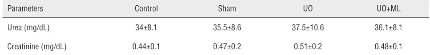
There was no significant difference for urea-creatinine levels between groups (Table-1).
Tissue MDA levels significantly increased in Group-2 compared with Groups-1, 3, and 4 ( p<0.001). Rats with ML administration (Group-4) showed reduced levels of lipid peroxidation as measured by MDA levels (Table-2). UUO also in-duced a significant increase in the tissue NO le-vels that have been prevented by ML (Table-2). The unilateral ureteral ligation was accompanied by a marked reduction in GSH level in the kidney tissues of rats ( p<0.001), and treatment with ML partially elevated the GSH levels (Table-2).
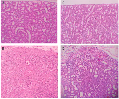
Histopathologic examination was normal in rats with only sham operation (Group-3) and in rats with no operation (Group-1) (Figures 1A and B). In rats with UUO, there were mild and severe tubular necrosis in the proximal tubules compared control and sham groups (Figure-1C). In rats tre-ated with UUO+ML, despite the presence of mild
Table 1 - Effects of UO alone and its combination with ML on plasma urea, creatinine levels in rats.
Parameters Control Sham UO UO+ML Urea (mg/dL) 34±8.1 35.5±8.6 37.5±10.6 36.1±8.1 Creatinine (mg/dL) 0.44±0.1 0.47±0.2 0.51±0.2 0.48±0.1
Table 2 - Effect of ML on the Levels of Malondialdehyde, Glutathione, and Nitric Oxide in Each Rat Group.
Parameters Kontrol Sham UO UO + MLS NO (nmol/g wet tissue) 30.3±9.3 30.3±10.2 63.2±15.2a 36.3±9.3b
MDA (nmol/g wet tissue) 2.6±0.7 2.7±0.7 5.1±1.2a 2.9±0.9b
GSH (umol/g wet tissue) 2.3±0.8 2.2±0.7 1.1±0.5c 2.1±0.7d Values are expressed as mean ± SD for eight rats in each group.
a Significantly different from sham (p<0.001); b Significantly different from UO group (p<0.001); c Significantly different from sham (p<0.05);
d Significantly different from UO group (p<0.05).
Abbreviations: NO = nitric oxide; MDA = malondialdehyde; GSH = reduced glutathione.
Figure 1 – A = normal tubulus and glomerules in kidney kortex H&Ex100 (control group). B = normal tubulus and glomerules in kidney kortex H&Ex100 (sham group). C = severe tubular necrosis, tubular degeneration and epithelial vacuolization in the proximal tubules H&Ex100(UUO group). D = mild epithelial vacuolization in the proximal tubules and normal glomerules H&Ex100 (UUO+ML treated group).
A C
B D
tubular degeneration and less severe tubular ne-crosis, glomeruli maintained a better morpholo-gy when compared with UUO group (Figure-1D). Severe leukocyte infiltration was observed in the periglomerular and peritubular interstitium of the kidneys of the rats in group-2 with UUO (Figures 2A and B).
Quantitative analysis of the focal leukocyte infiltration area in the interstitium showed that
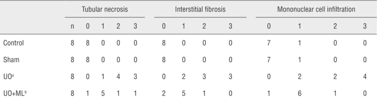
leukocyte infiltration was significantly reduced in rats that received UUO+ML (Group-4) (Figure-2C). UUO caused a significant interstitial fibrosis in rats that received no treatment (Group-2), and the percentage area of interstitial fibrosis in the kid-ney of rats with UUO that received no treatment was significantly greater than that of rats with UUO that received ML (Group-4) (Figures 3A, B and C). These changes are summarized in Table-3.
Figure 3 – A = Normal kidney morphology in a sham group. B = severe fibrosis was observed in the peritubular interstitium of the UUO. C = mild fibrosis was reduced in the ML-treated group (masson & trichrome,*400).
Figure 2 – A = Normal kidney morphology in a sham group. B = Leukocyte infiltration was observed in the peritubular interstitium of the UUO. C = Leukocyte infiltration was reduced in the ML-treated group (hematoxylin & eosin,*400).
A A C C B B
Table 3 - Semiquantitative analysis of tubular necrosis, interstitial fibrosis, mononuclear cell infiltration in control, Sham, UO, and UO+ML treated rats.
Tubular necrosis Interstitial fibrosis Mononuclear cell infiltration n 0 1 2 3 0 1 2 3 0 1 2 3 Control 8 8 0 0 0 8 0 0 0 7 1 0 0 Sham 8 8 0 0 0 8 0 0 0 7 1 0 0 UOa 8 0 1 4 3 0 2 3 3 0 2 2 4
UO+MLb 8 1 5 1 1 2 5 1 0 1 6 1 0 Score 0 = no degeneration, 1 = mild degeneration, 2 = moderate degeneration, and 3 = severe degeneration
a Statistical significant difference from the Sham group
DISCUSSION
The present study confirmed through a quantitative survey the protective role of ML on renal tissue damage after the induction of UUO in rats. Our results showed that the obstructed kidney had significantly higher tissue MDA, NO levels, and lower GSH levels along with more fibrosis. The cur-rent data demonstrate UUO structural and functio-nal alterations in the kidney with a concomitant in-crease in proinflammatory cytokines in the blood. The CysLT1 receptor antagonist montelukast, on the other hand, reduced the severity of injury, de-pressed the concentration of these cytokines and increased the antioxidative capacity.
Montelukast, one of the selective reversible CysLT1 receptor antagonists, is used for the main-tenance treatment of asthma and to relieve symp-toms of seasonal allergies(30). It is reported that montelukast can reduce eosinophilic inflammation in the airways (31-33).
Besides, CysLT1 receptor antagonists or biosynthesis inhibitors ameliorate ethanol-indu-ced gastric mucosal damage (13, 34), experimental colitis (35), and wound healing (36, 37).
Recently, Sener et al. (22) have reported that montelukast has protective effects on chronic renal failure-induced multiple organ injury. They attributed this to montelukast’s ability to inhibit neutrophil infiltration and apoptosis. They also suggested that montelukast balances the oxidant– antioxidant status and regulates the generation of proinflammatory mediators. In a different study, it has been shown that montelukast reversed ische-mia reperfusion-induced oxidant responses and improved microscopic damage and renal func-tions (31).
Apoptotic cell death has been reported to play an important role in UUO-induced renal da-mages (38). The lack of investigation on whether ML has affected the apoptotic cell death because of UUO may be a limitation of this study. However, in a recent study, curcumin and melatonine which is an antioxidant and antiinflammatory agent like ML, has been reported to prevent UUO-mediated apoptotic cell death and reduce the UUO related renal damage. While we believe that ML can re-duce the UUO-inre-duced renal damage by a similar
mechanism that prevents apoptosis-related cell death, there is a need for further study on that subject for verifications.
The pathogenesis of renal fibrosis cau-sed by UUO involves infiltration of the kidney by inflammatory cells including monocytes, ac-tivation and possible transformation of intrinsic renal cells, and interactions between infiltrating and resident cells. NF-kB is activated during renal obstruction, and inhibition of NF-kB activity has been demonstrated to prevent renal fibrosis indu-ced by obstruction (39, 40). In the present study, in agreement with these findings, ML treatment prevented renal fibrosis in UUO rats.
In obstructive nephropathy, it is known that particularly by treatment of UUO, some func-tions of the kidney can be regained. However, by recovery from the ureteral obstruction, reperfu-sion damage may occur in the renal tissue as a result of the elevated renal blood flow.
Ischemia-reperfusion (IR) injury is one of the underlying causes of acute renal failure and ROS along with NO, and it plays important ro-les in mediating cell damage during IR injury (41, 42). Inflammatory cells, neutrophils, are potent cells for production of ROS that are highly produ-ced during IR injury. Renal IR causes tissue injury by way of oxygen radicals and disturbs balance between oxidants and antioxidants in tissue (43). Rodriguez-Reynoso et al. found that exogenous melatonine (MLT) preserved renal function, incre-ased GSH levels, reduced lipid peroxidation, and prevented the rise in NO levels induced by renal IR (44).
They also indicated that MLT treatment reduced histological kidney injury. In accordance with the increase in toxic oxygen metabolites, the renal MDA level was also significantly increased, indicating the presence of enhanced lipid peroxi-dation due to IR injury, while the levels of tissue glutathione were declined, demonstrating the de-pletion of the antioxidant pool.
Several studies have demonstrated that IR in the kidney is associated with lipid peroxidation, which is an autocatalytic mechanism leading to oxidative destruction of cellular membranes (45-47). As montelukast reduced the oxidative injury on cellular structures, the level of the
intracellu-lar antioxidant glutathione, which is otherwise oxidized when inactivating free radicals, was not changed. Thus, it appears that the anti-oxidative effect of montelukast on lipid peroxidation does not involve the expenditure of tissue GSH stores, but the antioxidant pool is further supported by the action of montelukast. Moreover, IR-induced reduction in total antioxidant capacity was also reversed by montelukast treatment (31).
As shown in our study, ML has a protective role against both renal damage arising because of UUO and the reperfusion damage occurring as a result of the treatment of obstruction that gives way to elevated renal blood flow.
In conclusion, the results reported here indicate that ML exerts a preventive effect on UUO-induced kidney damage in rats by reducing oxidative stress. We therefore propose that ML supplementation therapy can be used for kidney protection in patients with UUO, such as ureteral stones. Hovewer, further animal and clinical stu-dies are needed to confirm our suggestion.
CONFLICT OF INTEREST
None declared.
REFERENCES
1. Sato S, Yamate J, Saito T, Hosokawa T, Saito S, Kurasaki M. Protective effect of taurine against renal interstitial fibrosis of rats induced by cisplatin.Naunyn Schmiedebergs Arch Pharmacol. 2002;365:277-83.
2. Vaughan ED Jr, Sorenson EJ, Gillenwater JY. The renal hemodynamic response to chronic unilateral complete ureteral occlusion. Invest Urol. 1970;8:78-90.
3. Schreiner GF, Harris KP, Purkerson ML, Klahr S. Immunological aspects of acute ureteral obstruction: immune cell infiltrate in the kidney. Kidney Int. 1988;34:487-93.
4. Ricardo SD, Ding G, Eufemio M, Diamond JR. Antioxidant expression in experimental hydronephrosis: role of mechanical stretch and growth factors. Am J Physiol. 1997;272:F789-98.
5. Saborio P, Krieg RJ Jr, Kuemmerle NB, Norkus EP, Schwartz CC, Chan JC. Alpha-tocopherol modulates lipoprotein cytotoxicity in obstructive nephropathy. Pediatr Nephrol. 2000;14:740-6.
6. Kawada N, Moriyama T, Ando A, Fukunaga M, Miyata T, Kurokawa K, et al. Increased oxidative stress in mouse kidneys with unilateral ureteral obstruction. Kidney Int. 1999;56:1004-13.
7. Klahr S. Urinary tract obstruction. Semin Nephrol. 2001;21:133-45.
8. Huang HS, Ma MC, Chen CF, Chen J. Changes in nitric oxide production in the rat kidney due to CaOx nephrolithiasis. Neurourol Urodyn. 2006;25:252-8.
9. Aviram M, Dornfeld L, Rosenblat M, Volkova N, Kaplan M, Coleman R, et al. Pomegranate juice consumption reduces oxidative stress, atherogenic modifications to LDL, and platelet aggregation: studies in humans and in atherosclerotic apolipoprotein E-deficient mice. Am J Clin Nutr. 2000;71:1062-76.
10. Schubert SY, Neeman I, Resnick N. A novel mechanism for the inhibition of NF-kappaB activation in vascular endothelial cells by natural antioxidants. FASEB J. 2002;16:1931-3.
11. Aviram M, Dornfeld L, Kaplan M, Coleman R, Gaitini D, Nitecki S, et al. Pomegranate juice flavonoids inhibit low-density lipoprotein oxidation and cardiovascular diseases: studies in atherosclerotic mice and in humans. Drugs Exp Clin Res.2002;28:49-62.
12. Wallace JL, MacNaughton WK, Morris GP, Beck PL. Inhibition of leukotriene synthesis markedly accelerates healing in a rat model of inflammatory bowel disease. Gastroenterology. 1989;96:29-36.
13. Wallace JL, McKnight GW, Keenan CM, Byles NI, MacNaughton WK. Effects of leukotrienes on susceptibility of the rat stomach to damage and investigation of the mechanism of action. Gastroenterology. 1990;98:1178-86.
14. Wallace JL, Beck PL, Morris GP. Is there a role for leukotrienes as mediators of ethanol-induced gastric mucosal damage? Am J Physiol. 1988;254:G117-23. 15. Damon M, Chavis C, Godard P, Michel FB, Crastes
de Paulet A. Purification and mass spectrometry identification of leukotriene D4 synthesized by human alveolar macrophages. Biochem Biophys Res Commun. 1983 Mar 16;111:518-24.
16. Williams JD, Czop JK, Austen KF. Release of leukotrienes by human monocytes on stimulation of their phagocytic receptor for particulate activators. J Immunol.1984 Jun;132:3034-40.
17. Aharony D. Pharmacology of leukotriene receptor antagonists . Am J Respir Crit Care Med. 1998;157:S214-9.
18. O’Byrne PM. Asthma treatment: antileukotriene drugs. Can Respir J. 1998;5 Suppl A:64A-70A.
19. Wenzel SE. Leukotriene receptor antagonists and related compounds. Can Respir J. 1999;6:189-93.
20. Carsin H, Bargues L, Stéphanazzi J, Paris A, Aubert P, Le Béver H. [Inflammatory reaction and infection in severe burns]. Pathol Biol (Paris). 2002;50:93-101.
21. Konturek SJ, Brzozowski T, Drozdowicz D, Beck G. Role of leukotrienes in acute gastric lesions induced by ethanol, taurocholate, aspirin, platelet-activating factor and stress in rats. Dig Dis Sci. 1988;33:806-13.
22. Sener G, Sakarcan A, Sehirli O, Ekşioğlu-Demiralp E, Sener E, Ercan F, et al. Chronic renal failure-induced multiple-organ injury in rats is alleviated by the selective CysLT1 receptor antagonist montelukast. Prostaglandins Other Lipid Mediat. 2007;83:257-67.
23. Wasowicz W, Nève J, Peretz A. Optimized steps in fluorometric determination of thiobarbituric acid-reactive substances in serum: importance of extraction pH and influence of sample preservation and storage. Clin Chem. 1993;39:2522-6.
24. Moron MS, Depierre JW, Mannervik B. Levels of glutathione, glutathione reductase and glutathione S-transferase activities in rat lung and liver. Biochim Biophys Acta. 1979;582:67-78. 25. Lowry OH, Rosebrough NJ, Farr AL, Randall RJ. Protein
measurement with the Folin phenol reagent. J Biol Chem. 1951;193:265-75.
26. Granger DL, Taintor RR, Boockvar KS, Hibbs JB Jr. Measurement of nitrate and nitrite in biological samples using nitrate reductase and Griess reaction. Methods Enzymol. 1996;268:142-51.
27. Sun Y, Oberley LW, Li Y. A simple method for clinical assay of superoxide dismutase. Clin Chem. 1988;34:497-500. 28. Allen CT. Laboratory methods in histochemistry. In: Prophet
EB, Mills B, Arrington JB, Sobin LH (eds). American registry of pathology, 1st edn. Washington DC. 1992; pp. 53. 29. Kinugasa F, Noto T, Matsuoka H, Urano Y, Sudo Y, Takakura
S, et al. Prevention of renal interstitial fibrosis via histone deacetylase inhibition in rats with unilateral ureteral obstruction. Transpl Immunol. 2010;23:18-23.
30. Canbay E, Agachan B, Ozturk T, Giris M, Asoglu O, Balik E, et al. Dual inhibition of wound healing and oxidative process by montelukast in experimental colon anastomoses. Surg Innov. 2010;17:248-55.
31. Sener G, Sehirli O, Velioğlu-Oğünç A, Cetinel S, Gedik N, Caner M, et al. Montelukast protects against renal ischemia/ reperfusion injury in rats. Pharmacol Res. 2006;54:65-71. 32. Damtew B, Marino JA, Fratianne RB, Spagnuolo PJ. Neutrophil
lipoxygenase metabolism and adhesive function following acute thermal injury. J Lab Clin Med. 1993;121:328-36.
33. Wallace JL, Beck PL, Morris GP. Is there a role for leukotrienes as mediators of ethanol-induced gastric mucosal damage? Am J Physiol. 1988;254(1 Pt 1):G117-23.
34. Carsin H, Bargues L, Stéphanazzi J, Paris A, Aubert P, Le Béver H. Inflammatory reaction and infection in severe burns. Pathol Biol (Paris). 2002;50:93-101.
35. Konturek SJ, Brzozowski T, Drozdowicz D, Beck G. Role of leukotrienes in acute gastric lesions induced by ethanol, taurocholate, aspirin, platelet-activating factor and stress in rats. Dig Dis Sci. 1988;33:806-13.
36. Kabasakal L, Sener G, Cetinel S, Contuk G, Gedik N, Yeğen BC. Burn-induced oxidative injury of the gut is ameliorated by the leukotriene receptor blocker montelukast. Prostaglandins Leukot Essent Fatty Acids. 2005;72:431-40.
37. Turtay MG, Firat C, Samdanci E, Oguzturk H, Erbatur S, Colak C. Effects of montelukast on burn wound healing in a rat model. Clin Invest Med. 2010;33:E413-21.
38. Hashem RM, Soliman HM, Shaapan SF. Turmeric-based diet can delay apoptosis without modulating NF-kappaB in unilateral ureteral obstruction in rats. J Pharm Pharmacol. 2008;60:83-9. 39. Cheung RT, Tipoe GL, Tam S, Ma ES, Zou LY, Chan PS.
Preclinical evaluation of pharmacokinetics and safety of melatonin in propylene glycol for intravenous administration. J Pineal Res. 2006;41:337-43.
40. Pignone AM, Rosso AD, Fiori G, Matucci-Cerinic M, Becucci A, Tempestini A, et al. Melatonin is a safe and effective treatment for chronic pulmonary and extrapulmonary sarcoidosis. J Pineal Res. 2006;41:95-100.
41. Basireddy M, Isbell TS, Teng X, Patel RP, Agarwal A. Effects of sodium nitrite on ischemia-reperfusion injury in the rat kidney. Am J Physiol Renal Physiol.2006;290:F779-86 42. Noiri E, Nakao A, Uchida K, Tsukahara H, Ohno M, Fujita T, et
al. Oxidative and nitrosative stress in acute renal ischemia. Am J Physiol Renal Physiol. 2001;281:F948-57.
43. Erdogan H, Fadillioglu E, Yagmurca M, Uçar M, Irmak MK. Protein oxidation and lipid peroxidation after renal ischemia-reperfusion injury: protective effects of erdosteine and N-acetylcysteine. Urol Res. 2006;34:41-6.
44. Rodríguez-Reynoso S, Leal C, Portilla-de Buen E, Castillo JC, Ramos-Solano F. Melatonin ameliorates renal ischemia/ reperfusion injury. J Surg Res. 2004;116:242-7.
45. Singh D, Chander V, Chopra K. Carvedilol attenuates ischemia-reperfusion-induced oxidative renal injury in rats. Fundam Clin Pharmacol. 2004;18:627-34.
46. Singh D, Chopra K. Effect of trimetazidine on renal ischemia/ reperfusion injury in rats. Pharmacol Res. 2004;50:623-9. Retraction in: Pharmacol Res. 2008;57:476.
47. Eschwège P, Paradis V, Conti M, Holstege A, Richet F, Detève J, et al. In situ detection of lipid peroxidation by-products as markers of renal ischemia injuries in rat kidneys. J Urol. 1999;162:553-7.
_______________________ Correspondence address:
Alper Otunctemur, MD Okmeydani Training and Research Hospital
Department of Urology 34384, Sisli, Istanbul, Turkey Fax: +90 212 314-5503 E-mail: [email protected]