1) Etimesgut Sait Ertürk State Hospital, Ankara 2) Section of Gastroenterology, Department of Internal Disease, and 3) Department of Medical Microbiology, Faculty of Medicine, Gazi University, Ankara;
4) Computer Engineering Department,
5) Genome and Stem Cell Center, Erciyes University, Kayseri;
6) Medipol University International Faculty of Medicine, Department of Medical Genetics, İstanbul; 7) Medipol University International School of Medicine, Department of Medical Microbiology, İstanbul, Turkey
Address for correspondence:
Sedat Yildiz
Etimesgut Sait Ertürk State Hospital
Dept. Internal Medicine Ankara, Turkey
Received: 04.06.2016 Accepted: 19.09.2016
Association of Enteric Protist Blastocystis spp. and Gut Microbiota
with Hepatic Encephalopathy
Sedat Yildiz1, İbrahim Doğan2, Funda Doğruman-Al3, Ufuk Nalbantoğlu4,5, Duran Üstek6, Fakhriddin Sarzhanov3, Süleyman
Yildirim7
INTRODUCTION
Liver cirrhosis is a common consequence of chronic liver disease characterized by recurrent parenchymal damage [1]. Hepatic encephalopathy (HE) is a serious neuropsychiatric sequela emerging in the advanced stages of cirrhosis [2] and therefore considered to be a spectrum of the mental disorder [3]. The earliest phase of this spectrum is minimal hepatic encephalopathyABSTRACT
Background & Aims: Hepatic encephalopathy (HE) is a serious neuropsychiatric sequela emerging in the
advanced stages of cirrhosis. The gut microbiota plays an important role in the development of HE. The aim of the study was to analyze the dynamic interplay between microbiota and Blastocystis in cirrhotic patients with or without encephalopathy.
Methods: The study was designed as cross-sectional study. A total of 37 patients from the Ankara city, admitted
to the University Hospital within a 6-month period prior to enrolment into the study were included in the study. After the regular health checks, clinical histories, clinical examinations, and Psychometric HE Score (PHES) points, patients’ MELD and CTP scores were recorded. The fecal microbiota configurations were characterized by targeting hypervariable regions V3 and V4 of the 16S rRNA gene using Illumina MiSeq System.
Results: Blastocystis spp. were detected in 21.6% (n = 8) of all cirrhotic patients. When those were analyzed
by subgroups, four of them were subtype 2, three were subtype 3 and one was subtype 1. Blastocystis spp. were not found in any of the patients with HE; however, they were detected in 38.1% of the patients without HE. Also the increase in the bacterial diversity was observed along with the absence of Blastocystis. It was suggested that there was an inverse relationship between Blastocystis spp. and advanced stages of HE and the structure and composition of gut microbiota.
Conclusion: The absence of Blastocystis spp. is associated with the HE severity and dysbiosis in the gut
microbiota.
Key words: Blastocystis – hepatic encephalopthy – minimal hepatic encephalopathy – gut microbiota – liver
cirrhosis
Abbreviations: Bl: Blastocystis; CTP: Child-Turcotte-Pugh; DNA: deoxyribonucleic acid; HE: hepatic
encephalopathy; IBD: inflammatory bowel disease; IBS: irritable bowel syndrome; MELD: Model for End Stage Liver Disease; MHE: Minimal hepatic encephalopathy; NASH: Non-alcoholic steatohepatitis; PBC: Primary Biliary Cirrhosis; PCR: Polymerase Chain Reaction; PHES: Psychometric Hepatic Encephalopathy Score; rRNA: ribosomal ribonucleic acid.
DOI: http://dx.doi.org/10.15403/jgld.2014.1121.254.yiz
(MHE), which may progress into coma and ultimately to death in the advanced stages [4]. While it is relatively easier to detect overt HE, tests are required for MHE [3]3. One of those tests is Psychometric Hepatic Encephalopathy Score (PHES) consisting of a series of tests (Number Connection Test-A, Number Connection Test-B, Digit Symbol Test, Serial Dotting Test, Line Drawing Test) [5]. Each test is scored between +1 and -3 standard deviations. Results are adjusted according to patients’ age and level of education. In total, a score is determined between +6 and -18. Individuals scored between -4 and -6 are diagnosed with MHE [3].
New studies have determined the hepatic damage and predicted the complications of cirrhosis such as HE, as these play an important role in patients’ mortality and morbidity. The
Child-Turcotte-Pugh (CTP) classification is the most widely used as being less complex and having a good predictive value [6]. It helps to predict complications in patients and also the response to invasive interventions [7].
Due to an increase in the need for liver transplants in recent years, the Model for End Stage Liver Disease (MELD) score has been developed to assess the short-term mortality in cirrhotic patients [8]. It is the best method to predict 3-month survival regardless of the etiology [8].
In healthy humans, nitrogenous metabolites generated by the gut bacteria from food, are transported by the portal vein to the liver, and metabolized through the urea cycle. Thus, one of the most important factors in the pathophysiology of HE is the gut microbiota [9]. The gut microbiota plays an important role in the development of HE by bacterial infections and the hyperdynamic circulatory state [9, 10]. In cirrhosis, reduced motility, decreased gastric acid secretion and pancreatobiliary secretions and portal hypertensive enteropathy affect the configuration of intestinal microbiota [10]. This alteration in gut microbiota is linked to HE [11].
Microbiota is a term used to describe the population of microorganisms living in the human body, especially in the gut and its communication with each other [11]. About 1014 bacterial cells live in the colon besides viruses and eukaryotic microorganisms [11]. Biochemical typing and culture have been used as gold standards to identify bacterial species; however, for the last two decades, culture-independent techniques have made enormous progress in the understanding of gut microbiota [11]. A class of these techniques depends on determining the sequence divergence of the small subunit ribosomal ribonucleic acid (16S rRNA) and can be used to profile diversity, qualitative and quantitative information on species and changes in the gut microbiota in health and disease [12, 13].
Publications in the past decade on gut microbiota have predominantly focused on the bacterial component of the community, which left gaps in our understanding of the role of other microorganisms including eukaryotic microorganisms and viruses [14]. One of the eukaryotic microorganisms in the gut microbiota is Blastocystis(Bl) [14]. Blastocystis, an anaerobic, single-celled stramenopile, is one of the most common intestinal protozoa in humans and animals [15]. There are four morphological forms described; vacuolar, granular, amoeboid and cystic [16-18]. It develops into vacuolar forms after the ingestion of cysts. Vacuolar forms may develop to amoeboid or granular forms in the large bowel [17]. At least 17 subtypes are determined by small subunit ribosomal RNA analysis [19]. Even though there is no consensus on Blastocystis’ role in the gut microbiota the subtype ST1-4 was found to be prevalent in humans [19]. It is still controversial if Blastocystis is commensal or pathogen [17]. In the last 6-10 years, researches about subset of Blastocystis showed that it is capable of long-term host colonization, suggesting that Blastocystis is a common member of the healthy gut microbiota [14]. But the prevalence of Blastocystis varies widely from country to country: it is 0.5-1% in Japan [20] and 3.3% in Singapore [21], which are developed countries. In contrast, the prevalence is much higher in developing countries such as Brazil (40.9%) [22] and Egypt (33.3%) [23]. It is 11.6% in the healthy individuals in Turkey [24].
Although in vitro and genomic studies supporting the pathogenicity of Blastocystis spp. have been published [25-28], the relationship between Blastocystis spp. and human diseases is poorly understood [27, 29-31]. Different Blastocystis spp. can occupy the healthy human gut asymptomatically [14]. New technologies and studies focusing on analyzing bacterial communities are opening new horizons for the role Blastocystis plays in the intestine and its interkingdom interactions [31].
A recent study regarding the bacterial component of microbiota made by Andersen et al. (2015), found that individuals with the microbiota dominated by Bacteroides were less inclined to Blastocystis host than individuals with
Ruminococcus and Prevotella enterotypes [31]. In that study,
associations between enterotypes and Blastocystis were investigated by using a metagenomics approach [31]. The negative correlation detected between Blastocystis and the
Bacteroides enterotype was able to point out a relationship
between bacterial and parasite components of the gut microbiota [31]. On the other hand, this could also show that a low richness gut microbiota reduces Blastocystis [31]. Thus Blastocystis is positively correlated with species’ richness and the Bacteroides enterotype is negatively correlated with richness [31].
The aim of our study was to analyze the dynamic interplay between microbiota and Blastocystis in cirrhotic patients with or without HE.
MATERIAL AND METHODS
Patients
A total of 37 patients (20 female, 17 male, median age 62 years) from the Ankara city, admitted to the University Hospital within a 6-month period prior to enrolment into the study (April-September 2014) were included in the study. After the regular health checks, clinical histories with data of recent travels, drugs used for chronic diseases, recent antibiotic use, gastrointestinal bleeding history, clinical examinations and Psychometric HE Score (PHES) points, MELD and CTP scores were recorded.
Stool specimens and microscopy
Stool samples collected from these patients were submitted to the laboratory for a fecal microscopy examination and a subsample of stools (500 mg) was stored immediately after collection and kept frozen at −200C until DNA extraction. Patients were grouped in two groups: with HE and without HE, based on the psychometric tests. Fresh stool specimens were examined immediately by performing formalin–ethyl acetate concentration techniques and iodine wet mounts, Trichrome-stained smears and modified acid-fast stains were performed in parallel [32]. Duplicate fresh samples from all patients were also cultivated in Jones’s medium at 370C for 48–72 h. Cultures were examined for the presence of Blastocystis by light microscopy [33]. Other parasites were not observed.
DNA extraction, PCR and Blastocystis subtyping From each frozen stool sample, 250 mg were used to extract genomic DNA using the QIAmp DNA Stool Mini Kit (Qiagen, Hilden, Germany) according to manufacturer’s instructions.
Genomic DNA was preserved (-80°C) until molecular analysis. The samples were verified for the presence of Blastocystis via PCR amplification of Blastocystis-specific SSU rRNA using the primers RD5 (5’-ATC TGG TTG ATC CTG CCAG T-3’) and BhRDr (5’-GAG CTT TTT AAC TGC AAC AAC G-3’) as recommended previously [34, 35]. PCR products were purified and sequenced by both strands using the dideoxy-terminal method (Macrogen, Korea). Sequences were edited in MEGA 4.0 and compared with reference sequences representing each ST in GenBank by BLAST queries.
16S Library preparation
The fecal microbiota configurations of 37 patients were characterized by targeting hypervariable regions V3 and V4 of the 16S rRNA gen using the Illumina MiSeq System. The 16S rRNA Metagenomic Sequencing Library Protocol of the Illumina MiSeq System was followed to prepare the library. Primers were designed by adding Illumina adapter overhang nucleotide sequences to the two universal primer pair Forward (5’-CCTACGGGNGGCWGCAG-3’ and Reverse (5’-GACTACHVGGGTATCTAATCC-3’). These primers (162S Amplicon F 5’-TCGTCGGCAGCGTCA GATGTGTATAAGAGACAGCCTACGGGNGGCWGCAG-3’ and 16S rRNA Amplicon R5’-GTCTCGTGGGCTCGGAGATG TGTATAAGAGACAGGACTACHVGGGTATCTAATCC-3’) were obtained from Integrated DNA Technologies (IDT) (Iowa, US).
DNA concentrations of 37 samples were measured in SpectraMax i3 platform (Molecular Devices, Sunnyvale, CA, US). 12.5 ng/μl template DNA was used for the amplification step of 16S rRNA V3 and V4 region in a total volume of 10 μl PCR reaction. For each sample, indices from the Nextera XT Index kit (FC-131-1001 or FC-131-1002) were added to the 5 μl of the volume. Biospeedy One DNA Polymerase (Bioeksen, Istanbul, Turkey) was used for all PCR reactions. For all other steps, Illumina protocol was strictly followed. In total, 20,562,042 high quality reads were obtained from 37 samples.
Bioinformatics analyses
Quality filtering and Amplicon assembly of 16S reads
After acquiring 37 16S rRNA amplicon sequencing samples in fast format from the MiSeq system, these samples were subjected to read quality filtering, dereplication, and amplicon assembly steps.
Maximum expected error filtering was applied to the reads assuming at most five expected errors in a read, following the procedure suggested by UPARSE36 program. Overlapping amplicon reads pairs were merged using semi-global pairwise alignment (i.e. modified Needleman-Wunsch algorithm) and consensus amplicons were generated by selecting the base with higher Phred score. Dereplication and filtering out redundant reads were performed using UPARSE pipeline and final high-quality 16S sequences were obtained. This preconditioning step resulted in filtering out a total of 36% of the generated sequences.
OTU clustering and taxonomy assignment
OTU clustering was performed using a 97% similarity threshold following the procedure of the UPARSE system. After determining the clusters and eliminating the singletons, each cluster centroid (median) was taken as the taxonomic
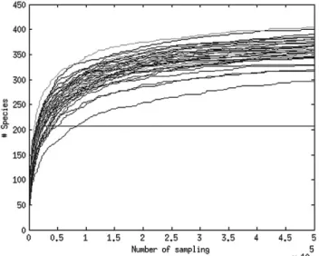
representative and used for being assigned to clade levels. RDP classifier [37] was used to classify the operational taxonomic units to bacterial taxa with a 95% confidence setting and taxonomic abundance profiles were generated for each sample. About 81.9% (SD. 4.9%) of the amplicons were assigned to known taxa successfully. A total of 217 genera and 74 families were identified. To observe the sampling depth sufficiency and metagenome coverage, rarefication curves were generated from species accumulation curves using subsampling without replacement (Fig. 1).
Fig. 1. Rarefaction curves of the sequenced 37 microbiome samples
Table I. Patients groups according to Blastocystis spp. growth and the presence of hepatic encephalopathy
Groups Blastocystis spp. growth Hepatic Encephalopathy Group 1 Blastocystis spp. positive negative
Group 2 Blastocystis spp. negative negative Group 3 Blastocystis spp. negative positive
Comparative analysis of metagenome samples
In order to capture the statistically meaningful microbiota composition differences, three main groups were compared in pairwise fashion (Table I). The tests were conducted at the clade levels of genus and family. Differentially abundant microbiota components were determined using LEfSe analysis [38].
Ethics statement
This study was approved by the Research Ethics Committee of Gazi University (date: 12/03/2014 and reference number 253) and written informed consent was obtained from all participants prior to the study. All the methods used in the study were carried out in accordance with the approved guidelines and Declaration of Helsinki, Ethical Principles for Medical Research Involving Human Subjects.
RESULTS
The mean age of the patients was 59.1 ± 13 (19-87) and 54.1% (n = 20) were female. The etiology of cirrhosis is shown in Fig. 2.
Fig. 2. Etiology of liver cirrhosis in the patients
included in the study. PBC: Primary Biliary Cirrhosis, Others (5) consist of non-alcoholic steatohepatitis (2), Budd-Chiari syndrome (1) and autoimmune hepatitis (2).
Blastocystis spp. were detected in 21.6% (n=8) of all
cirrhotic patients. When Blastocystis spp. were analyzed by subgroups, four of them were subtype 2, three were subtype 3 and one was subtype 1. Blastocystis spp. was not found in any of the patients with HE (none of 16 patients with either clinical or MHE); however, Blastocystis spp. were detected in 8 of 21 (38.1%) patients without HE (p = 0.006).
In none of the CTP class C patients, Blastocystis spp. were detected while 7 (36.8%) of the CTP A patients with mild cirrhosis and 1 (9.1%) of the CTP B patients were infected.
Likewise, when the patients were classified according to MELD score, Blastocystis spp. growth was 33.3% in the patients with score ≤ 8 and 6.7% in the patients with score 8-20. There was no growth in patients with score ≥20 (Table II).
The patients with encephalopathy were divided into subgroups as overt and minimal. The analyses were made according to these subgroups. There was a statistically significant difference between these groups and patients without encephalopathy for Blastocystis spp. growth (p=0.005 Pearson Chi-Square) (Table III). Also there was an inverse correlation between the presence of Blastocystis spp. and HE (Kendall’s correlation test, τ=-0.458, p=0.006) (Table IV). Decreasing trend in the frequency of Blastocystis spp. detection
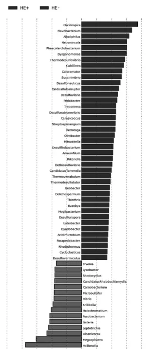
Fig. 3. Changes of some bacteria genera in cirrhotic patients with or
without encephalopathy sorted based on linear discriminant analysis scores.
Table III. Relationship of hepatic encephalopathy and Blastocystis spp. infection in cirrhotic patients
Hepatic encephalopathy Blastocystis spp. (-)
n (%) Blastocystis spp. (+) n (%)
Absent 13 (61.9%) 8 (38.1%)
Minimal 8 (100%) 0
Overt 8 100%) 0
Table II. Growth of Blastocystis spp. in patients classified according to MELD Score
MELD score n % Blastocystis spp. growth
≤8 21 56.8 33.3 %
8-20 15 40.5 6.7 %
≥20 1 2.7 0 %
was observed with cirrhotic stage progressing (τ= -0.366, p=0.021) (Table IV).
A percentage of 24.3% of the cirrhotic patients (n=9) use oral lactulose in order to reduce the effects of hyperammonemia. Lactulose use was significantly higher in the patients with a high CTP score (p=0.013) and MELD score (p=0.0001) than expected. Similarly, lactulose use was also significantly higher in the patients with HE (p=0.024). While there is such a relationship, there was not a significant relationship according to lactulose use between patients with or without Blastocystis spp. growth (p=0.649).
Profiles of bacteria populations of the cirrhotic patients are given in Fig. 3 when sorted according to the presence of HE.
These bacteria populations were determined according to density and diversity among three groups (Fig. 4). Increase in
the bacterial diversity was observed along with the absence of
Blastocystis. This connection was not statistically significant but
a trend could be seen. Lack of statistical significance is probably due to the low number of participants. This relationship could be detected in larger studies.
The groups were compared according to the two major phyla (Firmicutes and Bacteroidetes). The analysis showed that the rate of Bacteroidetes was higher when Blastocystis was absent and encephalopathy was arising (Kruskal-Wallis, p=0.03). On the other hand, there was no significant change in the Firmicutes (Kruskal-Wallis, p=0.11) (Fig. 5).
There was no correlation of Enterobactericeae with CTP and MELD scores (r = 0.1036, p-value= 0.547 and r = 0.1654, p= 0.33 respectively). However, we found that Enterobactericeae was enriched in HE patient samples (p= 0.0058). We observed a negative correlation of Ruminococcaceae with CTP and MELD (r = -0.3927, p= 0.018 and r = -0.3844, p= 0.02 respectively). At the same time, this relationship was also observed in
Ruminococaceae population between the analyzed groups (p=
0.0391, Kruskal-Wallis).
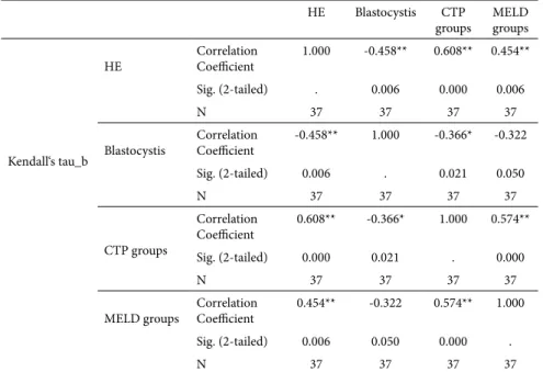
Table IV. Correlation between the specific groups.
HE Blastocystis CTP
groups MELD groups
Kendall‘s tau_b
HE Correlation Coefficient 1.000 -0.458** 0.608** 0.454** Sig. (2-tailed) . 0.006 0.000 0.006
N 37 37 37 37
Blastocystis Correlation Coefficient -0.458** 1.000 -0.366* -0.322 Sig. (2-tailed) 0.006 . 0.021 0.050 N 37 37 37 37 CTP groups Correlation Coefficient 0.608** -0.366* 1.000 0.574** Sig. (2-tailed) 0.000 0.021 . 0.000 N 37 37 37 37
MELD groups Correlation Coefficient 0.454** -0.322 0.574** 1.000 Sig. (2-tailed) 0.006 0.050 0.000 .
N 37 37 37 37
** Correlation is significant at the 0.01 level (2-tailed); * Correlation is significant at the 0.05 level (2-tailed). HE: hepatic encephalopathy.
Fi. 4. Bacterial density and diversity between the groups in Shannon
When bacterial abundance profiles were compared among the groups (Fig. 6), Alkaliphilus and Flavobacterium populations were found to be higher in Group 1. Conversely, Veillonella and
Streptococcus populations were lower in Group 1.
Lachnospiraceae population, especially Fecalibacterium,
was evaluated; analysis showed that there is no significant difference between the groups (Kruskal-Wallis, p= 0.0937).
However, there was not direct relationship between lactulose administration and Lactobacillus population (p= 0.11).
DISCUSSION
In this study we investigated the prevalence of Blastocystis spp. infection in cirrhotic patients and its correlation with HE and gut microbiota. The prevalence of Blastocystis spp. in all cirrhotic patients was 21.6%. Interestingly, Doğruman-Al et al. (2010) reported that the prevalence of Blastocystis spp. among healthy individuals in Ankara, Turkey was 11.6% [24]. In terms of subtypes of Blastocystis spp., subtype 2 was the most prevalent among the cohort in this study, followed by subtype 3. On the other hand, in 2008, Doğruman-Al et al. published a study about Blastocystis subtypes and their relationship with gastrointestinal symptoms [39]. It was found that subtype 3 was most common between symptomatic and asymptomatic groups, the second dominant genotype was subtype 1 in the symptomatic group and subtype 2 in the asymptomatic group [39]. It was suggested that subtype 1 is linked to elevated pathogenicity but subtype 2 is non-pathogenic [39]. As compared to our study, the domination of subtype 2 (mentioned as non-pathogenic) could play a key role in the configuration of the microbiota and development of HE in cirrhotic patients.
The relationship between Blastocystis and the bacterial component of microbiota has been investigated in a few cases, especially in the study by Nourisson et al. [40]. In this study, patients with Irritable Bowel Syndrome (IBS) were compared with controls. There was a significant difference between Blastocystis carriage and a negative correlation between Bifidobacteria and Blastocystis presence [40]. Also,
Faecalibacterium prausnitzii, which is an indicator of the
healthy gut [41], was found less often both in the Blastocystis-positive and IBS-C groups [40]. It was suggested that the decrease in Blastocystis presence could be related to the inflammatory environment. Change in microbiota, which has a key role in the protection against pathogens, could influence the presence of Blastocystis in the IBS patients. These data suggest that Blastocystis might have an impact on gut microbiota [42, 43]. Additionally, Blastocystis may cause changes in the composition of the gut microbiota by interacting with it [40]. Also, Verma et al. reported that the amount of the major genera of the gut microbiota was reduced after it was infected by Entamoeba histolytica [44].
In our study, we observed that the prevalence of Blastocystis spp. decreased as cirrhosis progressed. In cirrhotic patients, oral lactulose and antibiotic use increase with the progression of CTP stage and HE. It may reduce the incidence of Blastocystis spp. in gut microbiota by affecting the microbiota directly or indirectly. However, in our study there were no patients using antibiotics. Also, there was no significant relationship between the presence of Blastocystis spp. and oral lactulose use (p=0.379). Additionally, an increase in the bacterial diversity was observed along with the absence of Blastocystis. However, this was not statistically significant, probably due to the low number of participants. But this trend showed the effect of Blastocystis spp. on the microbiota. This knowledge contrasts with a recent study made by Audebert et al. [45], Fig. 6. Differences of bacterial populations between the groups.
These bacteria populations are considered as a part of healthy microbiota. These results support that the patients’ microbiotal environments were affected at a similar rate by the underlying disease.
We found that there were no differences in Lachnospiraceae abundance between the groups (r= 0.1668, p= 0.33 for CTP; r= 0.3133, p= 0.0628 for MELD). Ruminococaceae had a negative correlation with CTP score, MELD score, HE and the presence of Blastocystis spp. (r= -0.3927, p= 0.018 for CTP; r= -0.3844, p= 0.02 for MELD) (Fig. 7); Clostridiales Cluster XIV also had a weak negative correlation with CTP score (r= -0.3114, p= 0.0345) and MELD score (r= -0.2284, p= 0.018) (Fig. 7).
Although there was no significant difference of
Lactobacillus between the groups (Fig. 7), a negative correlation
who compared the bacterial diversity between Blastocystis-free and Blastocystis-colonized patients and found a higher bacterial diversity in Blastocystis-colonized patients. Sampling populations might be the reason of this difference. In our study, all the patients were cirrhotic. Our data suggests that the change in the bacterial diversity and the presence of Blastocystis could play a key role in the pathophysiology of HE and there is a need for further studies regarding this topic.
Dysbiosis has an important role in the pathogenesis of HE. Qin et al. observed that the MELD score was linked positively with Enterobacteriaceae and negatively with Ruminococcaceae [46]. In our study, we could not find a correlation between
Enterobactericeae with CTP and MELD scores. However,
there was a strong connection between Enterobactericeae and HE (p= 0.0058). In accordance with Qin’s study, we observed the negative correlation of Ruminococcaceae with CTP and MELD. At the same time, this relationship was also observed in Ruminococaceae population between the analyzed groups.
Populations of the other bacterial species were compared between the groups. Alkaliphilus and Flavobacterium population were higher in Group 1; on the other hand, the populations were becoming lower in Group 3. Conversely, when Veillonella and Streptococcus population were lower in Group 1, and higher in Group 2. The presence of Blastocystis spp. was variable between groups as correlated with these bacterial changes. Group 2 located between Group 1 and 3 was observed as a transition period.
Animal models are playing an important role in gathering information about the connection between HE, gut microbiota and Blastocystis spp. When considering changes in the gut microbiota and the high prevalence of Blastocystis spp. in IBS patients, Blastocystis spp. may be used as an indicator of the dysbiosis [40].
This hypothesis is supported by Scanlan & Stensvold [30].
Blastocystis was more common in patients with Prevotella and Ruminococcus enterotypes, on the other hand, it was seen rarely
in patients with Bacteroides enterotypes [31]. This relationship is not very clear, but likely a reduction in the bacterial variety in microbiota in association with an increase in a few bacterial species is a common situation in IBD patients [47, 48].
Bacterial taxa such as Lachnospiraceae, Ruminococcaceaea and Clostridiales Cluster XIV (consisting of some genera and species such as Fecaelibacterium) are often seen as the healthy microbiota [41, 49, 50]. Sokol et al. determined Fecalibacterium as an anti-inflammatory bacterium and with a potential to be a probiotic [41].
In our study, Lachnospiraceae population, especially
Fecalibacterium, was evaluated. The analysis showed that
there was no significant difference between the groups. This result may have occurred because of the sampling group. The sampling group consisted of cirrhotic patients and some of them had HE Therefore, the microbiotal environment was not healthy. This information also supports that the
Fecalibacterium population does not directly affect the presence
of encephalopathy. However, there will be more accurate data when compared to healthy individuals.
Ruminococaceae had a negative correlation with CTP
score, MELD score, HE and the presence of Blastocystis spp.;
Clostridiales Cluster XIV also had a weak negative correlation
with CTP score and MELD score.
Lactobacillus is used sometimes as a probiotic. And it
has been suggested that it reduces the strength of potentially harmful bacteria [51]. Lactulose administration increases the autochthonous bacteria such as Lactobacilllaceae [51]. Although there was not a significant difference of Lactobacillus between the groups, it was seen as a negative correlation Fig. 7. Population changes of Enterobacteriaceae, Ruminococaceae, Clostridiales Cluster XIV and Lactobacillus between
between Lactobacillus and Blastocystis spp. However, there was not a direct relationship between lactulose administration and
Lactobacillus population.
CONCLUSION
Because the major source of hyperammonia is the gut microbiota, there is a relationship between the gut microbiota and cirrhotic patients with HE. Our data suggests an inverse relationship between Blastocystis spp. and advanced stages of HE and that the structure and composition of gut microbiota significantly shifts. Thus, absence of Blastocystis spp. was associated with HE severity and dysbiosis in the gut microbiota. Further research is warranted to elucidate the potential role of this protist in HE.
Conflicts of interest: The authors declare no conflict of interest. Authors’ contributions: Y.S. planned the study, designed the patient
groups, sent samples to the laboratory, wrote the article. D.I. planned the study and wrote the article. D.F. examined microscopically the stool samples, isolated DNAs and edited the article. N.U. made the statistical analysis. U.D. made the Metagenomics analysis. S.F. collected the stool samples and isolated the DNAs. Y.S. edited the English typing.
Acknowledgement: Ö. Ufuk Nalbantoğlu was partially supported by
the Scientific and Technological Research Council of Turkey, Career Reintegration Grant TUBITAK-2232.
REFERENCES
1. Pinzani M, Rosselli M, Zuckermann M. Liver cirrhosis. Best Pract Res Clin Gastroenterol 2011; 25: 281-290. doi: 10.1016/j. bpg.2011.02.009
2. Felipo V. Hepatic encephalopathy: effects of liver failure on brain function. Nat Rev Neurosci 2013; 14: 851-858. doi: 10.1038/nrn3587
3. Kappus MR, Bajaj JS. Covert hepatic encephalopathy: not as minimal as you might think. Clin Gastroenterol Hepatol 2012; 10: 1208-1219. doi: 10.1016/j.cgh.2012.05.026
4. Zhan T, Stremmel W. The diagnosis and treatment of minimal hepatic encephalopathy. Dtsch Arztebl Int 2012; 109: 180-187. doi: 10.3238/ arztebl.2012.0180
5. Weissenborn K. Psychometric tests for diagnosing minimal hepatic encephalopathy. Metab Brain Dis 2013: 227-229. doi: 10.1007/s11011-012-9336-4
6. Pugh RN, Murray-Lyon IM, Dawson JL, Pietroni MC, Williams R. Transection of the oesophagus for bleeding oesophageal varices. Br J Surg 1973; 60: 646-649. doi: 10.1002/bjs.1800600817
7. de Franchis R, Primignani M. Why do varices bleed? Gastroenterol Clin North Am 1992; 21: 85-101.
8. Wiesner RH. Evidence-based evolution of the MELD/PELD liver allocation policy. Liver Transpl 2005; 11: 261-263. doi: 10.1002/ lt.20362
9. Garcovich M, Zocco MA, Roccarina D, Ponziani FR, Gasbarrini A. Prevention and treatment of hepatic encephalopathy: focusing on gut microbiota. World J Gastroenterol 2012; 18: 6693-6700. doi: 10.3748/ wjg.v18.i46.6693
10. Chen Y, Yang F, Lu H, et al. Characterization of fecal microbial communities in patients with liver cirrhosis. Hepatology 2011; 54: 562-572. doi: 10.1002/hep.24423
11. Rai R, Saraswat VA, Dhiman RK. Gut microbiota: its role in hepatic encephalopathy. J Clin Exp Hepatol 2015; 5(Suppl 1): S29-S36. doi:
10.1016/j.jceh.2014.12.003
12. Fraher MH, O’Toole PW, Quigley EMM. Techniques used to characterize the gut microbiota: a guide for the clinician. Nat Rev Gastroenterol Hepatol 2012; 9: 312-322. doi: 10.1038/nrgastro.2012.44
13. Goel A, Gupta M, Aggarwal R. Gut microbiota and liver disease. J Gastroenterol Hepatol. 2014; 29: 1139-1148. doi: 10.1111/jgh.12556
14. Scanlan PD, Stensvold CR, Rajilić-Stojanović M, et al. The microbial eukaryote Blastocystis is a prevalent and diverse member of the healthy human gut microbiota. FEMS Microbiol Ecol 2014; 90: 326-330. doi:
10.1111/1574-6941.12396
15. Silberman JD, Sogin ML, Leipe DD, Clark CG. Human parasite finds taxonomic home. Nature 1996; 380: 398. doi: 10.1038/380398a0
16. Stenzel DJ, Boreham PF. Blastocystis hominis revisited. Clin Microbiol Rev 1996; 9: 563-584.
17. Tan KS. New insights on classification, identification, and clinical relevance of Blastocystis spp. Clin Microbiol Rev 2008; 21: 639-665. doi: 10.1128/CMR.00022-08
18. Suresh K, Venilla GD, Tan TC, Rohela M. In vivo encystation of Blastocystis hominis. Parasitol Res 2009; 104: 1373-1380. doi: 10.1007/ s00436-009-1340-1
19. Alfellani MA, Jacob AS, Perea NO, et al. Diversity and distribution of Blastocystis sp. subtypes in non-human primates. Parasitology 2013; 140: 966-971. doi: 10.1017/S0031182013000255
20. Hirata T, Nakamura H, Kinjo N, et al. Prevalence of Blastocystis hominis and Strongyloides stercoralis infection in Okinawa, Japan. Parasitol Res 2007; 101: 1717-1719. doi: 10.1007/s00436-007-0712-7
21. Wong KH, Ng GC, Lin RT, Yoshikawa H, Taylor MB, Tan KS. Predominance of subtype 3 among Blastocystis isolates from a major hospital in Singapore. Parasitol Res 2008; 102: 663-670. doi: 10.1007/ s00436-007-0808-0
22. Aguiar JI, Gonçalves AQ, Sodré FC, et al. Intestinal protozoa and helminths among Terena Indians in the State of Mato Grosso do Sul: high prevalence of Blastocystis hominis. Rev Soc Bras Med Trop 2007; 40: 631-634. doi: 10.1590/S0037-86822007000600006
23. Rayan HZ, Ismail OA, El Gayar EK. Prevalence and clinical features of Dientamoeba fragilis infections in patients suspected to have intestinal parasitic infection. J Egypt Soc Parasitol 2007; 37: 599-608.
24. Dogruman-Al F, Simsek Z, Boorom K, et al. Comparison of methods for detection of Blastocystis infection in routinely submitted stool samples, and also in IBS/IBD Patients in Ankara, Turkey. PLoS One 2010; 5: e15484. doi: 10.1371/journal.pone.0015484
25. Puthia MK, Sio SW, Lu J, Tan KS. Blastocystis ratti induces contact-independent apoptosis, F-actin rearrangement, and barrier function disruption in IEC-6 cells. Infect Immun 2006; 74: 4114-4123. doi:
10.1128/IAI.00328-06
26. Puthia MK, Lu J, Tan KS. Blastocystis ratti contains cysteine proteases that mediate interleukin-8 response from human intestinal epithelial cells in an NF-kappaB-dependent manner. Eukaryot Cell 2008; 7: 435-443. doi: 10.1128/EC.00371-07
27. Tan KS, Mirza H, Teo JD, Wu B, Macary PA. Current Views on the Clinical Relevance of Blastocystis spp. Curr Infect Dis Rep 2010; 12: 28-35. doi: 10.1007/s11908-009-0073-8
28. Mirza H, Wu Z, Teo JD, Tan KS. Statin pleiotropy prevents rho kinase-mediated intestinal epithelial barrier compromise induced by
Blastocystis cysteine proteases. Cell Microbiol 2012; 14: 1474-1484. doi:
10.1111/j.1462-5822.2012.01814.x
29. Scanlan PD. Blastocystis: past pitfalls and future perspectives. Trends Parasitol 2012; 28: 327-334. doi: 10.1016/j.pt.2012.05.001
30. Scanlan PD, Stensvold CR. Blastocystis: getting to grips with our guileful guest. Trends Parasitol 2013; 29: 523-529. doi: 10.1016/j.pt.2013.08.006
31. Andersen LO, Bonde I, Nielsen HB, Stensvold CR. A Retrospective Metagenomics Approach to Studying Blastocystis. FEMS Microbiol Ecol 2015; 91: fiv072. doi: 10.1093/femsec/fiv072
32. Garcia LS. Commonly Asked Questions: Diagnostic Parasitology. 2012. 33. Suresh K, Smith H. Comparison of methods for detecting Blastocystis
hominis. Eur J Clin Microbiol Infect Dis 2004; 23: 509-511. doi: 10.1007/ s10096-004-1123-7
34. Scicluna SM, Tawari B, Clark CG. DNA barcoding of blastocystis. Protist 2006; 157: 77-85. doi: 10.1016/j.protis.2005.12.001
35. Stensvold CR. Blastocystis: Genetic diversity and molecular methods for diagnosis and epidemiology. Trop Parasitol 2013; 3: 26-34. doi:
10.4103/2229-5070.113896
36. Edgar RC. UPARSE: highly accurate OTU sequences from microbial amplicon reads. Nat Methods 2013; 10: 996-998. doi: 10.1038/ nmeth.2604
37. Wang Q, Garrity GM, Tiedje JM, Cole JR. Naive Bayesian classifier for rapid assignment of rRNA sequences into the new bacterial taxonomy. Appl Environ Microbiol 2007; 73: 5261-5267. doi: 10.1128/AEM.00062-07
38. Paulson JN, Stine OC, Bravo HC, Pop M. Differential abundance analysis for microbial marker-gene surveys. Nat Methods 2013; 10: 1200-1202. doi: 10.1038/nmeth.2658
39. Dogruman-Al F, Dagci H, Yoshikawa H, Kurt O, Demirel M. A possible link between subtype 2 and asymptomatic infections of Blastocystis hominis. Parasitol Res 2008; 103: 685-689. doi: 10.1007/s00436-008-1031-3
40. Nourrisson C, Scanzi J, Pereira B, et al. Blastocystis ıs associated with decrease of fecal microbiota protective bacteria: comparative analysis between patients with ırritable bowel syndrome and control subjects. PLoS One 2014; 9: e111868. doi: 10.1371/journal.pone.0111868
41. Sokol H, Pigneur B, Watterlot L, et al. Faecalibacterium prausnitzii is an anti-inflammatory commensal bacterium identified by gut microbiota analysis of Crohn disease patients. Proc Natl Acad Sci U S A 2008; 105: 16731-16736. doi: 10.1073/pnas.0804812105
42. Poirier P, Wawrzyniak I, Vivarès CP, Delbac F, El Alaoui H. New insights into Blastocystis spp.: a potential link with irritable bowel syndrome. PLoS Pathog 2012; 8: e1002545. doi: 10.1371/journal.ppat.1002545
43. Denoeud F, Roussel M, Noel B, et al. Genome sequence of the stramenopile Blastocystis, a human anaerobic parasite. Genome Biol 2011; 12: R29. doi: 10.1186/gb-2011-12-3-r29
44. Verma AK, Verma R, Ahuja V, Paul J. Real-time analysis of gut flora in Entamoeba histolytica infected patients of Northern India. BMC Microbiol 2012; 12: 183. doi: 10.1186/1471-2180-12-183
45. Audebert C, Even G, Cian A, et al. Colonization with the enteric protozoa Blastocystis is associated with increased diversity of human gut bacterial microbiota. Sci Rep 2016; 6: 25255. doi: 10.1038/srep25255
46. Qin N, Yang F, Li A, et al. Alterations of the human gut microbiome in liver cirrhosis. Nature 2014; 513: 59-64. doi: 10.1038/nature13568
47. Wohlgemuth S, Keller S, Kertscher R, et al. Intestinal steroid profiles and microbiota composition in colitic mice. Gut Microbes 2011; 2: 159-166. doi: 10.4161/gmic.2.3.16104
48. Smith PM, Howitt MR, Panikov N, et al. The microbial metabolites, short-chain fatty acids, regulate colonic Treg cell homeostasis. Science 2013; 341: 569-73. doi: 10.1126/science.1241165
49. Nava GM, Stappenbeck TS. Diversity of the autochthonous colonic microbiota. Gut Microbes 2011; 2: 99-104. doi: 10.4161/gmic.2.2.15416
50. Zoetendal EG, Ben-Amor K, Harmsen HJ, Schut F, Akkermans AD, de Vos WM. Quantification of uncultured Ruminococcus obeum-like bacteria in human fecal samples by fluorescent in situ hybridization and flow cytometry using 16S rRNA-targeted probes. Appl Environ Microbiol 2002;68:4225-4232. doi: 10.1128/AEM.68.9.4225-4232.2002
51. Riggio O, Varriale M, Testore GP, et al. Effect of lactitol and lactulose administration on the fecal flora in cirrhotic patients. J Clin Gastroenterol 1990; 12: 433-436.