TOPIC HIGHLIGHT
Ahmet Bilici, Department of Medical Oncology, Medical Fac-ulty, Istanbul Medipol University, 34214 Bagcilar, Istanbul, Turkey
Author contributions: Bilici A designed and wrote the high-light topic.
Correspondence to: Ahmet Bilici, MD, Department of Medi-cal Oncology, MediMedi-cal Faculty, Istanbul Medipol University, TEM Avrupa Otoyolu Goztepe Cikisi No: 1, 34214 Bagcilar, Istanbul, Turkey. [email protected]
Telephone: +90-532-5280486 Fax:+90-216-4422947 Received: October 26, 2013 Revised: January 27, 2014 Accepted: April 8, 2014
Published online: August 21, 2014
Abstract
The prognosis in patients with pancreatic cancer is poor and this cancer is the fourth leading cause of can-cer-related death worldwide. Although surgical resec-tion is the only curative treatment of choice for pan-creatic cancer, the majority of patients are diagnosed at an advanced stage, thus only 10%-15% of them are suitable for curative resection and the overall survival is less than 5%. Chemotherapy for metastatic disease is to palliate symptoms of patients and to improve sur-vival. Therefore, prognostic factors are important and a correct definition of poor prognostic factors may help to guide more aggressive adjuvant or aggressive treat-ment protocols in patients with pancreatic cancer. This article reviews the prognostic factors affecting survival of patients with pancreatic cancer in the light of recent advances in the literature.
© 2014 Baishideng Publishing Group Inc. All rights reserved. Key words: Pancreatic cancer; Prognostic factors;
Sur-vival; Carbohydrate antigen 19-9; Treatment
Core tip: The overall prognosis associated with
pancre-atic cancer has not improved over the last 20 years, even if new diagnostic and therapeutic strategies have emerged. Thus, investigations on predictive factors in pancreatic cancer are needed because these fac-tors should have predictive value in relation to longer survival after surgery than after palliative treatment. Prognostic factors are important and a correct defini-tion of poor prognostic factors may help to guide more aggressive adjuvant or aggressive treatment protocols in patients with pancreatic cancer.
Bilici A. Prognostic factors related with survival in patients with pancreatic adenocarcinoma. World J Gastroenterol 2014; 20(31): 10802-10812 Available from: URL: http://www.wjgnet. com/1007-9327/full/v20/i31/10802.htm DOI: http://dx.doi. org/10.3748/wjg.v20.i31.10802
INTRODUCTION
Pancreatic adenocarcinoma still remains a major public health issue and is the fourth leading cause of
cancer-related death worldwide[1]. Although surgical resection is
the only curative treatment of choice for pancreatic can-cer, unfortunately, the majority of patients are diagnosed at an advanced stage, and thus only 10%-15% of them are suitable for curative resection and the overall survival
is less than 5%[2,3]. Chemotherapy is used in the adjuvant
setting and in the treatment of locally advanced inoper-able and metastatic disease.
The primary goals of chemotherapy for metastatic
disease are palliation and improved survival[4,5].
There-fore, identifying poor prognostic factors that may pre-dict the tumor recurrence and prognosis of patients is important for selecting appropriate treatment protocols. So it is important to determine new biological or patho-logical indicators related to survival in addition to
well-WJG 20th Anniversary Special Issues (14): Pancreatic cancer
Prognostic factors related with survival in patients with
pancreatic adenocarcinoma
Ahmet Bilici
known prognostic factors such as clinical and
pathologi-cal stage, performance status, and surgipathologi-cal margin[6]. In
this article, the prognostic factors affecting survival of patients with pancreatic cancer were reviewed.
SURGICAL AND PATHOLOGICAL
FACTORS
The primary surgical or pathological factors that influ-ence prognosis are whether the tumor is localized at the pancreas and whether the tumor has spread to lymph
nodes or distant organs[1] because the highest cure rate
occurs if the tumor is truly localized to the pancreas. In the present TNM staging system, tumor size, peripan-creatic extension, and vascular involvement are used. Traditionally, TNM staging, especially in the presence of metastasis (advanced stage), has been found to be an important prognostic factor in patients with pancreatic cancer for survival[7-9].
Surgical margin
Surgical resection is the only potentially curative option for treatment of pancreatic cancer and the nature of sur-gery for resectable tumors depends on the tumor local-ization and size. The incidence of R1 resection has been indicated as being 20% in the literature, but the improve-ment of pathological work-up procedures has increased
the rate of R1 resection up to 80%[10,11]. Menon et al[12]
reported that of 27 patients with pancreatic cancer, 22 patients underwent R1 resection and the median sur-vival rate for patients with R1 resection was significantly
worse than that of patients with R0 resection (14 mo vs
not reached). In a study performed by Raut et al[13], they
reported that the rate of R1 resection was 16.7% and patients who underwent an R1 resection had a median overall survival (OS) of 21.5 mo compared with 27.8 mo in patients who underwent an R0 resection. In addi-tion, multivariate analysis showed that high mean opera-tive blood loss and large tumor size were independent predictors of an R1 resection, but margin status did not independently influence survival.
Another study including 265 pancreatic carcinoma pa-tients who had undergone surgical resection reported that R1 resection in 49 patients (51%) and R2 resection in four
patients (4%) were performed[14]. The R1-positive margin
was localized at the retroperitoneal resection margin in 76% and at the trans-section margin in 14% of tumors. Median survival time was better in R0-resected patients
compared with R1-resected patients (22 mo vs 15 mo).
A positive resection margin after pancreatic resection is considered to be a poor prognostic factor, and some have proposed that an R1 margin may be a biologic predictor of more aggressive disease. On the other hand, whether these patients with pancreatic carcinoma who underwent margin-positive resection have to be managed with ag-gressive treatment modalities has not been described.
Lymph nodes status and lymph node ratio
Lymph node ratio (LNR) may be more useful than nodal (N) status in prognostic subclassification of pancreatic adenocarcinomas after pancreatoduodenectomy. Recent studies have suggested that LNR may also be an
impor-tant prognostic factor in pancreatic cancer[15-17]. In the
TNM staging system, the number of resected lymph nodes may be very important, but node-positive patients are not a homogenous group, because stage migration may occur in resected pancreatic cancer patients. To re-solve these limitations, recently LNR was proposed as a new prognostic factor by several authors to prevent the ‘stage migration’ phenomenon[15-17]. Riediger et al[17], in 204
resected patients, reported that LNR was the strongest predictor of survival and they concluded that the routine estimation of the LNR may be helpful not only for the individual prediction of prognosis but also for the indi-cation of adjuvant therapy. The analysis of Surveillance, Epidemiology, and End Results and MGH (Massachusetts General Hospital) in 10254 and 827 resected patients, respectively, showed that higher LNR (> 0.2) was as-sociated with worse survival by univariate analysis, and in addition the hazard ratio (HR) raised proportionally when more lymph nodes were examined in multivariate analysis. This study concluded that while the contribu-tion of the number of positive nodes to survival was relatively small, LNR was strongly associated with sur-vival, and thus, LNR provided a stronger and more ac-curate predictor of survival than the number of positive nodes[18].
Perineural and blood vessel invasion
Both perineural (PNI) and blood vessel invasion (BVI) have been previously investigated in patients with pan-creatic cancer and found to be important prognostic indicators for survival[14,19,20]. Lee et al[19] showed that PNI
was an important adverse prognostic factor for patients with surgical resection, as was pN stage. In a study
per-formed by Chatterjee et al[21], PNI and BVI were found
to be associated with the OS and lymph node status in patients who were treated with neoadjuvant treatment. The median OS for patients with PNI was worse than
that of patients without PNI (22 mo vs 36 mo).
More-over, the median OS was better in patients without BVI
compared with patients with BVI (34 mo vs 22 mo).
They detected that retroperitoneal resection margin was correlated with the presence of both BVI and PNI. The authors concluded that PNI and BVI were significantly poor prognostic indicators.
Tumor localization
Some studies have investigated the prognostic signifi-cance of tumor localization in pancreatic signifi-cancer patients, but there is currently no consensus[7-9,19,22]. In a study
per-formed by Park et al[8], univariate analysis indicated that
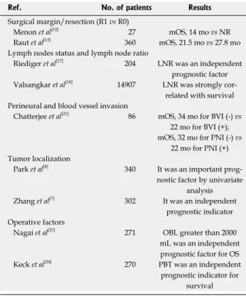
Table 1 Surgical and pathological factors in pancreatic cancer
OS, but the significance of tumor site as an independent prognostic indicator could not be proved in the
multi-variate analysis. Lee et al[22] showed that high CEA level
was significantly correlated with tumor location. In the patients with elevated CEA level, tumors were located mostly at the pancreas body and tail. The authors could not show that tumor location was a prognostic factor by multivariate analysis, although in the univariate analysis it was detected as being a prognostic factor. However, in
another study carried out by Zhang et al[7], localization
of the primary tumor was found to be an independent prognostic factor. In other words, the mortality risk was increased for tumors located at the body and tail of the pancreas compared to the tumors located at the head and neck of the pancreas.
Operative factors
An influence of operative blood loss (OBL) on survival in patients with pancreatic cancer after curative
resec-tion has been investigated. Nagai et al[23] retrospectively
analyzed 271 patients and found that the OS was sig-nificantly affected by the amount of OBL. The median survival times were 26.0, 15.3, and 8.7 mo for OBL less than 1000, 1000 to 2000, and greater than 2000 mL,
respectively (< 1000 mL vs 1000-2000 mL, p = 0.019;
1000-2000 mL vs > 2000 mL, p < 0.0001). Moreover,
OBL greater than 2000 mL was also detected to be an independent prognostic factor in multivariate analysis
(HR = 2.55) and OBL of 2010 mL was found to be an appropriate cut-off level to predict early mortality within 6 mo after resection. Male sex, year of resection, and plexus invasion were independently associated with OBL greater than 2000 mL. In light of these results, the authors concluded that excessive OBL was found to be a prognostic determinant of survival and it can be used to stratify the risk for pancreatic cancer mortality after surgery for pancreatic cancer. On the other hand, prog-nostic significance of perioperative blood transfusion (PBT) has also been reported. In a study performed by
Keck et al[24], PBTs were given in 46% of 270 pancreatic
cancer patients. Univariate analysis showed that PBT was related with poorer survival, as were positive margins, more than one involved node, and poorer grading. In addition, they found that PBT was an independent prog-nostic indicator for survival by multivariate analysis after resection. The authors thought that impact of PBT was independent of the perioperative complications or resec-tion type. Table 1 shows selected trials of surgical and pathological prognostic factors in pancreatic cancer.
CLINICAL FACTORS
Performance status
Some studies have evaluated the impact of performance status (PS) on survival for patients with pancreatic ad-enocarcinoma, but the results are conflicting. In a study
carried out by Sezgin et al[25], the authors reported that
only PS was an independent prognostic factor for OS in patients with advanced pancreatic cancer. Similarly, Tas et al[26] found that initial poor PS (PS 2-4) was
sig-nificantly associated with worse survival for patients with all stages of pancreatic cancer. In addition, poor PS remained as an independent prognostic indicator for survival by multivariate analysis and in patients with poor PS, severe weight loss (> 10%), large tumor di-ameter (> 3 cm), and especially metastatic disease was related with significantly shorter OS. On the other hand, in another study, although an influence of PS on survival was detected in the univariate analysis, its prognostic significance was lost in multivariate analysis[8]. Lee et al[22]
showed that in the elevated CA19-9 level group (≥ 37
U/mL), PS was significantly higher compared with the
normal CA19-9 group. Furthermore, PS (0 vs 1-2) was
found to be an important prognostic factor in the uni-variate analysis for OS.
Diabetes mellitus, obesity and jaundice
Diabetes mellitus (DM) is commonly diagnosed in pancre-atic cancer patients, but the significance of new-onset DM as a cause of underlying pancreatic cancer is unknown. Some studies have investigated the prognostic significance
of DM in pancreatic cancer[8,25,27], but an impact of DM
on survival could not be proved.
Cachexia is a known characteristic of pancreatic can-cer with detects as 80% of patients cachexic at diagnosis. Therefore, measurement of body mass index (BMI) at
Ref. No. of patients Results
Surgical margin/resection (R1 vs R0)
Menon et al[12] 27 mOS, 14 mo vs NR
Raut et al[13] 360 mOS, 21.5 mo vs 27.8 mo
Lymph nodes status and lymph node ratio
Riediger et al[17] 204 LNR was an independent
prognostic factor Valsangkar et al[18] 14907 LNR was strongly
cor-related with survival Perineural and blood vessel invasion
Chatterjee et al[21] 86 mOS, 34 mo for BVI (-) vs
22 mo for BVI (+); mOS, 32 mo for PNI (-) vs
22 mo for PNI (+) Tumor localization
Park et al[8] 340 It was an important
prog-nostic factor by univariate analysis Zhang et al[7] 302 It was an independent
prognostic indicator Operative factors
Nagai et al[23] 271 OBL greater than 2000
mL was an independent prognostic factor for OS Keck et al[24] 270 PBT was an independent
prognostic indicator for survival mOS: Median overall survival; NR: Not reach; LNR: Lymph node ratio; BVI: Blood vessel invasion; PNI: Perineural invasion; OBL: Operative blood loss; PBT; Perioperative blood transfusion.
the time of diagnosis does not provide accurate
repre-sentation of a patient’s long-term exposure to obesity[28].
However, some studies have shown that high BMI is associated with increased risk of pancreatic cancer
inci-dence and mortality[29,30]. On the other hand, studies of
obesity and survival in patients with pancreatic cancer are notably controversial. In a population-based study
in-cluding 510 patients with pancreatic cancer, Gong et al[31]
indicated that elevated HR of 1.3 was detected for obese
(BMI ≥ 30) compared with normal range BMI (< 25)
patients. But, the relation between OS and BMI could not be found. Similarly, recent study evaluated the as-sociation of BMI with the risk of death from pancreatic cancer in a pooled analysis of data from Asia Cohort
Consortium[32]. It did not support an relation between
BMI and risk of death from pancreatic cancer. As a
dif-ferent these studies, in a study carried out by Yuan et al[33]
the association of prediagnostic BMI with pancreatic cancer survival was analyzed. Higher prediagnostic BMI was associated with more advanced stage at diagnosis, with 72.5% of obese patients presenting with metastatic disease versus 59.4% of healthy-weight patients. Fur-thermore, higher baseline BMI was associated with re-duced survival. HR for death was 1.53, comparing BMI
≥ 35 kg/m2 with BMI < 25 kg/m2 (p = 0.001).
In a study performed by Smith et al[34], the presence
of preoperative jaundice was found to be associated with poor survival in patients with pancreatic cancer. Another study showed that preoperative jaundice was the only independent prognostic factor for pancreatic cancer pa-tients[19]. On the other hand, Perini et al[35] demonstrated
that both preoperative DM and jaundice had no adverse effect on survival for curative resection in pancreatic
cancer patients. Recently, Strasberg et al[36] analyzed 400
patients with resected pancreatic cancer, and preopera-tive jaundice was found to be a significant indicator of poor outcome in the multivariate analysis. Moreover, the relationship was detected between jaundice and nodal status, and jaundiced patients who underwent preopera-tive stenting had a survival advantage. The underlying mechanism related with the influence of jaundice on survival is unknown and additional studies are required to determine the exact mechanism for this effect.
Treatment and gemcitabine
Chemotherapy is only modestly effective in advanced dis-ease but has a significant impact in the adjuvant setting, with 5-fluorouracil and gemcitabine both having efficacy in a subgroup of patients and increasing 5-year survival
from 10%-15% with surgery alone to 20%-25%[37-40].
Park et al[8] analyzed 340 patients with pancreatic cancer
and of 141 stage Ⅲ patients, 57 received supportive
care (BSC) only, 25 received chemotherapy (CT), and 59 received concurrent chemoradiotherapy (CCRT); of the
199 stage Ⅳ patients, 119 were treated with BSC only
and 80 received CT. Univariate analysis showed that CT and CCRT were significant prognostic indicators for OS
in stage Ⅲ patients compared with patients that received
BSC only (11.3 mo vs 10.4 mo vs 6.4 mo, respectively; p
< 0.001). Similarly, in stage Ⅳ patients, median OS for patients who were treated with CT was significantly bet-ter than that of patients who received BSC only (6.4 mo vs 3.1 mo, p < 0.001). In addition, initial treatment effect remained an independent prognostic factor compared to
BSC only in the multivariate analysis[8].
In a study performed by Lee et al[19], gemcitabine
chemotherapy was found to be the only independent prognostic indicator for OS in advanced or unresectable pancreatic cancer patients who had undergone palliative
surgical by pass. Moreover, Zhang et al[7] evaluated 302
all-stage pancreatic cancer and found that the median OS of patients who did not receive any treatment or those treated with BSC only was 1.3 mo, while the me-dian OS for patients who had undergone surgery, CT, biliary drainage therapy, arterial interventional CT, and comprehensive CT was 11.0, 7.3, 3.5, 9.0, and 11.0 mo,
respectively (p < 0.05). In the multivariate analysis, the
presence of treatment vs no therapy or BSC only was an
independent prognostic factor (HR = 13.93, p = 0.000).
However, platinum combination CT was significantly as-sociated with improved OS compared to non-platinum
CT regimen (HR = 0.56, p = 0.011). Selected trials
re-lated with clinical prognostic factors are summarized in Table 2.
LABORATORY AND MOLECULAR
FACTORS
Prognostic role of carbohydrate antigen 19-9 levels
Serum carbohydrate antigen (CA) 19-9, the sialylated Lewis blood group antigen defined by the monoclonal antibody 1116 NS 19-9, is a tumor-associated antigen
synthesized by normal pancreatic and ductal cells[41].
CA19-9 is considered to be the standard serum marker of pancreatic cancer due to its high sensitivity of 70%-90%
and specificity of around 90%[42]. Serum CA19-9 levels
have been found to be a useful tumor marker in differ-entiating benign from malignant pancreatic lesions, and
to monitor tumor response to treatment[42,43]. Previous
studies suggested that preoperative CA19-9 levels could
predict the resectability of pancreatic cancer[44,45], and
other studies reported that pretreatment CA19-9 level was an important prognostic factor in patients with
pan-creatic cancer who received CT or CCRT[8,9,45,46].
Park et al[8] reported that elevated CA19-9 levels (>
670 U/mL) were found to have prognostic significance for OS by univariate analysis, while it was an indepen-dent prognostic factor for OS in the multivariate analysis. Furthermore, another study found similar findings. The median OS time for patients with high CA19-9 level was worse than that of patients with normal CA19-9 level (3.8 vs 5.0 mo), which was not significant, but multivariate analysis indicated that it was an independent prognostic
indicator for OS (HR = 4.54, p = 0.033)[7]. Recently, in a
study by Humphris et al[47], low postoperative CA19-9 at
indepen-Table 2 Clinical prognostic factors in pancreatic cancer in selected trials
dent prognostic factors (median OS; 25.6 mo vs 14.8 mo,
p = 0.0052) in 260 patients with pancreatic cancer who underwent surgical resection. Patients with postoperative CA19-9 levels > 90 U/mL did not benefit from adju-vant chemotherapy compared with those with a CA19-9
level of ≤ 90 U/mL (median OS 26.0 mo vs 16.7 mo,
p = 0.0108). Normalization of CA19-9 within 6 mo of resection was also an independent favorable prognostic
factor (median OS: 29.9 mo vs 14.8 mo, p = 0.0004) and
normal perioperative CA19-9 levels were identified as being a good prognostic group, which was associated with a 5-year survival of 42%.
Other tumor markers
Carcinoembryonic antigen (CEA) is the standard tumor marker and is commonly used for predicting treatment response and prognosis of patients with colorectal
cancer[48]. In contrast to the CA19-9 level, an impact of
CEA on survival of pancreatic cancer patients has not yet been determined, but CEA might be beneficial in
predicting pancreatic cancer. Zhang et al[7] in their study
including 302 patients with pancreatic cancer reported that the patients with high CEA levels had a median sur-vival of 2.0 mo compared to patients with normal levels (5.0 mo). This difference was statistically significant (HR
= 1.43, p = 0.030). However, the significance of CEA
levels as an independent prognostic factor could not be proved in the multivariate analysis. In a study carried out
by Lee et al[22], they retrospectively analyzed 187
pancre-atic cancer patients, and reported that the median OS time for patients with normal CEA levels was signifi-cantly better than that of patients with high CEA levels
(16.3 mo vs 10.2 mo, p = 0.004). In addition, elevated
CEA levels were found to be an independent prognostic factor in the multivariate analysis.
Despite these findings, to detect whether CEA can be applicable as a prognostic marker of pancreatic can-cer, it should be evaluated in a large number of patients with all stages of pancreatic cancer. Various tumor
mark-ers such as CA125, CA15-3, CA72-4, and CA242 have also been analyzed, but their importance as independent prognostic indicators could not be definitively demon-strated[7,49].
Hematological parameters
Platelet, lymphocyte, and neutrophil counts, mean platelet volume, and the ratios of various hematologic cells have been shown to be valuable prognostic factors in various malignancies, such as renal, gynecological, and colorectal
cancers[50-53]. Schwarz et al[54] demonstrated that
preop-erative platelet count predicts survival after resection of pancreatic adenocarcinoma. On the other hand, in a study comprising 205 patients performed by Domínguez et al[55], there was no evidence to support preoperative
platelet count as either an adverse or favorable prognostic factor in pancreatic ductal adenocarcinoma, which was
not compatible with a study of Zhang et al[7]. Despite
conflicting results regarding platelet counts, white blood cells (WBCs) were found to be an independent prog-nostic factor for OS in patients with pancreatic cancer
in two studies[7,46]. Although low hemoglobin levels were
associated with poorer OS time, the significance as an in-dependent prognostic marker could not be proved by the multivariate analysis[7].
The prognostic value of pretreatment platelet to lym-phocyte ratio (PLR) and neutrophil to lymlym-phocyte ratio (NLR) in patients with pancreatic cancer has also been
evaluated[56,57]. Preoperative PLR has been defined as an
independent significant prognostic marker by Smith et al[58]
in resected pancreatic ductal adenocarcinoma. In the same study, the median overall survival in patients with a PLR of 150 or less was 19.7 mo, 13.7 mo in those with a PLR of 151-300, and 5.8 mo in patients with a value of
> 300. Aliustaoglu et al[57] showed that there was no
sta-tistically significant difference between cases with PLR
values ≤ 160 and > 160. However, they analyzed NLR
in the same patients with pancreatic cancer. Patients with a NLR value of < 5 had a significantly higher median
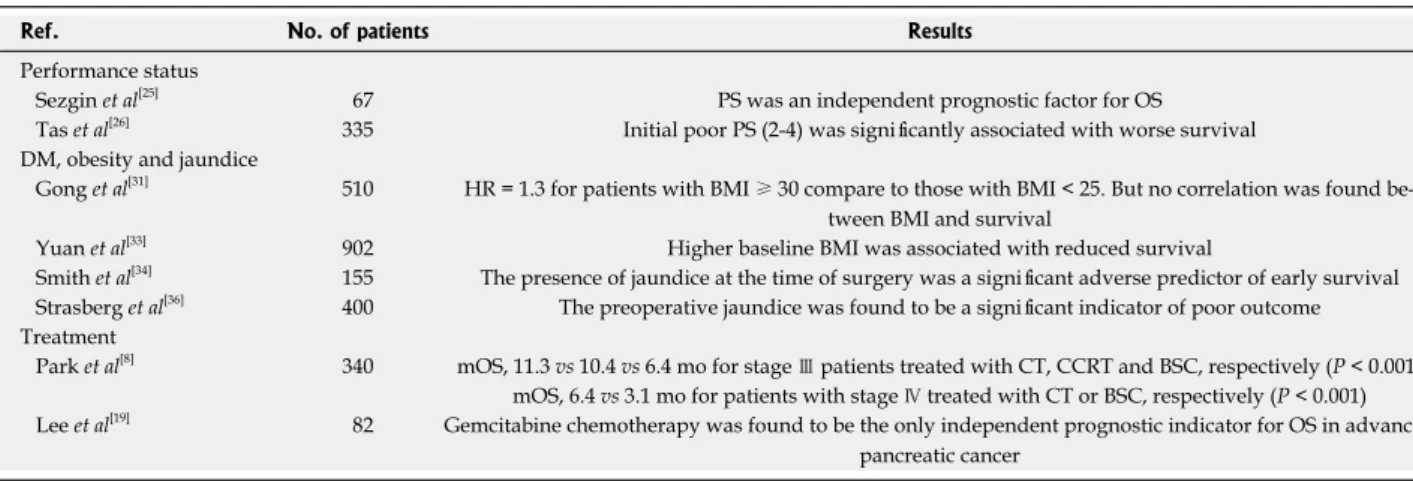
Ref. No. of patients Results
Performance status
Sezgin et al[25] 67 PS was an independent prognostic factor for OS
Tas et al[26] 335 Initial poor PS (2-4) was significantly associated with worse survival
DM, obesity and jaundice
Gong et al[31] 510 HR = 1.3 for patients with BMI ≥ 30 compare to those with BMI < 25. But no correlation was found
be-tween BMI and survival
Yuan et al[33] 902 Higher baseline BMI was associated with reduced survival
Smith et al[34] 155 The presence of jaundice at the time of surgery was a significant adverse predictor of early survival
Strasberg et al[36] 400 The preoperative jaundice was found to be a significant indicator of poor outcome
Treatment
Park et al[8] 340 mOS, 11.3 vs 10.4 vs 6.4 mo for stage Ⅲ patients treated with CT, CCRT and BSC, respectively (P < 0.001)
mOS, 6.4 vs 3.1 mo for patients with stage Ⅳ treated with CT or BSC, respectively (P < 0.001) Lee et al[19] 82 Gemcitabine chemotherapy was found to be the only independent prognostic indicator for OS in advanced
pancreatic cancer
DM: Diabetes mellitus; mOS: Median overall survival; PS:Performance status, BMI: Body mass index; CT: Chemotherapy; CCRT: Concurrent chemoradio-therapy; BSC: Best supportive care.
OS time compared to those with a NLR value of ≥ 5
(p = 0.015). Recently, Stotz et al[56] evaluated NLR in 371
patients with primary operable and inoperable pancreatic
cancer. They reported that multivariate analysis
identi-fied increased NLR as an independent prognostic
fac-tor for inoperable PC patients (HR = 2.53, p < 0.001)
and surgically resected pancreatic cancer patients (HR
= 1.61, p = 0.039). Furthermore, in inoperable
pancre-atic cancer patients, the modified Glasgow prognostic score was associated with poor cancer-specific survival only in univariate analysis (HR = 1.44). In light of these findings, the authors concluded that risk prediction for cancer-related end points using NLR does add indepen-dent prognostic information to other well-established prognostic factors in patients with pancreatic cancer, regardless of the undergoing therapeutic modality. Thus, the NLR should be considered for future individual risk assessment in pancreatic cancer patients.
Biochemical parameters
Some serum chemistry markers such as albumin, lactate dehydrogenase (LDH), bilirubin, creatinine, and blood urea nitrogen (BUN) have previously been tested, but the prognostic role of these markers has not yet been fully defined. Serum albumin and BUN levels were found to be independent prognostic factors for predic-tion of survival in pancreatic cancer, while total bili-rubin, direct bilibili-rubin, glutamic-pyruvic transaminase, glutamic-oxalacetic transaminase, serum creatinine, and
LDH were not[7]. However, the patients with high serum
LDH levels had poor prognosis compared to those with
normal levels (4.3 mo vs 7.0 mo) by univariate analysis.
Tas et al[59] demonstrated that high serum LDH levels
were significantly associated with tumor burden and re-flected tumor growth and invasion potential in patients with pancreatic cancer. Similarly, Stocken et al[46], in their
study including 653 pancreatic cancer patients, detected that albumin, alkaline phosphatase (ALP), LDH, BUN, and aspartate aminotransferase (AST) were independent prognostic indicators for survival in patients with ad-vanced pancreatic cancer. A recent study conducted by
Haas et al[60] showed that in univariate analysis,
pretreat-ment LDH (HR = 2.04) levels were significantly associ-ated with time-to progression (TTP). Regarding OS, baseline LDH (HR = 2.07), C-reactive protein (CRP) (HR = 1.69), and bilirubin (HR = 1.62) were significant prog-nostic factors. In the multivariate analyses, pre-treatment bilirubin and CRP for OS had an independent prognos-tic value. They concluded that CRP, LDH, and bilirubin can also provide prognostic information on TTP and OS. Table 3 indicates selected trials of laboratory factors in pancreatic cancer.
Molecular markers
Gemcitabine is transported into the cell mainly by hu-man equilibrative nucleoside transporter 1 (hENT1) (also known as SLC29A1). hENT1 has been investigated as a predictive biomarker of gemcitabine efficacy, mostly in pancreatic cancer, and populations of cells with lower hENT1 expression may be relatively gemcitabine resis-tant due to reduced intracellular accumulation of the
drug[61]. Previous studies suggest that hENT1 protein
expression is associated with increased OS and DFS in
pancreatic cancer patients who received gemcitabine[62,63].
Recently, in patients who were included in the ESPAC 1-3 trials and were treated with adjuvant gemcitabine or
Table 3 Selected trials of laboratory prognostic factors in pancreatic cancer
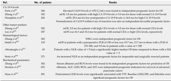
Ref. No. of patients Results
CA 19-9 levels
Park et al[8] 340 Elevated CA19-9 levels (> 670 U/mL) were found to independent prognostic factor for OS
Zhang et al[7] 302 mOS, 3.8 mo for patients with high CA 19-9 levels vs 5.0 mo for those with normal CA 19-9 levels
Humphris et al[47] 260 mOS, 25.6 mo for low postoperative CA 19-9 levels vs 14.8 mo for high CA 19-9 levels
Normalization of CA19-9 within 6 mo of resection was also an independent favorable prognostic factor Other tumor markers
Zhang et al[7] 302 mOS, 2.0 mo for patients with high CEA levels vs 5.0 mo for those with normal CEA levels
Lee et al[22] 187 mOS was 16.3 and 10.2 mo for patients with normal CEA vs high CEA levels, repsectively
Hematological factors
Zhang et al[7] 302 WBCs were independent prognostic factor for OS
Smith et al[58] 110 mOS in patients with a preoperative PLR of 150 or less was 19.7 mo, 13.7 mo in those with a PLR of
151-300, and 5.8 mo in patients with a value of > 300
Aliustaoglu et al[57] 65 Patients with a NLR value of < 5 had a significantly higher median OS time compared to those with a NLR
value of ≥ 5
Stotz et al[56] 371 An increased NLR as an independent prognostic factor for inoperable and surgically resected patients
Biochemical parameters
Zhang et al[7] 302 Serum albumin and BUN levels were found to be independent prognostic factors for prediction of OS
Stocken et al[46] 653 Albumin, ALP, LDH, BUN, and AST were independent prognostic indicators for survival of advanced
pancreatic cancer
Haas et al[60] 291 Pretreatment LDH levels were significantly associated with TTP. Baseline LDH,CRP, and bilirubin were
significant prognostic factors for OS
mOS: Median overall survival; WBC: White blood cell; PLR: Platelet to lymphocyte ratio; NLR: Neutrophil to lymphocyte ratio; BUN: Blood urea nitrogen; LDH: Lactate dehydrogenase; AST: Aspartate aminotransferase; ALP: Alkaline phosphatase; TTP: Time to progression; CRP: C-reactive protein; CEA: Car-cinoembryonic antigen.
5-fluorouracil (5-FU), the results of tissue microarrays for hENT1 was presented at the 2013 ASCO annual
meeting[64]. The median OS time for patients with high
hENT1 expression who received gemcitabine was sig-nificantly better than that of patients with low hENT1
expression (26.2 mo vs 17.1 mo, p = 0.002). However,
there was no difference among patients treated with 5-FU with respect to hENT1 expression. The authors concluded that patients with high hENT1 expression might benefit more from gemcitabine treatment.
SPARC (secreted protein and rich in cysteine), a ma-tricellular protein found to be under-expressed in certain cancers, has emerged as a multifunctional protein ca-pable of inhibiting the growth of pancreatic, colorectal,
and ovarian cancers[65,66]. The significance of expression
of SPARC as a prognostic factor in the stroma of
pan-creatic tumors has been shown[67]. In a study performed
by Sinn et al[68], immunohistochemistry in the tissue
sam-ple for expression of SPARC in the stroma around the tumor, but also in the tumor cell, of patients from the Charité Onkologie (CONKO)-001 study was carried out and their results were presented at the 2013 ASCO an-nual meeting. Patients who received gemcitabine as adju-vant treatment had a longer DFS and OS when stromal and cytoplasmic expression of SPARC was not-strong or negative, respectively, compared with strong expression of SPARC. Thus, SPARC expression estimation, both in the tumor or its stroma, seems to be a valuable prognos-tic factor in patients receiving gemcitabine as adjuvant therapy in patients with pancreatic cancer.
The prognostic significance of circulating tumor cells (CTCs) has been investigated and patients who had CTCs (more than 1 in 7.5 mL) before curative surgery, or after therapy initiation, has a trend towards poorer OS or PFS[69]. Bidard et al[70] prospectively analyzed patients
with locally advanced unresectable pancreatic cancer before and after 2 mo of chemotherapy for CTCs. More than one tumor cell in 7.5 mL was considered as posi-tive. Before treatment, 5% of patients had positive de-tection of CTCs and 9% at the end of 2 mo of therapy. This positivity was found to be associated with poor tumor differentiation and the OS was shorter in these positive patients. The determination of CTCs in patients with pancreatic cancer seems to have a negative
prog-nostic role[71]. There is a significant relationship between
the amount of peritumoral CD4+ and CD8+ T-cells and survival in patients with pancreatic cancer and it was
found to be an independent prognostic factor for OS[71].
Transforming growth factor β (TGF-β) acts as sup-pressor and promoter of cancer progression. Intracellular Smad proteins (common mediator SMAD4) play a piv-otal role in mediating antimitogenic and proapoptotic
ef-fects of TGF-β[72]. In 55% of pancreatic tumors SMAD4
alterations are found and it is inactivated in the majority
of pancreatic adenocarcinoma with concurrent
muta-tional inactivation of the INK4A/ARF tumor suppressor
locus and activation of the KRAS oncogene[73]. Previous
reports revealed unclear results related with SMAD4 as
a predictor of survival in pancreatic cancer[74-76].
Black-ford et al[76] reported that SMAD4 gene inactivation was
associated with poorer prognosis in resected pancreatic adenocarcinoma. In other words, median survival time in patients without SMAD4 gene inactivation was
sig-nificantly better than those with inactivation (14.2 mo vs
11.5 mo, p = 0.006). Recent study showed a significant
relationship was found between SMAD4 expression and
tumor size (p = 0.006), lymphatic invasion (p = 0.033),
and lymph node metastasis (p = 0.006)[77]. Moreover, loss
of SMAD4 expression was significantly associated with shorter OS and it was found to be an independent prog-nostic factor for both OS and DFS by multivariate
analy-sis. Similarly, another study has confirmed these results[78].
Novel prognostic biomarkers
Hypoxia-inducible factor 1 alpha (HIF1α) has been found to be an unfavorable prognostic indicator in many cancers and is known to regulate some genes in the angiogenesis
pathway[79]. Some studies have previously been showed
that HIF1α had a strong impact on the prognosis of
patients with pancreatic adenocarcinoma[80-82]. NEDD9,
a focal adhesion scaffolding protein, has been recently proposed to regulate invasion and metastasis in some cancer types[83-85]. In a study performed by Xue et al[86],
they investigated the expression and prognostic signifi-cance of NEDD9 in patients with pancreatic signifi-cancer. NEDD9 protein and mRNA levels were elevated in pancreatic carcinoma lesions compared with noncan-cerous tissues. A high NEDD9 expression level was significantly correlated with clinical staging, lymph node metastasis, and histological differentiation. The median survival time for patients with a higher NEDD9 expres-sion was significantly shorter than that of patients with lower NEDD9 expression. In addition, the multivariate analysis revealed that NEDD9 was an independent fac-tor of poor prognosis.
FOXM1 (Forkhead box M1) is a typical prolifera-tion-related transcription factor and is also intimately involved in tumorigenesis. It induces cell proliferation and cell cycle progression by promoting the entry into S-phase and M-phase[87]. Xia et al[88] in their study,
evalu-ated correlation between FoxM1 expression level and survival of patients with pancreatic adenocarcinoma. They showed that a high level of expression of FoxM1 was significantly correlated with clinical staging, lymph node metastasis, and histological differentiation. Fur-thermore, patients with a higher FoxM1 expression had a significantly shorter survival time compared to patients with lower FoxM1 expression and FoxM1 was found to be an independent factor for survival.
Recent study indicated that B7H4, HSP27 and DJ-1 protein expressions in the tissue specimens of 41 pa-tients with resected pancreatic cancer were independent-ly associated with a negative impact of chemotherapy
with gemcitabine on patient’s survival[89]. In addition,
patients who overexpressed B7H4 had worse prognosis than patients without overexpression. In a study carried
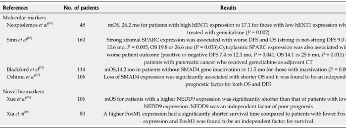
Table 4 Molecular and novel biomarkers as prognostic factors in pancreatic cancer
out by Perini et al[90], prognostic significance of
epider-mal growth factor receptor (EGFR) overexpression in pancreas cancer was investigated. Univariate analysis showed that positive EGFR expression in tumor tissue had worse survival, as were male gender, portal vein re-section, perineural, lymphovascular and peri-pancreatic invasion, positive margins, however, prognostic signifi-cance of positive EGFR expression as an independent prognostic factor could not be confirmed in the multiva-riate analysis. Selected studies associated with molecular and novel biomarkers are listed in Table 4.
CONCLUSION
The overall prognosis associated with pancreatic cancer has not improved over the last 20 years, even if new diagnostic and therapeutic strategies have emerged. So, investigations on predictive factors in pancreatic cancer are needed because these factors should have predictive value in relation to longer survival after surgery than af-ter palliative treatment. In addition to some well-known prognostic factors such as tumor stage, surgical margin, perineural invasion, PS, treatment effect, and CA19-9, recently new prognostic indicators that have an impact on survival of patients with pancreatic cancer have ap-peared. The prognostic value of operative factors includ-ing OBL and PBT, NLR, and molecular markers such as SPARC, hENT1, SMAD4, CTCs, HIF1α, NEDD9 and FOXM1 has recently been shown. Prognostic factors are important and a correct definition of poor prognostic factors may help to guide more aggressive adjuvant or aggressive treatment protocols in patients with pancre-atic cancer.
REFERENCES
1 Siegel R, Naishadham D, Jemal A. Cancer statistics, 2013.
CA Cancer J Clin 2013; 63: 11-30 [PMID: 23335087 DOI:
10.3322/caac.21166]
2 Alexakis N, Halloran C, Raraty M, Ghaneh P, Sutton R, Neoptolemos JP. Current standards of surgery for pancre-atic cancer. Br J Surg 2004; 91: 1410-1427 [PMID: 15499648 DOI: 10.1002/bjs.4794]
3 Michaud DS. Epidemiology of pancreatic cancer. Minerva
Chir 2004; 59: 99-111 [PMID: 15238885]
4 Lockhart AC, Rothenberg ML, Berlin JD. Treatment for pancreatic cancer: current therapy and continued progress.
Gastroenterology 2005; 128: 1642-1654 [PMID: 15887156 DOI:
10.1053/j.gastro.2005.03.039]
5 Shimada K, Sakamoto Y, Sano T, Kosuge T. Prognostic factors after distal pancreatectomy with extended lymph-adenectomy for invasive pancreatic adenocarcinoma of the body and tail. Surgery 2006; 139: 288-295 [PMID: 16546491 DOI: 10.1016/j.surg.2005.08.004]
6 Boggi U, Del Chiaro M, Croce C, Vistoli F, Signori S, Moretto C, Amorese G, Mazzeo S, Cappelli C, Campani D, Mosca F. Prognostic implications of tumor invasion or adhesion to peri-pancreatic vessels in resected peri-pancreatic cancer. Surgery 2009; 146: 869-881 [PMID: 19744432 DOI: 10.1016/j.surg.2009.04.029] 7 Zhang DX, Dai YD, Yuan SX, Tao L. Prognostic factors in
patients with pancreatic cancer. Exp Ther Med 2012; 3: 423-432 [PMID: 22969906 DOI: 10.3892/etm.2011.412]
8 Park JK, Yoon YB, Kim YT, Ryu JK, Yoon WJ, Lee SH. Sur-vival and prognostic factors of unresectable pancreatic can-cer. J Clin Gastroenterol 2008; 42: 86-91 [PMID: 18097296 DOI: 10.1097/01.mcg.0000225657.30803.9d]
9 Wentz SC, Zhao ZG, Shyr Y, Shi CJ, Merchant NB, Wash-ington K, Xia F, Chakravarthy AB. Lymph node ratio and preoperative CA 19-9 levels predict overall survival and recurrence-free survival in patients with resected pancreatic adenocarcinoma. World J Gastrointest Oncol 2012; 4: 207-215 [PMID: 23444312 DOI: 10.4251/wjgo.v4.i10.207]
10 Willett CG, Lewandrowski K, Warshaw AL, Efird J, Comp-ton CC. Resection margins in carcinoma of the head of the pancreas. Implications for radiation therapy. Ann Surg 1993; 217: 144-148 [PMID: 8094952]
11 Butturini G, Stocken DD, Wente MN, Jeekel H, Klinkenbijl JH, Bakkevold KE, Takada T, Amano H, Dervenis C, Bassi C, Büchler MW, Neoptolemos JP. Influence of resection mar-gins and treatment on survival in patients with pancreatic cancer: meta-analysis of randomized controlled trials. Arch
Surg 2008; 143: 75-83; discussion 83 [PMID: 18209156 DOI:
10.1001/archsurg.2007.17]
12 Menon KV, Gomez D, Smith AM, Anthoney A, Verbeke CS. Impact of margin status on survival following
pancreatoduo-References No. of patients Results
Molecular markers
Neoptolemos et al[64] 48 mOS, 26.2 mo for patients with high hENT1 expression vs 17.1 for those with low hENT1 expression who
treated with gemcitabine (P = 0.002)
Sinn et al[68] 160 Strong stromal SPARC expression was associated with worse DFS and OS (strong vs not-strong DFS 9.0 vs
12.6 mo, P = 0.005; OS 19.8 vs 26.6 mo (P = 0.033).Cytoplasmic SPARC expression was also associated with worse patient outcome (positive vs negative DFS 7.4 vs 12.1 mo, P = 0.041; OS 14.1 vs 25.6 mo, P = 0.011) in
patients with pancreatic cancer who received gemcitabine as adjuvant CT
Blackford et al[76] 114 mOS,14.2 mo in patients without SMAD4 gene inactivation vs 11.5 mo for those with inactivation (P = 0.006)
Oshima et al[77] 106 Loss of SMAD4 expression was significantly associated with shorter OS and it was found to be an independent
prognostic factor for both OS and DFS Novel biomarkers
Xue et al[86] 106 mOS for patients with a higher NEDD9 expression was significantly shorter than that of patients with lower
NEDD9 expression. NEDD9 was an independent factor of poor prognosis
Xia et al[88] 80 A higher FoxM1 expression had a significantly shorter survival time compared to patients with lower FoxM1
expression and FoxM1 was found to be an independent factor for survival
mOS: Median overall survival; DFS: Disease-free survival; hENT1: Human equilibrative nucleoside transporter 1; SPARC: Secreted protein and rich in cys-teine; CT: Chemotherapy; FOXM1: Forkhead box M1.
denectomy for cancer: the Leeds Pathology Protocol (LEEPP).
HPB (Oxford) 2009; 11: 18-24 [PMID: 19590619 DOI: 10.1111/
j.1477-2574.2008.00013.x]
13 Raut CP, Tseng JF, Sun CC, Wang H, Wolff RA, Crane CH, Hwang R, Vauthey JN, Abdalla EK, Lee JE, Pisters PW, Ev-ans DB. Impact of resection status on pattern of failure and survival after pancreaticoduodenectomy for pancreatic ad-enocarcinoma. Ann Surg 2007; 246: 52-60 [PMID: 17592291 DOI: 10.1097/01.sla.0000259391.84304.2b]
14 Andrén-Sandberg A. Prognostic factors in pancreatic can-cer. N Am J Med Sci 2012; 4: 9-12 [PMID: 22393541 DOI: 10.4103/1947-2714.92893]
15 Berger AC, Watson JC, Ross EA, Hoffman JP. The metastat-ic/examined lymph node ratio is an important prognostic factor after pancreaticoduodenectomy for pancreatic adeno-carcinoma. Am Surg 2004; 70: 235-240; discussion 240 [PMID: 15055847]
16 Pawlik TM, Gleisner AL, Cameron JL, Winter JM, Assump-cao L, Lillemoe KD, Wolfgang C, Hruban RH, Schulick RD, Yeo CJ, Choti MA. Prognostic relevance of lymph node ratio following pancreaticoduodenectomy for pancreatic cancer.
Surgery 2007; 141: 610-618 [PMID: 17462460 DOI: 10.1016/
j.surg.2006.12.013]
17 Riediger H, Keck T, Wellner U, zur Hausen A, Adam U, Hopt UT, Makowiec F. The lymph node ratio is the stron-gest prognostic factor after resection of pancreatic cancer. J
Gastrointest Surg 2009; 13: 1337-1344 [PMID: 19418101 DOI:
10.1007/s11605-009-0919-2]
18 Valsangkar NP, Bush DM, Michaelson JS, Ferrone CR, War-go JA, Lillemoe KD, Fernández-del Castillo C, Warshaw AL, Thayer SP. N0/N1, PNL, or LNR? The effect of lymph node number on accurate survival prediction in pancreatic ductal adenocarcinoma. J Gastrointest Surg 2013; 17: 257-266 [PMID: 23229885 DOI: 10.1007/s11605-012-1974-7]
19 Lee SR, Kim HO, Son BH, Yoo CH, Shin JH. Prognostic fac-tors associated with long-term survival and recurrence in pancreatic adenocarcinoma. Hepatogastroenterology 2013; 60: 358-362 [PMID: 23574658 DOI: 10.5754/hge12727]
20 Wang PH, Song N, Shi LB, Zhang QH, Chen ZY. The rela-tionship between multiple clinicopathological features and nerve invasion in pancreatic cancer. Hepatobiliary Pancreat
Dis Int 2013; 12: 546-551 [PMID: 24103287 DOI: 10.1016/
S1499-3872(13)60086-7]
21 Chatterjee D, Katz MH, Lee JE, Wolf RA, Varadhachary GR, Pisters PW. Perineural and blood vessel invasion identified after neoadjuvant treatment correlates with poor prognosis in patients with pancreatic ductal adenocarcinoma. Ameri-can Pancreas Club: 45th Annual Meeting, 2011: May 6-7 22 Lee KJ, Yi SW, Chung MJ, Park SW, Song SY, Chung JB, Park
JY. Serum CA 19-9 and CEA levels as a prognostic factor in pancreatic adenocarcinoma. Yonsei Med J 2013; 54: 643-649 [PMID: 23549809 DOI: 10.3349/ymj.2013.54.3.643]
23 Nagai S, Fujii T, Kodera Y, Kanda M, Sahin TT, Kanzaki A, Yamada S, Sugimoto H, Nomoto S, Takeda S, Morita S, Na-kao A. Impact of operative blood loss on survival in invasive ductal adenocarcinoma of the pancreas. Pancreas 2011; 40: 3-9 [PMID: 20881897 DOI: 10.1097/MPA.0b013e3181f7147a] 24 Keck T, Wellner U, Sick O, Hopt UT. Makowiec
Periopera-tive blood transfusions may influence prognosis after sur-gery for pancreatic cancer independent of complications or body mass index: Multivariate analysis of 270 resected pa-tients. American Pancreas Club: 45th Annual Meeting, 2011: May 6-7
25 Sezgin C, Karabulut B, Uslu R, Sanli UA, Goksel G, Yuzer Y, Goker E. Gemcitabine treatment in patients with inoperable locally advanced/metastatic pancreatic cancer and prognos-tic factors. Scand J Gastroenterol 2005; 40: 1486-1492 [PMID: 16293561 DOI: 10.1080/00365520510023819]
26 Tas F, Sen F, Odabas H, Kılıc L, Keskın S, Yıldız I. Perfor-mance status of patients is the major prognostic factor at all
stages of pancreatic cancer. Int J Clin Oncol 2013; 18: 839-846 [PMID: 22996141 DOI: 10.1007/s10147-012-0474-9]
27 Kang SP, Saif MW. Clinical outcome of pancreatic cancer patients with diabetes mellitus: is diabetes a poor prognos-tic factor? Highlights from the “2010 ASCO Annual Meet-ing”. Chicago, IL, USA. June 4-8, 2010. JOP 2010; 11: 334-335 [PMID: 20601806]
28 Pannala R, Leibson CL, Rabe KG, Timmons LJ, Ransom J, de Andrade M, Petersen GM, Chari ST. Temporal association of changes in fasting blood glucose and body mass index with diagnosis of pancreatic cancer. Am J Gastroenterol 2009; 104: 2318-2325 [PMID: 19513024 DOI: 10.1038/ajg.2009.253] 29 Larsson SC, Orsini N, Wolk A. Body mass index and
pan-creatic cancer risk: A meta-analysis of prospective studies.
Int J Cancer 2007; 120: 1993-1998 [PMID: 17266034]
30 Stolzenberg-Solomon RZ, Graubard BI, Chari S, Limburg P, Taylor PR, Virtamo J, Albanes D. Insulin, glucose, insulin resistance, and pancreatic cancer in male smokers. JAMA 2005; 294: 2872-2878 [PMID: 16352795]
31 Gong Z, Holly EA, Bracci PM. Obesity and survival in popula-tion-based patients with pancreatic cancer in the San Francisco Bay Area. Cancer Causes Control 2012; 23: 1929-1937 [PMID: 23015286 DOI: 10.1007/s10552-012-0070-3]
32 Lin Y, Fu R, Grant E, Chen Y, Lee JE, Gupta PC, Ramadas K, Inoue M, Tsugane S, Gao YT, Tamakoshi A, Shu XO, Ozasa K, Tsuji I, Kakizaki M, Tanaka H, Chen CJ, Yoo KY, Ahn YO, Ahsan H, Pednekar MS, Sauvaget C, Sasazuki S, Yang G, Xiang YB, Ohishi W, Watanabe T, Nishino Y, Matsuo K, You SL, Park SK, Kim DH, Parvez F, Rolland B, McLerran D, Sinha R, Boffetta P, Zheng W, Thornquist M, Feng Z, Kang D, Potter JD. Association of body mass index and risk of death from pancreatic cancer in Asians: findings from the Asia Cohort Consortium. Eur J Cancer Prev 2013; 22: 244-250 [PMID: 23044748 DOI: 10.1097/CEJ.0b013e3283592cef] 33 Yuan C, Bao Y, Wu C, Kraft P, Ogino S, Ng K, Qian ZR,
Ru-binson DA, Stampfer MJ, Giovannucci EL, Wolpin BM. Pre-diagnostic body mass index and pancreatic cancer survival. J
Clin Oncol 2013; 31: 4229-4234 [PMID: 24145341 DOI: 10.1200/
JCO.2013.51.7532]
34 Smith RA, Dajani K, Dodd S, Whelan P, Raraty M, Sutton R, Campbell F, Neoptolemos JP, Ghaneh P. Preoperative resolu-tion of jaundice following biliary stenting predicts more fa-vourable early survival in resected pancreatic ductal adenocar-cinoma. Ann Surg Oncol 2008; 15: 3138-3146 [PMID: 18787902 DOI: 10.1245/s10434-008-0148-z]
35 Perini MV, Montagnini AL, Jukemura J, Penteado S, Abdo EE, Patzina R, Cecconello I, Cunha JE. Clinical and patho-logic prognostic factors for curative resection for pancreatic cancer. HPB (Oxford) 2008; 10: 356-362 [PMID: 18982152 DOI: 10.1080/13651820802140752]
36 Strasberg SM, Gao F, Sanford D, Linehan DC, Hawkins WG, Fields R, Carpenter DH, Brunt EM, Phillips C. Jaundice: an important, poorly recognized risk factor for diminished sur-vival in patients with adenocarcinoma of the head of the pan-creas. HPB (Oxford) 2014; 16: 150-156 [PMID: 23600768 DOI: 10.1111/hpb.12094]
37 Neoptolemos JP, Stocken DD, Friess H, Bassi C, Dunn JA, Hickey H, Beger H, Fernandez-Cruz L, Dervenis C, Lacaine F, Falconi M, Pederzoli P, Pap A, Spooner D, Kerr DJ, Büchler MW. A randomized trial of chemoradiotherapy and chemo-therapy after resection of pancreatic cancer. N Engl J Med 2004; 350: 1200-1210 [PMID: 15028824 DOI: 10.1056/NEJ-Moa032295]
38 Regine WF, Winter KA, Abrams RA, Safran H, Hoffman JP, Konski A, Benson AB, Macdonald JS, Kudrimoti MR, Fromm ML, Haddock MG, Schaefer P, Willett CG, Rich TA. Fluorouracil vs gemcitabine chemotherapy before and after fluorouracil-based chemoradiation following resection of pancreatic adenocarcinoma: a randomized controlled trial.
jama.299.9.1019]
39 Thomas A, Dajani K, Neoptolemos JP, Ghaneh P. Adjuvant therapy in pancreatic cancer. Dig Dis 2010; 28: 684-692 [PMID: 21088421 DOI: 10.1159/000320099]
40 Neoptolemos JP, Stocken DD, Bassi C, Ghaneh P, Cunning-ham D, Goldstein D, Padbury R, Moore MJ, Gallinger S, Mar-iette C, Wente MN, Izbicki JR, Friess H, Lerch MM, Dervenis C, Oláh A, Butturini G, Doi R, Lind PA, Smith D, Valle JW, Palmer DH, Buckels JA, Thompson J, McKay CJ, Rawcliffe CL, Büchler MW. Adjuvant chemotherapy with fluorouracil plus folinic acid vs gemcitabine following pancreatic cancer resection: a randomized controlled trial. JAMA 2010; 304: 1073-1081 [PMID: 20823433 DOI: 10.1001/jama.2010.1275] 41 Bünger S, Laubert T, Roblick UJ, Habermann JK. Serum
biomarkers for improved diagnostic of pancreatic cancer: a current overview. J Cancer Res Clin Oncol 2011; 137: 375-389 [PMID: 21193998 DOI: 10.1007/s00432-010-0965-x]
42 Duffy MJ, Sturgeon C, Lamerz R, Haglund C, Holubec VL, Klapdor R, Nicolini A, Topolcan O, Heinemann V. Tumor markers in pancreatic cancer: a European Group on Tumor Markers (EGTM) status report. Ann Oncol 2010; 21: 441-447 [PMID: 19690057 DOI: 10.1093/annonc/mdp332]
43 Shah UA, Saif MW. Tumor markers in pancreatic cancer: 2013. JOP 2013; 14: 318-321 [PMID: 23846917]
44 Kim YC, Kim HJ, Park JH, Park DI, Cho YK, Sohn CI, Jeon WK, Kim BI, Shin JH. Can preoperative CA19-9 and CEA levels predict the resectability of patients with pancreatic adenocarcinoma? J Gastroenterol Hepatol 2009; 24: 1869-1875 [PMID: 19686409 DOI: 10.1111/j.1440-1746.2009.05935.x] 45 Koom WS, Seong J, Kim YB, Pyun HO, Song SY. CA 19-9 as
a predictor for response and survival in advanced pancre-atic cancer patients treated with chemoradiotherapy. Int J
Radiat Oncol Biol Phys 2009; 73: 1148-1154 [PMID: 18760544
DOI: 10.1016/j.ijrobp.2008.06.1483]
46 Stocken DD, Hassan AB, Altman DG, Billingham LJ, Bram-hall SR, Johnson PJ, Freemantle N. Modelling prognostic factors in advanced pancreatic cancer. Br J Cancer 2008; 99: 883-893 [PMID: 19238630 DOI: 10.1038/sj.bjc.6604568] 47 Humphris JL, Chang DK, Johns AL, Scarlett CJ, Pajic M,
Jones MD, Colvin EK, Nagrial A, Chin VT, Chantrill LA, Samra JS, Gill AJ, Kench JG, Merrett ND, Das A, Musgrove EA, Sutherland RL, Biankin AV. The prognostic and predic-tive value of serum CA19.9 in pancreatic cancer. Ann Oncol 2012; 23: 1713-1722 [PMID: 22241899 DOI: 10.1093/annonc/ mdr561]
48 Carriquiry LA, Piñeyro A. Should carcinoembryonic anti-gen be used in the management of patients with colorectal cancer? Dis Colon Rectum 1999; 42: 921-929 [PMID: 10411440] 49 Louhimo J, Alfthan H, Stenman UH, Haglund C. Serum
HCG beta and CA 72-4 are stronger prognostic factors than CEA, CA 19-9 and CA 242 in pancreatic cancer. Oncology 2004; 66: 126-131 [PMID: 15138364 DOI: 10.1159/000077438] 50 Donskov F, von der Maase H. Impact of immune
param-eters on long-term survival in metastatic renal cell carcino-ma. J Clin Oncol 2006; 24: 1997-2005 [PMID: 16648500 DOI: 10.1200/JCO.2005.03.9594]
51 Zhang L, Conejo-Garcia JR, Katsaros D, Gimotty PA, Mas-sobrio M, Regnani G, Makrigiannakis A, Gray H, Schlienger K, Liebman MN, Rubin SC, Coukos G. Intratumoral T cells, recurrence, and survival in epithelial ovarian cancer. N Engl
J Med 2003; 348: 203-213 [PMID: 12529460 DOI: 10.1056/
NEJMoa020177]
52 Fumagalli LA, Vinke J, Hoff W, Ypma E, Brivio F, Nespoli A. Lymphocyte counts independently predict overall survival in advanced cancer patients: a biomarker for IL-2 immuno-therapy. J Immunother 2003; 26: 394-402 [PMID: 12973028] 53 Sierko E, Wojtukiewicz MZ. Platelets and angiogenesis in
malignancy. Semin Thromb Hemost 2004; 30: 95-108 [PMID: 15034801 DOI: 10.1055/s-2004-822974]
54 Schwarz RE, Keny H. Preoperative platelet count predicts
survival after resection of periampullary adenocarcinoma.
Hepatogastroenterology 2001; 48: 1493-1498 [PMID: 11677994]
55 Domínguez I, Crippa S, Thayer SP, Hung YP, Ferrone CR, Warshaw AL, Fernández-Del Castillo C. Preoperative plate-let count and survival prognosis in resected pancreatic duc-tal adenocarcinoma. World J Surg 2008; 32: 1051-1056 [PMID: 18224462 DOI: 10.1007/s00268-007-9423-6]
56 Stotz M, Gerger A, Eisner F, Szkandera J, Loibner H, Ress AL, Kornprat P, AlZoughbi W, Seggewies FS, Lackner C, Stojakovic T, Samonigg H, Hoefler G, Pichler M. Increased neutrophil-lymphocyte ratio is a poor prognostic factor in patients with primary operable and inoperable pancreatic cancer. Br J Cancer 2013; 109: 416-421 [PMID: 23799847 DOI: 10.1038/bjc.2013.332]
57 Aliustaoglu M, Bilici A, Seker M, Dane F, Gocun M, Konya V, Ustaalioglu BB, Gumus M. The association of pre-treatment peripheral blood markers with survival in patients with pan-creatic cancer. Hepatogastroenterology 2010; 57: 640-645 [PMID: 20698242]
58 Smith RA, Bosonnet L, Raraty M, Sutton R, Neoptolemos JP, Campbell F, Ghaneh P. Preoperative platelet-lymphocyte ra-tio is an independent significant prognostic marker in resect-ed pancreatic ductal adenocarcinoma. Am J Surg 2009; 197: 466-472 [PMID: 18639229 DOI: 10.1016/j.amjsurg.2007.12.057] 59 Tas F, Aykan F, Alici S, Kaytan E, Aydiner A, Topuz E. Prog-nostic factors in pancreatic carcinoma: serum LDH levels predict survival in metastatic disease. Am J Clin Oncol 2001; 24: 547-550 [PMID: 11801751]
60 Haas M, Heinemann V, Kullmann F, Laubender RP, Klose C, Bruns CJ, Holdenrieder S, Modest DP, Schulz C, Boeck S. Prognostic value of CA 19-9, CEA, CRP, LDH and bili-rubin levels in locally advanced and metastatic pancreatic cancer: results from a multicenter, pooled analysis of pa-tients receiving palliative chemotherapy. J Cancer Res Clin
Oncol 2013; 139: 681-689 [PMID: 23315099 DOI: 10.1007/
s00432-012-1371-3]
61 Damaraju VL, Damaraju S, Young JD, Baldwin SA, Mackey J, Sawyer MB, Cass CE. Nucleoside anticancer drugs: the role of nucleoside transporters in resistance to cancer chemo-therapy. Oncogene 2003; 22: 7524-7536 [PMID: 14576856 DOI: 10.1038/sj.onc.1206952]
62 Farrell JJ, Elsaleh H, Garcia M, Lai R, Ammar A, Regine WF, Abrams R, Benson AB, Macdonald J, Cass CE, Dicker AP, Mackey JR. Human equilibrative nucleoside transporter 1 levels predict response to gemcitabine in patients with pan-creatic cancer. Gastroenterology 2009; 136: 187-195 [PMID: 18992248 DOI: 10.1053/j.gastro.2008.09.067]
63 Xiao JC, Zhang TP, Zhao YP. Human equilibrative nucleo-side transporter 1 (hENT1) predicts the Asian patient re-sponse to gemcitabine-based chemotherapy in pancreatic cancer. Hepatogastroenterology 2013; 60: 258-262 [PMID: 23574652 DOI: 10.5754/hge12687]
64 Neoptolemos JP, Greenhalf W, Ghaneh P, Palmer DH, Cox TF, Garner E, Campbell F, Mackey JR, Moore MJ, Valle JW, Mcdonald A, Tebbutt NC, Dervenis C, Glimelius B, Charn-ley RM, Lacaine F, Mayerle J, Rawcliffe CL, Bassi C, Buchler MV.HENT1 tumor levels to predict survival of pancreatic ductal adenocarcinoma patients who received adjuvant gemcitabine and adjuvant 5FU on the ESPAC trials. J Clin
Oncol 2013; (Suppl): Abstract 4006
65 Puolakkainen PA, Brekken RA, Muneer S, Sage EH. En-hanced growth of pancreatic tumors in SPARC-null mice is associated with decreased deposition of extracellular matrix and reduced tumor cell apoptosis. Mol Cancer Res 2004; 2: 215-224 [PMID: 15140943]
66 Tang MJ, Tai IT. A novel interaction between procaspase 8 and SPARC enhances apoptosis and potentiates chemother-apy sensitivity in colorectal cancers. J Biol Chem 2007; 282: 34457-34467 [PMID: 17897953 DOI: 10.1074/jbc.M704459200] 67 Hidalgo M, Von Hoff DD. Translational therapeutic
opportu-nities in ductal adenocarcinoma of the pancreas. Clin Cancer Res 2012; 18: 4249-4256 [PMID: 22896691 DOI: 10.1158/1078-0432. CCR-12-1327]
68 Sinn M, Sinn BV, Striefler JK, Lindner JL, Stieler JM, Loh-neis P, Bischoff S, Bläker H, Pelzer U, Bahra M, Dietel M, Dörken B, Oettle H, Riess H, Denkert C. SPARC expression in resected pancreatic cancer patients treated with gem-citabine: results from the CONKO-001 study. Ann Oncol 2014; 25: 1025-1032 [PMID: 24562449]
69 Negin BP, Meropol NJ, Alpaugh RK, Ruth K, McAleer C, Halbherr T, Bingham C, Fittipaldi P, Cohen SJ. Characteriza-tion and prognostic significance of circulating tumor cells in the peripheral blood of patients with metastatic pancreatic cancer. J Clin Oncol 2010; (Suppl): Abstract 4127
70 Bidard FC, Huguet F, Louvet C, Mineur L, Bouché O, Chibau-del B, Artru P, Desseigne F, Bachet JB, Mathiot C, Pierga JY, Hammel P. Circulating tumor cells in locally advanced pancre-atic adenocarcinoma: the ancillary CirCe 07 study to the LAP 07 trial. Ann Oncol 2013; 24: 2057-2061 [PMID: 23676420] 71 Oikonomopoulos GM, Syrigos KN, Saif MW. Prognostic
factors in pancreatic cancer. JOP 2013; 14: 322-324 [PMID: 23846918 DOI: 10.6092/1590-8577/1644]
72 Fink SP, Mikkola D, Willson JK, Markowitz S. TGF-beta-induced nuclear localization of Smad2 and Smad3 in Smad4 null cancer cell lines. Oncogene 2003; 22: 1317-1323 [PMID: 12618756]
73 Hahn SA, Schutte M, Hoque AT, Moskaluk CA, da Costa LT, Rozenblum E, Weinstein CL, Fischer A, Yeo CJ, Hruban RH, Kern SE. DPC4, a candidate tumor suppressor gene at human chromosome 18q21.1. Science 1996; 271: 350-353 [PMID: 8553070]
74 Tascilar M, Skinner HG, Rosty C, Sohn T, Wilentz RE, Of-ferhaus GJ, Adsay V, Abrams RA, Cameron JL, Kern SE, Yeo CJ, Hruban RH, Goggins M. The SMAD4 protein and prog-nosis of pancreatic ductal adenocarcinoma. Clin Cancer Res 2001; 7: 4115-4121 [PMID: 11751510 DOI: 10.1158/1078-0432. CCR-09-0227]
75 Biankin AV, Morey AL, Lee CS, Kench JG, Biankin SA, Hook HC, Head DR, Hugh TB, Sutherland RL, Henshall SM. DPC4/Smad4 expression and outcome in pancreatic ductal adenocarcinoma. J Clin Oncol 2002; 20: 4531-4542 [PMID: 12454109 DOI: 10.1200/JCO.2002.12.063]
76 Blackford A, Serrano OK, Wolfgang CL, Parmigiani G, Jones S, Zhang X, Parsons DW, Lin JC, Leary RJ, Eshleman JR, Goggins M, Jaffee EM, Iacobuzio-Donahue CA, Maitra A, Cameron JL, Olino K, Schulick R, Winter J, Herman JM, La-heru D, Klein AP, Vogelstein B, Kinzler KW, Velculescu VE, Hruban RH. SMAD4 gene mutations are associated with poor prognosis in pancreatic cancer. Clin Cancer Res 2009; 15: 4674-4679 [PMID: 19584151]
77 Oshima M, Okano K, Muraki S, Haba R, Maeba T, Suzuki Y, Yachida S. Immunohistochemically detected expression of 3 major genes (CDKN2A/p16, TP53, and SMAD4/DPC4) strongly predicts survival in patients with resectable pan-creatic cancer. Ann Surg 2013; 258: 336-346 [PMID: 23470568 DOI: 10.1097/SLA.0b013e3182827a65]
78 Singh P, Srinivasan R, Wig JD. SMAD4 genetic alterations predict a worse prognosis in patients with pancreatic ductal adenocarcinoma. Pancreas 2012; 41: 541-546 [PMID: 22504380 DOI: 10.1097/MPA.0b013e318247d6af]
79 Couvelard A, O’Toole D, Turley H, Leek R, Sauvanet A, Degott C, Ruszniewski P, Belghiti J, Harris AL, Gatter K, Pezzella F. Microvascular density and hypoxia-inducible factor pathway in pancreatic endocrine tumours: negative
correlation of microvascular density and VEGF expression with tumour progression. Br J Cancer 2005; 92: 94-101 [PMID: 15558070 DOI: 10.1038/sj.bjc.6602245]
80 Sun HC, Qiu ZJ, Liu J, Sun J, Jiang T, Huang KJ, Yao M, Huang C. Expression of hypoxia-inducible factor-1 alpha and associated proteins in pancreatic ductal adenocarcinoma and their impact on prognosis. Int J Oncol 2007; 30: 1359-1367 [PMID: 17487356]
81 Tao J, Li T, Li K, Xiong J, Yang Z, Wu H, Wang C. Effect of HIF-1alpha on VEGF-C induced lymphangiogenesis and lymph nodes metastases of pancreatic cancer. J Huazhong
Univ Sci Technolog Med Sci 2006; 26: 562-564 [PMID: 17219968]
82 Hoffmann AC, Mori R, Vallbohmer D, Brabender J, Klein E, Drebber U, Baldus SE, Cooc J, Azuma M, Metzger R, Hoelscher AH, Danenberg KD, Prenzel KL, Danenberg PV. High expres-sion of HIF1a is a predictor of clinical outcome in patients with pancreatic ductal adenocarcinomas and correlated to PDGFA, VEGF, and bFGF. Neoplasia 2008; 10: 674-679 [PMID: 18592007] 83 Little JL, Serzhanova V, Izumchenko E, Egleston BL, Parise
E, Klein-Szanto AJ, Loudon G, Shubina M, Seo S, Kurokawa M, Ochs MF, Golemis EA. A requirement for Nedd9 in luminal progenitor cells prior to mammary tumorigenesis in MMTV-HER2/ErbB2 mice. Oncogene 2014; 33: 411-420 [PMID: 23318423 DOI: 10.1038/onc.2012.607]
84 Kim M, Gans JD, Nogueira C, Wang A, Paik JH, Feng B, Brennan C, Hahn WC, Cordon-Cardo C, Wagner SN, Flotte TJ, Duncan LM, Granter SR, Chin L. Comparative oncoge-nomics identifies NEDD9 as a melanoma metastasis gene.
Cell 2006; 125: 1269-1281 [PMID: 16814714 DOI: 10.1016/
j.cell.2006.06.008]
85 Kondo S, Iwata S, Yamada T, Inoue Y, Ichihara H, Kichi-kawa Y, Katayose T, Souta-Kuribara A, Yamazaki H, Ho-sono O, Kawasaki H, Tanaka H, Hayashi Y, Sakamoto M, Kamiya K, Dang NH, Morimoto C. Impact of the integrin signaling adaptor protein NEDD9 on prognosis and meta-static behavior of human lung cancer. Clin Cancer Res 2012; 18: 6326-6338 [PMID: 23037767 DOI: 10.1158/1078-0432. CCR-11-2162]
86 Xue YZ, Sheng YY, Liu ZL, Wei ZQ, Cao HY, Wu YM, Lu YF, Yu LH, Li JP, Li ZS. Expression of NEDD9 in pancreatic ductal adenocarcinoma and its clinical significance.
Tu-mour Biol 2013; 34: 895-899 [PMID: 23247867 DOI: 10.1007/
s13277-012-0624-8]
87 Wierstra I. FOXM1 (Forkhead box M1) in tumorigenesis: overexpression in human cancer, implication in tumorigen-esis, oncogenic functions, tumor-suppressive properties, and target of anticancer therapy. Adv Cancer Res 2013; 119: 191-419 [PMID: 23870513 DOI: 10.1016/B978-0-12-407190-2.00016-2] 88 Xia JT, Wang H, Liang LJ, Peng BG, Wu ZF, Chen LZ, Xue
L, Li Z, Li W. Overexpression of FOXM1 is associated with poor prognosis and clinicopathologic stage of pancreatic ductal adenocarcinoma. Pancreas 2012; 41: 629-635 [PMID: 22249132 DOI: 10.1097/MPA.0b013e31823bcef2]
89 Tsiaousidou A, Lambropoulou M, Chatzitheoklitos E, Tripsi-anis G, Tsompanidou C, Simopoulos C, Tsaroucha AK. B7H4, HSP27 and DJ-1 molecular markers as prognostic factors in pancreatic cancer. Pancreatology 2013; 13: 564-569 [PMID: 24280570 DOI: 10.1016/j.pan.2013.10.005]
90 Perini MV, Montagnini AL, Coudry R, Patzina R, Penteado S, Abdo EE, Diniz A, Jukemura J, da Cunha JE. Prognostic significance of epidermal growth factor receptor overexpres-sion in pancreas cancer and nodal metastasis. ANZ J Surg 2013; Epub ahead of print [PMID: 24112413 DOI: 10.1111/ ans.12399]
P- Reviewer: Mino-Kenudson M, Smith RC S- Editor: Ma YJ L- Editor: A E- Editor: Liu XM