T
URKISHJ
OURNAL ofO
NCOLOGYCoexistence of Three Malignancies: Two Different Lung
Cancers Synchronous with Lymphoma
Received: October 06, 2017 Accepted: October 06, 2017 Online: October 26, 2017 Accessible online at: www.onkder.org
Turk J Oncol 2017;32(3):120–122 doi: 10.5505/tjo.2017.1624
CASE REPORT
Nalan OGAN,1 Ayşe BAHA,1 Egemen AKINCIOĞLU,2 Tevfik KAPLAN,3 Handan DOĞAN,4 Meral GÜLHAN,5
1Department of Chest Diseases, Ufuk University, Faculty of Medicine, Ankara-Turkey 2Department of Pathology, Gülhane Education and Reseach Hospital, Ankara-Turkey 3Department of Chest Surgery, Ufuk University, Faculty of Medicine, Ankara-Turkey 4Department of Pathology, Ufuk University, Ankara-Turkey
5Department of Chest Diseases, Hitit University, Ankara-Turkey
SUMMARY
Epithelial tumors synchronous with hematologic malignancies are very rare. The hystopathologic type and stage of synchronous tumors are very important for prognosis. A 77-year-old male patient was di-agnosed with lymphoma after an excisional biopsy was taken from the retroauricular region. Positron emission tomography, performed for lymphoma staging, revealed a positive solid nodule (SUVmax: 24.3) in the posterobasal segment of the right lower lobe and a negative subsolid nodule in the anterior segment of the upper lobe. Right lower lobectomy and wedge resection for subsolide nodule in the upper lobe were performed. Histopathological examination revealed mildly differentiated squamous cell carcinoma for the solid nodule and mildly differentiated adenocarcinoma with lepidic pattern for the subsolid nodule. The patient was treated for only lymphoma according to the decision of the tumor board, and his condi-tion is stable for 1 year without any evidence of lung cancer recurrence. This case presents the treatment approach and the fact that triple synchronic malignant cases are rare in the literature.
Keywords: Adenocarcinoma; lymphoma; multidisciplinary approach; squamous cell carcinoma; synchronous tumors.
Copyright © 2017, Turkish Society for Radiation Oncology
Introduction
Multiple lung malignancies are cancers that require a harder and specialized approach for diagnosis and treat-ment. Their incidence has increased with the progress in imaging and other techniques. They can be syn-chronous or metasyn-chronous. Metasyn-chronous cases are more frequent because of the increased risk caused by antitumoral treatments.[1] Hematologic malignan-cies synchronous with epithelial lung malignanmalignan-cies are rare, as case reports in the literature. It is very important to define the type and stage of every single tumor for treatment strategy and prognosis in patients with mul-tiple synchronous tumors. In this case report, a diffuse large B-cell lymphoma synchronous with two different
(squamous and adenocarcinoma) tumors of the lung is presented because it is very rarely seen in the literature.
Case Report
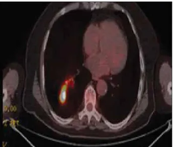
A 77-year-old male patient was referred to our center after being diagnosed with a pulmonary lesion with suspicious tumoral activity. He was diagnosed with ana-plastic large cell lymphoma after a retroauricular lymph node biopsy was performed in the first center. He had a history of smoking 50 packets/year. Positron emission tomography (PET) was performed for lymphoma stag-ing, which revealed an SUVmax of 24.3 at the right lung superior segment that had a 17-mm nodule (Fig. 1). Pul-monary function test results were as follows: forced vital
Dr. Nalan Ogan
Ufuk Üniversitesi Tıp Fakültesi Göğüs Hastalıkları Kliniği, Ankara-Turkey
121
bronchoscopy revealed an irregular mucosa in the pos-terior segment of the right lower lobe. A biopsy result revealed squamous cell carcinoma. The patient was evaluated by the tumor board, and lung resection fol-lowed by lymphoma treatment was planned. Right lower lobectomy, upper lobe wedge resection (due to limited lung function), and mediastinal lymph node dissection were performed. Pathology results were consistent with mildly differentiated squamous cell carcinoma in the lobectomy specimen and mildly differentiated adeno-carcinoma with lepidic pattern in the wedge resection specimen. There was no mediastinal lymph node metas-tasis. Visceral pleural invasion was detected in the lesion on the anterior segment of the upper lobe. The tumor on the lower lobe was graded as IA, whereas the tumor on the anterior segment of the upper lobe was graded as 1B (Fig. 3). The patient was reevaluated by the tumor board postoperatively; adjuvant therapy was not deemed ben-eficial, and chemotherapy for lymphoma was planned by the medical oncology department.
Discussion
The incidence of synchronous lung cancer is 0.2%–20%. [2] PET-CT, which has become widely used, plays an im-portant role in the diagnosis and grading of such cases. Although rare, false negativity is possible.[3] PET scan was negative for the nodule located on the right lung up-per lobe; however, our patient was diagnosed with ad-enocarcinoma after wedge resection. False negativity for subsolid nodules in PET-CT, like our case, is common.
Hematologic malignancy synchronous with lung cancer is very rare. Its etiology and pathophysiology is still unclear.[4] Coexistence with lung cancers in known cases is usually metachronous or diagnosed by biopsy from intrathoracic residue after lymphoma treatment. The risk of a second primary cancer increases after che-motherapy, radiotherapy, or combination therapy for malignancies with high therapeutic response such as Hodgkin’s lymphoma or testicular cancer.[1] Pulmo-capacity (FVC): 84% (2.42 L), forced expiratory volume
in the first second (FEV1): 71% (1.53 L), and FEV1/FVC, 63. Chest computed tomography (CT) revealed a nodule that had a 12-mm circumference located in the right up-per lobe anterior segment with a ground-glass density, another calcified nodule that had a 6-mm circumfer-ence at the same localization, and an additional 22×18-mm nodule located at the right lower lobe posterobasal segment with a spiculated contour (Fig. 2). Fiberoptic
Turk J Oncol 2017;32(3):120–122 doi: 10.5505/tjo.2017.1624
Fig. 3. A 12-mm diameter pulmonary nodule with
sub-pleural placement in the anterior segment of the right upper lobe.
Fig. 2. A 12-mm diameter pulmonary nodule with
sub-pleural placement in the anterior segment of the right upper lobe.
Fig. 1. Increased pathologic involvement in the 17-mm
nodule in the superior segment of the right lower lobe of the lung (SUVmax: 24.3).
122
nary carcinoma synchronous with lymphoma cases are reported in the literature. We described two separate lung malignancies synchronous with lymphoma in this case report.[5,6]
Five-year survival rates of patients with synchronous tumors are lower than those with metachronous tumors. In a study by Marcel et al., patients who underwent re-section for synchronous lung cancers were compared to patients with lung cancers, which had a single primary, and it was shown that approximately half of the syn-chronous tumors were bilateral (51%) and 5-year sur-vival was 23% in the early stages. A study reports that in synchronous tumors, the tumor with the most advanced stage is the best predictor for survival.[7]
Studies state that multiple pulmonary lesions have to be elucidated histopathologically because of the risk of synchronous tumors, and aggressive surgery in the early stages is necessary.[8] The American College of Chest Physicians guidelines for synchronous lung can-cers state that following invasive mediastinal staging and extrathoracic imaging, every single tumor should be re-moved unless there is enough pulmonary reserve and no N2 involvement. Lobectomy, bilobectomy, or pneu-monectomy is recommended for synchronous tumors of the same lung, while consecutive surgical resection in 4–6 weeks is recommended for bilateral synchronous tumors.[9] A study states that pneumonectomy should be avoided because of poor postoperative prognosis.[10] Every case should be evaluated according to the patient’s functional status. Lobectomy for the tumor in the lower right lobe and limited (wedge) resection was performed in our case because of the patient’s advanced age and co-morbidities. Lesions were evaluated separately postop-eratively, and adjuvant therapy for lung cancer was not planned, but lymphoma treatment without delay was performed.
Conclusion
As a result, separate diagnosis, differentiation between a metastasis and a synchronous tumor, in addition to a thorough staging is necessary for patients with multiple tumors. A multidisciplinary approach and an evalua-tion by a tumor board are important for the prognosis of these patients, and curative treatment for the coexis-tence of two different solid tumors with lymphoma was thus possible.
Disclosures Statement
The authors declare no conflicts of interest.
Ethics Committee Approval: This study was conducted
in-accordance with local ethical rules.
Peer-review: Externally peer-reviewed.
Conflict of Interest: None declared.
Authorship contributions: Concept – N. O, M. G; Design –
A. B, T. K; Supervision – M. G; Materials – Egemen Akıncıoğlu; Handan Doğan; T. K; Data collection &/or processing – N. O, A. B; Analysis and/or interpretation – M. G; Literature search – N. O, A. B; Writing – N. O; Critical review – M. G
References
1. van Leeuwen FE, Travis LB. Second cancers. In: DeVita VT Jr, Hellman S, Rosenberg SA, editors. Cancer: Prin-ciples and Practice of Oncology. 6th ed. Philadelphia: Lippincott Williams&Wilkins; 2001. p. 2939–64. 2. Jung EJ, Lee JH, Jeon K, Koh WJ, Suh GY, Chung MP, et
al. Treatment outcomes for patients with synchronous multiple primary non-small cell lung cancer. Lung Can-cer 2011;73(2):237–42.
3. Long NM, Smith CS. Causes and imaging features of false positives and false negatives on F-PET/CT in on-cologic imaging. Insights Imaging 2011;2(6):679–98. 4. Ichihara E, Tabata M, Takigawa N, Sato Y, Kondo E, Aoe
M, et al. Synchronous pulmonary MALT lymphoma and pulmonary adenocarcinoma after metachronous gastric MALT lymphoma and gastric adenocarcinoma. J Thorac Oncol 2008;3(11):1362–3.
5. Rothenburger M, Semik M, Hoffmeier A, Baba H, Kamanabrou D, Roos N, et al. Coexistence of non-Hodgkin’s lymphoma and non-small cell lung carcino-ma: diagnosis and treatment. Thorac Cardiovasc Surg 2002;50(1):59–61.
6. Zheng JX, Li XZ, Xiang RL, Gao Z. Synchronous prima-ry pulmonaprima-ry lymphoma presenting with pulmonaprima-ry adenocarcinoma: A case report and literature review. Indian J Cancer 2015;52 Suppl 1:e37–40.
7. van Rens MT, Zanen P, Brutel de La Rivière A, Elbers HR, van Swieten HA, van Den Bosch JM. Survival in synchronous vs. single lung cancer: upstaging better re-flects prognosis. Chest 2000;118(4):952–8.
8. Uçgun I, Akçayir Sahin I, Metintaş M, Alataş F, Erginel S, Dündar E. Synchronous primary lung cancers: due to the four cases. Tuberk Toraks 2004;52:262–7.
9. Detterbeck FC, Postmus PE, Tanoue LT. The stage clas-sification of lung cancer: Diagnosis and management of lung cancer, 3rd ed: American College of Chest Physi-cians evidence-based clinical practice guidelines. Chest 2013;143(5 Suppl):e191S–e210S.
10. Liu M, He W, Yang J, Jiang G. Surgical treatment of syn-chronous multiple primary lung cancers: a retrospective analysis of 122 patients. J Thorac Dis 2016;8(6):1197– 204.
Ogan et al. Coexistence of Three Malignancies