Effects of different pulmonary surfactants in the
prevention of postoperative intraabdominal
adhesion formation
☆
Yavuz Yilmaz
a,⁎
, Istemi H. Celik
b, Arzu Pampal
c, Gamze Demirel
b, Ferda Topal
d,
Serife S. Oguz
b, Sibel S. Kilicoglu
e, Ibrahim O. Ozen
f, Ugur Dilmen
ga
Department of Pediatric Surgery, Neonatal Intensive Care Unit, Zekai Tahir Burak Maternity Teaching Hospital 06230 Cebeci, Ankara, Turkey
b
Department of Neonatology, Neonatal Intensive Care Unit, Zekai Tahir Burak Maternity Teaching Hospital 06230 Cebeci, Ankara, Turkey
c
Department of Pediatric Surgery, Ufuk University, Faculty of Medicine, 06520, Balgat, Ankara, Turkey
d
Department of Histology and Embryology, Ankara University, Faculty of Medicine, 06100 Sihhiye, Ankara, Turkey
e
Department of Histology and Embryology, Ufuk University, Faculty of Medicine, 06520, Balgat, Ankara, Turkey
f
Department of Pediatric Surgery, Gazi University, Faculty of Medicine, 06500 Emek, Ankara, Turkey
g
Department of Pediatrics and Neonatology, Yildirim Beyazit University, Faculty of Medicine, 06800 Bilkent, Ankara, Turkey
Received 29 September 2011; revised 20 January 2012; accepted 22 January 2012
Key words: Postoperative adhesion; Surface-active phospholipids; Pulmonary surfactant; Beractant; Poractant Abstract
Background: After abdominal surgery, the formation of postoperative adhesion is a serious problem. The aim of the study is to evaluate the efficacy of 2 different pulmonary surfactants, poractant and beractant, on adhesion prevention in an experimental model.
Materials and Methods: An experimental intraabdominal adhesion model was created in 18 adult female rats by cecal abrasion. The rats were randomly assigned to 3 groups. Group I received no further treatment, whereas groups II and III received intraperitoneal poractant and beractant, respectively, before closing the incision. On the 15th postoperative day, all rats underwent relaparotomy, intraabdominal adhesions were scored macroscopically according to Canbaz scoring system, and the cecum in each animal was evaluated microscopically.
Results: The median adhesion scores of group II and III rats were significantly lower when compared with group I (P = .02). Group III had a lower median adhesion score than did group II, but this did not reach significance (PN .05).
Conclusion: These observations suggest that intraperitoneal instillation of both pulmonary surfactants is associated with lower adhesion scores, higher adhesion-free cases, and improved histologic findings. © 2012 Elsevier Inc. All rights reserved.
Postoperative adhesion formation is an important clinical problem after abdominal surgery[1]. Chronic abdominal pain, feeding intolerance, bowel obstruction, need for reoperative
☆ Nofinancial assistance and funding were received to support this
study. The authors have no conflict of interest to disclose. This study is an experimental study.
⁎ Corresponding author. Tel.: +905333843149; fax: +90 312 309 47 02. E-mail address:[email protected](Y. Yilmaz).
www.elsevier.com/locate/jpedsurg
0022-3468/$– see front matter © 2012 Elsevier Inc. All rights reserved. doi:10.1016/j.jpedsurg.2012.01.080
surgery, and female infertility are the most common complications related to postoperative intraabdominal adhe-sions[2-4].
Perioperative mechanical peritoneal damage, tissue ische-mia, or the presence of foreign material may cause the adhesion formation [5]. Various approaches to reduce inflammatory response and fibrin deposition including improved surgical techniques, pharmacologic interventions, and agents that provide a physical barrier have been evaluated, but unfortunately, no efficient solution has been achieved. Numerous agents such as phospholipase inhibitors, dextran, corticosteroids, phospholipids, methylene blue, anti-inflam-matory drugs, polysaccharides, bioresorbable membranes, tissue plasminogen activator, and Ankaferd blood stopper have been investigated to prevent adhesion formation[6-8].
Surface-active phospholipids, which have excellent release and lubricant properties, were identified as a surfactant-like substance in the effluent of peritoneal dialysis [9]. Intraab-dominal administration of phospholipids showed a significant reduction of adhesion formation[10-14]. Pulmonary surfac-tant, a multicomponent complex of several phospholipids, neutral lipids, and specific proteins, is synthesized and secreted into alveolar spaces by type II epithelial cells[15]. The main functions of pulmonary surfactant are reducing the collapsing force in the alveolus, conferring mechanical stability to the alveoli, and maintaining the alveolar surface relatively free of liquid. We hypothesized that pulmonary surfactants including phospholipid components and phospha-tidylcholine might be effective in the prevention of postoperative intraabdominal adhesions. The aim of the study is to evaluate and compare the efficacy and safety of 2 different animal-derived pulmonary surfactants in an exper-imental model of postoperative intraabdominal adhesion.
1. Materials and methods
The study protocol was approved by the Animal Ethics Committee and performed according to the guidelines of the Research Committee of Faculty of Medicine at Gazi University. Adult female Wistar-Albino rats weighing 240 to 265 g were maintained in cages in an environment with controlled temperature (21°C ± 2°C) and humidity (55.5%) with 14-hour light and 10-hour dark cycle. They were fed with commercial food and water ad libitum. All animals received humane care in compliance with the Principles of Laboratory Animal Care formulated by the National Society for Medical Research and Guide for the Care and Use of Laboratory Animals prepared by the Institute of Laboratory Animal Resources[16].
The study included 18 adult female rats randomly assigned to 3 groups of 6 rats in each group. Control group (group I): surgical induction of intraabdominal adhesion and intraperitoneal administration of 2 mL 0.9% NaCl.
Poractant group (group II): surgical induction of intraabdominal adhesion and intraperitoneal administration of I00 mg/kg
poractant (Curosurf; Chiesi Farmaceutici, Parma, Italy). Por-actant (80 mg/mL) was diluted with 0.9% NaCl to 2 mL. Beractant group (group III): surgical induction of intraabdom-inal adhesion and intraperitoneal administration of I00 mg/kg beractant (Survanta; Abbott Laboratories, Chicago, IL). Berac-tant (25 mg/mL) was diluted with 0.9% NaCl to 2 mL.
Rats were humanely killed on the 15th postoperative day, and macroscopic evaluation and adhesion scoring were performed according to the Canbaz scoring system. The cecum of each rat was taken for histologic evaluation.
1.1. Surgical technique
Intraabdominal adhesion model was created as described by Hemadeh et al[17]. The operations were performed under 50 mg/kg ketamine hydrochloride (Ketalar; Eczacibasi, Istanbul, Turkey) and 5 mg/kg xylazine hydrochloride (Alfazyne; Ege Vet, Izmir, Turkey) administration intramuscularly. All surgical procedures were performed by the same 2 surgeons in sterile conditions. During the first operation, after a 3.0-cm-long abdominal midline incision, the terminal ileum and cecum were exteriorized. The anterior surface of the cecum was abraded with a dry gauze until its serosal shine was lost and punctuate hemorrhagic spots were observed. To stimulate the adhesion formation, a drop of absolute alcohol was applied to the hemorrhagic areas of the cecum before replacing the cecum and ileum in the abdomen. The abdominal incision was closed in 2 layers with a continuous 3-0 silk suture. In group I, the midline incision was closed after instillation of 0.9% NaCl. After adhesion formation as noted above, rats in groups II and III received poractant and beractant in the abdominal cavity, respectively. After 15 days, all animals were humanely killed by cardiac blood withdrawal under anesthesia. The abdominal cavity was inspected after performing a reverse U-shaped incision. Adhesions were evaluated and scored by one of the authors blinded to group assignment. The scoring was performed as described by Canbaz et al [18] (Table 1). The macroscopic evaluation was obtained by calculating the medians of adhesion scores of each group. Each cecum was excised en bloc and underwent a standard histologic evaluation. Exclusion criteria were the presence of intraabdominal adhesion at the first operation and postoperative problems including wound infection, peritonitis, perforation, or death before the 15th postoperative day.
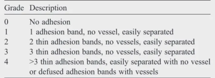
Table 1 Canbaz adhesion severity scoring method Grade Description
0 No adhesion
1 1 adhesion band, no vessel, easily separated 2 2 thin adhesion bands, no vessels, easily separated 3 3 thin adhesion bands, no vessels, easily separated 4 N3 thin adhesion bands, easily separated with no vessel
1.2. Histologic examination
For light microscope analysis, tissue samples from the cecum were obtained from all animals. To avoid mucosal injury, the intestinal lumen was carefully cannulated and gently washed with normal saline solution before the sampling. The cecum samples were fixed in 10% neutral-buffered formalin solution for 2 days. The specimens were fixed in paraffin after dehydration with rising concentrations of ethanol and xylene. Embedded tissues of 5 μm in thickness were sectioned by Leica RM 2125 RT (Bensheim, Germany). Randomly selected slices were stained with hematoxylin and eosin. Histopathologic examinations were performed by 2 pathologists blinded to the groups and photographed with a Nikon eclipse E 600 (Japan). Histologic features of mucosal, submucosal, and serosal architecture; edema; proliferation of capillary and fibroblast; collagen formation; and leukocyte infiltration were evaluated.
1.3. Statistical analysis
Statistical analyses were performed using the SPSS statistical package (version 15.0; SPSS, Chicago, IL). Categorical variables between groups were analyzed using Kruskal-Wallis and Jonckheere-Terpstra tests. Comparison of adhesion score between 2 groups was examined using the Mann-Whitney U test, where the data were nonnormal. A P value of less than .05 indicated statistical significance.
2. Results
All rats completed the study. The distribution of animals according to adhesion formation and median adhesion scores of each group is listed in Table 2. The adhesion scores of groups II and III were significantly lower when compared with group I (P = .02). Group III had lower adhesion scores than did group II (PN .05).
In all group I specimens, the histologic features showed regular appearance of mucosal architecture including the surface and crypt epithelia, lamina propria constituents, and muscularis mucosae (FigA and A1). In certain areas of the submucosa, disintegration of the tissue and edema was observed. When we evaluated the specimens in a systematic fashion, that is, assessment of mucosa, submucosa, muscu-laris, and serosa structures, the thickness of the serosa was well marked. In the serosal layer, we observed edema in certain areas (FigA and A1). Group I presented no alteration to normal tissue. In the specimens of group II (FigB), the covering epithelial tissue was intact with the enterocytes and goblet cells. Mild congestion and edema were present in the lamina propria and submucosa. A clear increment of the serosal layer thickness was observed. Proliferation of capillaries was evident. The qualitative increase in the number offibroblasts was seen. Polymorphonuclear leuko-cyte infiltrations—especially eosinophiles—were wide-spread in the serosal layer (Fig B). The mesothelial cells were intact. Group III specimens (FigC, C1) represented a mild edema in the lamina propria and submucosa similar to the group II. Capillary proliferation was markedly decreased in contrast to group II. Regression of leukocyte infiltrations was observed. The collagen was tightly organized, and the edema of the serosal layer was not observed.
3. Discussion
In the present study, we evaluated the efficacy of different pulmonary surfactants on the formation of postoperative abdominal adhesions. This study is the first to evaluate the effect of different animal-derived pulmonary surfactants (which are usually used to treat respiratory distress syndrome [RDS] in the prevention of postoperative intraabdominal adhesions.
Peritoneal injury related to surgical trauma, ischemia, desiccation, thermal injury, foreign body, and infection may lead to adhesion formation by increasing vascular perme-ability and secretion offibrin-reach exudate. Reduced tissue plasminogen activity and elevated inflammatory cytokines such as tumour necrosis factor, interleukins 1 and 6, and plasminogen activator inhibitors 1 and 2 are related to adhesion formation [19,20]. As fibrinolytic activity is suppressed, thefibrin deposition persists. Once fibrin bands are infiltrated with fibroblasts, they become organized into adhesions[21].
Studies have focused on the various steps of this pathophysiologic process. Anti-inflammatory agents, anti-oxidants, anticoagulants, fibrinolytics, and bioresorbable physical barriers have been used in an attempt to prevent adhesions[22]. Pulmonary surfactants are used to treat RDS in preterm infants. They contain several phospholipids, neutral lipids, and specific proteins. It has been speculated that phospholipids acts as both a superb boundary (solid to solid) lubricant and a release (antistick) agent. Chen and
Table 2 Distribution of rats according to adhesion between groups
Adhesion score Group I (control; n = 6) Group II (poractant; n = 6) Group III (beractant; n = 6) 0 – 2 3 1 2 1 3 2 3 3 – 3 – – – 4 1 – – Median (minimum-maximum) 2 (1-4) 1.5 (0-2)⁎ 0.5 (0-1)⁎,#
⁎P b .05 when compared with group I.
Hills [23] showed the absorption of 1,2-dipalmitoylpho-sphatidylcholine, the major constituent of pulmonary surfactants, by mesothelium and the formation of a layer on the peritoneum afterward. This newly formed oligola-mellar layer is shown to cover the whole surface of the visceral and parietal peritoneum with a small amount offluid during the healing of the serosal defects[24,25]. Pulmonary surfactants can be used for postoperative adhesion preven-tion as off-label drugs with the lubricanting, releasing, and formatting of a layer activity of phospholipids, especially phosphatidylcholine.
Previous studies have also shown the beneficial effects of phospholipids in the prevention of abdominal adhesion. Ar'Rajab et al[11,12]found phospholipids at a dose of 20 to 40 mg/rat effective in reducing adhesions. Jansen et al[26]
and Muller et al[13,27,28]studied various dosage regimens of phospholipids from 30 to 70 mg/kg with rat and rabbit adhesion models and showed beneficial effects of
phospho-lipids. Our study showed that adhesion scores of the poractant and beractant groups were significantly lower than those of the control group, whereas the beractant group had lower adhesion score than did the poractant group, but the difference did not reach significance. Surfactant groups had a higher number of adhesion-free cases than did the control group. Chen et al [29] evaluated the properties of phospholipids derived from patients receiving peritoneal dialysis and showed that phosphatidylcholine deficiency was related to adhesion formation and suggested that surface active phospholipids for adhesion prevention should mainly include phosphatidylcholine. Chen and Hills[23]noted that 25 mg/kg phospholipid as dry powder containing phospha-tidylcholine and phosphatidylglycerol with 7:3 ratio was effective in the prevention of adhesions in a rat model. Bhandarkar et al[30]studied 35 to 50 mg/kg phospholipid as dry powder containing phosphatidylcholine and phos-phatidylglycerol with 7:3 ratio in a rabbit adhesion model
Fig Cecum micrographs stained with hematoxylin and eosin. A, Micrographs on thefirst line represents group I. B, Micrographs on the second line represents group II. C, Micrographs on the third line represents group III. The magnifications of objectives are as follows; A, B, and C: ×20; A1 and C1: ×40; and B1: ×60. A, The structure of cecum with mucosa (Mc), crypts of Lieberkühn (Lc), muscularis mucosae (Mm), submucosae (SM), muscular layer (M), and edema (e) in the serosa (S). B, Polymorphonuclear leukocyte infiltration (⁎) and the capillaries (c). C, Morphology close to the regular structure of cecum.
and showed a beneficial effect of phospholipid. Phospho-lipid volume was used as 10 mL/kg in these studies. In our study, we used a drug volume of 2 mL/rat (about 8 mL/kg). We used 100 mg/kg poractant and 100 mg/kg beractant in study groups II and III. These are the standard doses used to treat RDS. Either of the doses contains 25 mg phospholipids including 9 mg phosphatidylcholine in poractant and 11 to 15 mg phosphatidylcholine in beractant [31]. Poractant is prepared from pig lung, whereas beractant is prepared from bovine lung. In our study, both phospholipid and phospha-tidylcholine doses, and drug volume administration were lower than other studies. However, this study showed the effectiveness of these doses of phospholipid and phospha-tidylcholine in terms of prevention of adhesion formation. The macroscopic evaluation revealed better adhesion scores for either surfactant therapy when compared with the control group. Beractant and poractant are widely available drugs, and this is another advantage of these drugs from other previously used pharmacologic agents, especially experi-mental agents for preventing postoperative adhesions.
In this study, we also found that beractant is more effective than poractant, with thefindings of lower adhesion scores, higher adhesion-free cases, and better histologic findings. Because either drug consists of the same proportion of the phospholipids, this affect might be associated with higher phosphatidylcholine dosage and drug volume of beractant, and this properties could be associated with better layer formation activity over the visceral peritoneum.
Macroscopic and histopathologic evaluation showed that poractant and beractant had better histologic findings than did the control group, whereas beractant had better histologic results than did poractant. Microscopic evaluation of group I showed disintegration of the tissue and edema in the submucosa, whereas it was edema in the serosa layer, indicating the success of the adhesion-producing model. In group II, only mild edema was seen in the serosal layer with clear increment. Proliferation of capillaries, fibroblasts, and leukocyte were observed in the serosal layer. Group III had lower capillary and fibroblast proliferation, leukocyte infiltration, and edema. These results showed efficacy of pulmonary surfactants in the prevention of postoperative adhesions. Serosal interaction with the drug may be better in group III than in group II because of drug volume, had a higher phosphatidylcholine content of beractant, and may be associated with better histologic results.
One interesting finding of the study is the probable fibrinolytic effect of surfactants in preventing intraabdominal adhesions. Reduced fibrinolytic activity was found in the lungs of newborns with surfactant deficiency[32]. It is not known whether or not surfactant therapy has any effects on reduced fibrinolytic activity. Reduced fibrinolytic activity seems to be associated with the formation of postoperative intraabdominal adhesions. Surfactant also has an anti-inflammatory effect in the treatment of RDS. Owing to the findings observed in this study, it can be speculated that pulmonary surfactant might improve fibrinolytic and
anti-inflammatory activity, which may play a role in preventing adhesion formation.
We showed that pulmonary surfactants, especially beractant, were effective in preventing adhesions in an experimental model. Lower adhesion scores and 50% adhesion-free cases were noted in the beractant-treated group. Further animal and clinical studies are needed to corroborate thesefindings and determine the efficacy, safety, and most appropriate dosage of pulmonary surfactants in the prevention of postoperative adhesions.
References
[1] Ergul E, Korukluoglu B. Peritoneal adhesions: facing the enemy. Int J Surg 2008;6:253-60.
[2] Holmdahl L, Risberg B, Beck DE, et al. Adhesions: pathogenesis and prevention-panel discussion and summary. Eur J Surg 1997(Suppl): 56-62.
[3] Ellis H, Moran BJ, Thompson JN, et al. Adhesion-related hospital readmissions after abdominal and pelvic surgery: a retrospective cohort study. Lancet 1999;353:1476-80.
[4] Liakakos T, Thomakos N, Fine PM, et al. Peritoneal adhesions: etiology, pathophysiology, and clinical significance. Recent advances in prevention and management. Dig Surg 2001;18:260-73.
[5] Reijnen MM, Bleichrodt RP, van Goor H. Pathophysiology of intra-abdominal adhesion and abscess formation, and the effect of hyaluronan. Br J Surg 2003;90:533-41.
[6] Mahdy T, Mohamed G, Elhawary A. Effect of methylene blue on intra-abdominal adhesion formation in rats. Int J Surg 2008;6:452-5. [7] DeCherney AH, diZerega GS. Clinical problem of intraperitoneal
postsurgical adhesion formation following general surgery and the use of adhesion prevention barriers. Surg Clin North Am 1997;77: 671-88.
[8] Turkcapar AG, Ozarslan C, Erdem E, et al. The effectiveness of low molecular weight heparin on adhesion formation in experimental rat model. Int Surg 1995;80:92-4.
[9] Hills BA, Burke JR, Thomas K. Surfactant barrier lining peritoneal mesothelium: lubricant and release agent. Perit Dial Int 1998;18: 157-65.
[10] Treutner KH, Bertram P, Lerch MM, et al. Prevention of postoperative adhesions by single intraperitoneal medication. J Surg Res 1995;59: 764-71.
[11] Ar'Rajab A, Ahren B, Rozga J, et al. Phosphatidylcholine prevents postoperative peritoneal adhesions: an experimental study in the rat. J Surg Res 1991;50:212-5.
[12] Ar'Rajab A, Snoj M, Larsson K, et al. Exogenous phospholipid reduces postoperative peritoneal adhesions in rats. Eur J Surg 1995;161:341-4.
[13] Muller SA, Treutner KH, Tietze L, et al. Influence of intraperitoneal phospholipid dosage on adhesion formation and wound healing at different intervals after surgery. Langenbecks Arch Surg 2001;386: 278-84.
[14] Muller SA, Treutner KH, Haase G, et al. Effect of intraperitoneal antiadhesive fluids in a rat peritonitis model. Arch Surg 2003;138: 286-90.
[15] Goerke J. Pulmonary surfactant: functions and molecular composition. Biochim Biophys Acta 1998;1408:79-89.
[16] National Research Council of The National Academias. Guide for the Care and Use of Laboratory Animals. http://grants.nih.gov/grants/ olaw/Guide-for-the-Care-and-Use-of-Laboratory-Animals.pdf. [17] Hemadeh O, Chilukuri S, Bonet V, et al. Prevention of peritoneal
adhesions by administration of sodium carboxymethyl cellulose and oral vitamin E. Surgery 1993;114:907-10.
[18] Canbaz MA, Ustun C, Kocak I, et al. The comparison of gonadotropin-releasing hormone agonist therapy and intraperitoneal Ringer's lactate solution in prevention of postoperative adhesion formation in rat models. Eur J Obstet Gynecol Reprod Biol 1999;82:219-22. [19] van Goor H, de Graaf JS, Grond J, et al. Fibrinolytic activity in the
abdominal cavity of rats with faecal peritonitis. Br J Surg 1994;81: 1046-9.
[20] Ivarsson ML, Falk P, Holmdahl L. Response of visceral peritoneum to abdominal surgery. Br J Surg 2001;88:148-51.
[21] Gomel V, Urman B, Gurgan T. Pathophysiology of adhesion formation and strategies for prevention. J Reprod Med 1996;41:35-41. [22] Yetkin G, Uludag M, Citgez B, et al. Prevention of peritoneal adhesions by intraperitoneal administration of vitamin E and human amniotic membrane. Int J Surg 2009;7:561-5.
[23] Chen Y, Hills BA. Surgical adhesions: evidence for adsorption of surfactant to peritoneal mesothelium. Aust N Z J Surg 2000;70:443-7. [24] Beavis J, Harwood JL, Coles GA, et al. Synthesis of phospholipids by
human peritoneal mesothelial cells. Perit Dial Int 1994;14:348-55. [25] Hills BA. Graphite-like lubrication of mesothelium by oligolamellar
pleural surfactant. J Appl Physiol 1992;73:1034-9.
[26] Jansen M, Treutner KH, Jansen PL, et al. Phospholipids reduce the intraperitoneal adhesion of colonic tumor cells in rats and adhesion on extracellular matrix in vitro. Int J Colorectal Dis 2004;19: 525-32.
[27] Muller SA, Treutner KH, Jorn H, et al. Phospholipids reduce adhesion formation in the rabbit uterine horn model. Fertil Steril 2002;77: 1269-73.
[28] Muller SA, Treutner KH, Tietze L, et al. Efficacy of adhesion prevention and impact on wound healing of intraperitoneal phospho-lipids. J Surg Res 2001;96:68-74.
[29] Chen Y, Hills BA, Hills YC. Unsaturated phosphatidylcholine and its application in surgical adhesion. ANZ J Surg 2005;75:1111-4. [30] Bhandarkar DS, Nathanson LK, Hills BA. Spray of phospholipid
powder reduces peritoneal adhesions in rabbits. Aust N Z J Surg 1999;69:388-90.
[31] Young TE. Respiratory drugs. In: Young TE, editor. Neofax. Montvale: Thomson Reuters; 2010. p. 284-7.
[32] Cederqvist K, Siren V, Petaja J, et al. High concentrations of plasminogen activator inhibitor-1 in lungs of preterm infants with respiratory distress syndrome. Pediatrics 2006;117:1226-34.