i
BIOACTIVE GLYCOPEPTIDE NANOFIBERS FOR
TISSUE REGENERATION APPLICATIONS
A THESIS SUBMITTED TO
THE GRADUATE SCHOOL OF ENGINEERING AND SCIENCE OF BILKENT UNIVERSITY
IN PARTIAL FULFILLMENT OF THE REQUIREMENTS FOR THE DEGREE OF
MASTER OF SCIENCE IN
MATERIALS SCIENCE AND NANOTECHNOLOGY
By
ÖZÜM ŞEHNAZ ÇALIŞKAN
May 2016
ii
BIOACTIVE GLYCOPEPTIDE NANOFIBERS FOR TISSUE
REGENERATION APPLICATIONS By Özüm Şehnaz Çalışkan
May 2016
We certify that we have read this thesis and that in our opinion it is fully adequate, in scope and in quality, as a thesis for the degree of Master of Science.
Mustafa Özgür Güler (Advisor)
Ayşe Begüm Tekinay (Co-Advisor)
Hüseyin Özkan
Tarık Baytekin
Approved for the Graduate School of Engineering and Science:
Levent Onural
iii
ABSTRACT
BIOACTIVE GLYCOPEPTIDE NANOFIBERS FOR TISSUE
REGENERATION APPLICATIONS
Özüm Şehnaz Çalışkan
M.S.in Materials Science and Nanotechnology Advisor: Mustafa Özgür Güler
Co-Advisor: Ayse Begüm Tekinay May, 2016
Natural extracellular matrix (ECM) is rich in glycopeptides and glycosaminoglycans, which function in controlling cellular processes. In this thesis, glycopeptide molecules that mimic natural glycopeptides and glycosaminoglycans were designed and synthesized and it was demonstrated that they induce directed differentiation of mesenchymal stem cells into chondrogenic and adipogenic lineages.
In the first part of the study, hyaluronic acid (HA)-mimicking glycopeptide amphiphile molecules were synthesized to induce chondrogenic differentiation of mesenchymal stem cells (MSC). HA is the most abundant glycosaminoglycan (GAG) found in hyaline cartilage ECM. Peptide amphiphiles were synthesized by solid phase peptide synthesis method and used to form self-assembled bioactive glycopeptide nanofibers which mimic fibrous morphology of the ECM. Scanning electron microscopy (SEM), transmission electron microscopy (TEM), and circular
iv
dichroism (CD) were used for morphology and secondary structure analyses of the obtained nanofibers. It was demonstrated that glycopeptide amphiphiles create fibrous structure formed by nanofibers. Morphological changes, GAG production (Safranin-O staining and DMMB analysis), and chondrogenic gene marker expressions (qRT-PCR) of MSCs cultured on HA-mimetic nanofibers were analyzed. It was shown that HA-mimetic glycopeptide nanofibers induce early differentiation of MSCs into hyaline like chondrocytes.
In the second part of the study, it was demonstrated that minor changes on glycopeptide backbone can create specific glycopeptides which induce differentiation of MSCs into brown adipocytes. Brown fat adipocytes do not store chemical energy as fat but dissipates it as heat and so they have emerged as promising anti-obesity agents. Lipid droplet accumulation (Oil Red-O staining) and adipogenic gene marker expression analyses (qRT-PCR) showed that the new glycopeptide nanofiber scaffold is a specific inducer of differentiation of MSCs into brown fat adipocytes.
Keywords: Peptide amphiphile, peptide nanofiber, glycosaminoglycan, glycopeptide, mesenchymal stem cell, extracellular matrix, hyaluronic acid, mesenchymal stem cell differentiation, cartilage tissue, tissue regeneration, brown fat tissue, adipogenesis.
v
ÖZET
BİYOAKTİF GLİKOPEPTİT NANOFİBERLERİN DOKU
REJENERASYONU UYGULAMALARI
Özüm Şehnaz Çalışkan
Malzeme Bilimi ve Nanoteknoloji, Yüksek Lisans Tez Danışmanı: Mustafa Özgür Güler Tez Eşdanışmanı: Ayşe Begüm Tekinay
Mayıs, 2016
Doğal hücreler arası matris (HAM), hücresel olayları kontrol eden glikopeptit ve glikozaminoglikanlar bakımından zengin bir yapıdır. Bu tez çalışmasında, doğal glikopeptitleri ve glikozaminoglikanları taklit eden ve mezenkimal kök hücrelerin (MKH) kıkırdak ve yağ hücrelerine farklılaşmasını tetikleyen glikopeptit moleküller tasarlanmış ve sentezlenmiştir.
Çalışmanın ilk bölümünde, hyaluronik asidi (HA) taklit eden glikopeptit amfifil molekülleri MKH’lerin kıkırdak hücre hattına farklılaşmasını tetiklemek amacıyla sentezlenmiştir. HA, camsı kıkırdak HAM’inde en fazla miktarda bulunan glikozaminoglikandır. Peptit amfifil molekülleri katı fazlı peptit sentezi yöntemi ile sentezlenmiş ve HAM’in fibröz yapısını taklit etmek amacıyla kendiliğinden bir araya gelen biyoaktif glikopeptit nanofiberler oluşturulması için kullanılmışlardır.
vi
Taramalı ve geçirimli elektron mikroskopisi ve dairesel dikorizm yöntemleri ile elde edilen nanofiberlerin ikincil yapıları ve morfolojileri incelenmiş, glikopeptit amfifillerin nanofiberlerden oluşan boşluklu fibröz bir yapı oluşturduğu gösterilmiştir. HA’yı taklit eden nanofiberler üzerinde kültürlenen MKH’lerin morfolojik değişimleri, GAG üretim miktarları (Safranin-O boyaması ve DMMB analizi) ve kondrojenik gen işaretleyicilerinin ifade seviyeleri (gerçek zamanlı RT-PZR) analiz edilmiştir. Analizler sonucunda HA-taklidi glikopeptit nanofiberlerin MKH’lerin camsı kıkırdak hücrelere farklılaşmasını sağladığı ve bunu erken dönemde başlattığı gösterilmiştir.
Çalışmanın ikinci kısmında, glikopeptit omurgasında yapılan kimyasal değişimler sayesinde MKH’lerin kahverengi yağ hücrelerine dönüşümü gösterilmiştir. Kahverengi yağ hücreleri kimyasal enerjiyi yağ olarak depolamaz; bunun yerine ısı olarak harcarlar. Bu sayede umut vaat eden anti-obezite ajanları olarak kabul edilmektedirler. Lipit damla birikimi (Oil-Red O boyası) ve adipojenik gen işaretleyicilerinin ifade seviyelerinin incelenmesi (gerçek zamanlı RT-PZR) ile elde ettiğimiz sonuçlar, bu glikopeptitnanofiber iskelenin MKH’lerin kahverengi yağ hücrelerine dönüşmesinin spesifik tetikleyicisi olduğunu göstermiştir.
Anahtar Sözcükler: Peptit amfifil, peptit nanofiberler, glikozaminoglikan, glikopeptit, mezenkimal kök hücre, hücrelerarası iskele, hyaluronik asit, mezenkimal kök hücre farklılaşması, kıkırdak doku, doku rejenerasyonu, kahverengi yağ dokusu, adipojenez.
vii
Acknowledgement
Time I spent at UNAM has been one of the important periods of my life. I felt this time course scientifically and personally developed me. I would like to express my appreciations to all people who contribute this process either by their experiences, knowledge, help or their mental and emotional support.
I would like to express the deepest gratitude to my advisor Prof. Mustafa Özgür Güler and to my co-advisor Prof. Ayşe Begüm Tekinay who are the leading roles of this period. They gained me different scientific perspectives, let me be part of their research groups, and patiently followed my maturation and completion of this work. Their immense knowledge, encouragement and guidance are invaluable.
I would like to thank our old post-doc Dr. Ashif Shaikh for sharing his knowledge, valuable collaboration and for supplying me GlcNAc-PA. I would like to express my sincere appreciations to Dr. Seher Üstün Yaylacı, who taught me in vitro experiments for her valuable supports. I want to express my special thanks to our cartilage sub-group members Çağla Eren, İbrahim Çelik, Özge Uysal, and separately to Elif Arslan who is old-hand member of our group for sharing her experienced knowledge. We shared lots of knowledge, experiences and help as cartilage group. I would like to express my special thanks to Dr. Gözde Uzunallı. She taught, supervised and helped me a lot although she was also new in her chondrogenesis studies.
I learnt something from every people that I communicated through this period but I owe more special thanks to some of them who shared a lot their valuable time
viii
with me and contributed to this study. I express my sincere appreciation to Melike Sever for her kind help for qRT-PCR experiments and analyses. I owe her a lot. I am grateful to Meryem Hatip for going through all TEM images for selection and her kind support. I appreciate Merve Şen for her help in primer design. I would like to express my special thanks to Gökhan Günay and Seren Hamsici, who also make this master period enjoyable, for their help in FACS analyses and friendship. I would also thank to our new post-doc Bhavna Rajasekaran for her kind help about confocal microscopy. I also acknowledge Yasin Tümtaş and Öncay Yaşa, whom I learnt experimental details about adipogenesis study for their help and legating their materials to me.
I owe very special thanks to two people; Zeynep Orhan and Nurcan Haştar, for their excellent collaborations in projects that we took part together. This thesis study would not be completed without their help; their efforts are invaluable. I would like to express my most sincere, deepest gratitude to them.
I would like to give my special thanks to Göksu Çınar, Egemen Deniz Eren, and Hatice Kübra Kara for their warm friendship, and I would also like to acknowledge my old and present lab and office members Gülistan Tansık, M. Aref Khalily, Dr. Ruslan Garifullah, Gülcihan Gülseren, Nuray Gündüz, Berna Şentürk, Dr. Rashad Mammadov, Dr. Büşra Mammadov, Melis Göktaş, Dr. Özlem Erol, Dr. Hakan Ceylan, Oya İlke Şentürk, Begüm Kocatürk, Dr. Aslı Çelebioğlu, Zeynep Aytaç, Yelda Ertaş, Dr. Fatma Kayacı, Dr. Okan Öner Ekiz, Seylan Ayan, Sude Selin Su Yirmibeşoğlu, Hepi Hari Susapto, İ. Ceren Garip, Dilek Sezer, İkra Gizem Yıldız, Elif Ergül, Aygül Zengin, Mevhibe Geçer, Mustafa Beter, Canelif Yılmaz, Idil Uyan, Fatih Yergöz, Şehmus Tohumeken, Oğuz Tuncay, Ahmet Emin Topal, Alper Devrim
ix
Özkan and Göksemin Şengül for creating a peaceful, warm and so an excellent working environment.
And Melis Şardan Ekiz, a new PhD, a wise supervisor, a good collaborator and a dear friend; she deserves the sincerest gratitude. I am really grateful for her help, the PAs that she supplied me and for her emotional support.
My special thanks are to Zeynep Erdoğan, whom I learnt a lot and to Mustafa Güler, for their technical helps. I would also like to thank our janitorial laborers for providing us a clean environment and doing it in a cheerful way. I especially thank to Suna Abla and Hatice Abla for spreading their happiness with their good-humor.
I would like to thank the National Nanotechnology Research Center (UNAM) and Bilkent University for providing pleasant facilities and latest equipments, and to thank The Scientific and Technological Research Council of Turkey (TÜBİTAK BIDEB 2210-C, 213M406, 113T045) for partial financial support.
I would like to express my appreciations to MBG department of Bilkent University and to İhsan Gürsel for letting me use their departmental and private centrifuges.
I am grateful to my old and gold friends Betül Savaş, Selen Güzel, Seda Karakaş, Özge Taşdan, Nazlı Sezek and Gülşah Zorgör who had always been there for me. I also convey my thanks to my dear undergraduate departmental friends Gizem Güneş, Ayça Yörükoğlu and Burcu Nur Keçeli who are thousands of miles away but pursuing a similar career for their warm friendship. They are the ones who can understand the feelings of a graduate student.
x
I would like to thank my whole family; from grandmothers to aunts they wish me the best. I would like to give my sincere gratitude to my new family, the Çalışkans, for their warm and peaceful support and encouragement.
I would like to express my gratitude to my mother, my father and my little sister separately. My mother who always believes in me, whom I know will always be there for me unconditionally, my father who was being with me during his short winter break by coming Ankara, cooking and carrying the dinner for me and being with me here while I performing experiments, and my little sister who is my emotional support in Ankara, who is always humorous just the way she is, they gave their best encouragements and supports me in the way I am. Their presence in my life is priceless.
His support, encouragement and help to complete my thesis are invaluable but I want to give my deepest, most amative thanks to Okan Çalışkan, my beloved one, for the hearty laughter that we had, for his excellent perception which can always intuit my feelings, and for the miles that we took separately but to reach each other.
xi
Contents
ABSTRACT ... iii ÖZET... v CHAPTER 1 ... 1 INTRODUCTION ... 11.1 A brief introduction to tissue regeneration ... 2
1.2 Tissue Types ... 4
1.2.1 Cartilage Tissue 5
1.2.2 Adipose Tissue 8
1.3 Glycosaminoglycans (GAGs): Chemistry, Functions, Regenerative Potentials, and Applications ... 9
1.3.1 Chemical and functional properties of glycosaminoglycans 11 1.3.2 GAG types: a more detailed explanation 12 1.3.3 Roles of GAGs in biology 15 1.3.4 GAG-protein interactions 19 1.4 Peptide Amphiphiles ... 20
CHAPTER 2 ... 22
EXPERIMENTAL ... 22
2.1 Materials ... 23
2.2 α-N-Acetyglucosamine Carboxylic Acid Synthesis ... 24 2.2.1 Synthesis of Allyl-N-Acetyl-α-D-Glucopyronoside-3,4,6 Triacetate .. 24
xii
2.2.2 Synthesis of N-Acetyl-α-D-Glucopyronoside-3,4,6 Triacetate
Carboxylic Acid 25
2.3 Synthesis, Identification and Purification of Peptide Amphiphile Molecules ... 25
2.3.1 Synthesis of Peptide Amphiphile Molecules 25 2.3.2 Characterization of Peptide Amphiphile Molecules 28 2.3.3 Purification of Peptide Amphiphile Molecules 28 2.4 Formation of Peptide Amphiphile Nanostructures, Spectroscopy and Morphological Characterizations ... 29
2.4.1 Formation of Self-Assembled PA Nanostructures 29
2.4.2 Circular Dichorism 29
2.4.3 Scanning Electron Microscopy Imaging 30
2.4.4 Transmission Electron Microscopy Imaging 31
2.5 Cell Culture and Maintenance ... 31 2.6 Cell viability, Adhesion and Proliferation Analyses ... 31
2.6.1 Viability Assay: MTT assay 31
2.6.2 Cell Adhesion 32
2.6.3 Cell Proliferation 33
2.7 Cell Differentiation Analyses ... 33 2.7.1 Sulfated Glycosaminoglycan Production Analyses 33
2.7.2 Oil Red O Staining 35
2.7.3 Real-Time Gene Expression Analysis 35
xiii
CHAPTER 3 ... 38 GLYCOPEPTIDE NANOFIBERS FOR CELL DIFFERENTIATION INTO HYALINE CHONDROCYTES ... 38
3.1 INTRODUCTION ... 39 3.2 RESULTS ... 40 3.2.1 Synthesis and Characterization of α-N-acetylglucosamine Allyl
Glycoside 40
3.2.2 Synthesis and Characterization of Peptide Amphiphile Molecules 43
3.2.3 PA Nanofiber Formation 48
3.2.4 Characterizations of PA Nanofibers 50
3.2.5 Biocompatibility of Peptide Amphiphile Nanofiber Networks 53 3.2.6 Proliferation Analyses of rMSCs on PA Nanofiber Network 55 3.2.7 Adhesion Analyses of rMSCs on PA Nanofiber Network 56
3.2.8 sGAG Production Analyses 57
3.2.9 Gene Expression Analyses of Chondrogenic Differentiation Gene
Markers 63
3.3 DISCUSSION ... 72 3.4 CONCLUSION ... 76 CHAPTER 4 ... 77 MONITORING EFFECTS OF SINGLE AMINO ACID CHANGE ON DIFFERENTIATION FATE OF rMSCs ... 77
4.1 INTRODUCTION ... 78 4.2 RESULTS ... 80
xiv
4.2.1 Synthesis and Characterization of Peptide Amphiphile Molecules 80
4.2.2 PA Nanofiber Formation 86
4.2.3 PA Nanofiber Characterizations 87
4.2.4 Cell Viability on Peptide Amphiphile Nanofiber Networks 90 4.2.5 Adhesion Profile of rMSCs on PA Nanofibers 91 4.2.6 Effects of PA Nanofiber Network on Cell Proliferation 93 4.2.7 Investigation the Differentiation Response of rMSCs to Different
Glucose Bearing Nanofiber Scaffolds 94
4.3 DISCUSSION ... 104
4.4 CONCLUSION ... 110
CHAPTER 5 ... 111
CONCLUSIONS AND FUTURE PERSPECTIVES ... 111
xv
List of Figures
Figure 2.1 Synthesis of 2-Acetamido 2-deoxy-a-D-glucopyranose 3,4,6 triacetate carboxylic acid derivative ... 24 Figure 2.2 Schematic representation of the sequential synthesis of amphiphilic glycopeptide.. ... 27 Figure 3.1 1H-NMR spectra of Allyl-N-Acetyl-α-D-Glucopyronoside-3,4,6 triacetate. ... 41 Figure 3.2 Mass spectrum of Allyl-N-Acetyl-α-D-Glucopyronoside-3,4,6 triacetate.. ... 41 Figure 3.3 1H-NMR spectra of N-Acetylglucosamine-E-PA. N-Acetyl-α-D-Glucopyronoside-3,4,6 triacetate carboxylic acid coupled to the Lauryl-VVAGEK-Am backbone from the side chain of lysine. ... 42 Figure 3.4 Mass spectrum of N-Acetyl-α-D-Glucopyronoside-3,4,6 triacetate carboxylic acid ... 42 Figure 3.5 Chemical structures of the peptide amphiphiles ... 44 Figure 3.6 Liquid chromatography-mass spectrometry (LC-MS) analysis of GlcNAc-PA ... 45 Figure 3.7 Liquid chromatography-mass spectrometry (LC-MS) analysis of Glc-PA. ... 46 Figure 3.8 Liquid chromatography-mass spectrometry (LC-MS) analysis of K-PA. 47 Figure 3.9 Liquid chromatography-mass spectrometry (LC-MS) analysis of E-PA. 48 Figure 3.10 CD spectra of peptide amphiphile nanofibers at physiological pH. ... 50 Figure 3.11 CD spectra of single peptide amphiphile solutions at physiological pH.. ... 51
xvi
Figure 3.12 SEM micrographs of PA scaffolds ... 52 Figure 3.13 TEM (a,b) and STEM (c,d) micrographs of PA nanofibers. ... 53 Figure 3.14 Relative cellular viability of rMSCs cultured on PA nanofiber networks and on bare surface after 12 h incubation. ... 54 Figure 3.15 Relative proliferation of rMSCs cultured on PA nanofiber networks and on bare surface after 48 h incubation. ... 55 Figure 3.16 Relative adhesion of rMSCs cultured on PA nanofiber networks and on bare surface after 5 h incubation.. ... 57 Figure 3.17 Safranin-O staining of rMSCs in growth medium at day 3, showing the extent of sulfated glycosaminoglycan incorporation. ... 59 Figure 3.18 Safranin-O staining of rMSCs in growth medium at day 7, showing the extent of sulfated glycosaminoglycan incorporation. ... 59 Figure 3.19 Safranin-O staining of rMSCs in growth medium at day 14, showing the extent of sulfated glycosaminoglycan incorporation. ... 60 Figure 3.20 sGAG levels of rat mesenchymal stem cells at day 3, analyzed by DMMB assay ... 61 Figure 3.21 sGAG levels of rat mesenchymal stem cells at day 7, analyzed by DMMB assay. ... 62 Figure 3.22 sGAG levels of rat mesenchymal stem cells at day 14, analyzed by DMMB assay ... 62 Figure 3.23 Gene expression analyses of rMSCs cultured with growth medium at different time points.. ... 65 Figure 3.24 Sox9 gene expression analyses of rMSCs cultured with growth medium at different time points. ... 66
xvii
Figure 3.25 Gene expression analyses of rMSCs cultured with growth medium at day 3. ... 69 Figure 3.26 Gene expression analyses of rMSCs cultured with growth medium at day 7. ... 70 Figure 3.27 Gene expression analyses of rMSCs cultured with growth medium at day 14.. ... 71 Figure 4.1 Chemical structures of the peptide amphiphiles. ... 81 Figure 4.2 Liquid chromatography-mass spectrometry (LC-MS) analysis of E-Glc-PA.. ... 83 Figure 4.3 Liquid chromatography-mass spectrometry (LC-MS) analysis of K-Glc-PA.. ... 84 Figure 4.4 Liquid chromatography-mass spectrometry (LC-MS) analysis of K-PA..85 Figure 4.5 Liquid chromatography-mass spectrometry (LC-MS) analysis of E-PA.. 86 Figure 4.6 CD spectra of peptide amphiphile nanofibers at physiological pH.. ... 88 Figure 4.7 CD spectra of single peptide amphiphile solutions at physiological pH. . 88 Figure 4.8 SEM micrographs of PA scaffolds.. ... 89 Figure 4.9 TEM (a,b) and STEM (c,d) micrographs of PA nanofibers. ... 90 Figure 4.10 Relative cellular viability of rMSCs cultured on PA nanofiber networks and on bare surface after 12 h incubation.. ... 91 Figure 4.11 Relative adhesion of rMSCs cultured on PA nanofiber networks and on bare surface after 5 h incubation.. ... 92 Figure 4.12 Relative proliferation of rMSCs cultured on PA nanofiber networks and on bare surface after 48 h incubation. ... 94
xviii
Figure 4.13 Optical microscope images of rat mesenchymal stem cells cultured on E-Glc-PA/K-PA nanonetwork after 7 days of incubation.. ... 95 Figure 4.14 Oil Red-O staining of cells in growth medium at day 7, showing the lipid droplet accumulation ... 97 Figure 4.15 Oil Red-O staining of cells in growth medium at day 11, showing the lipid droplet accumulation... 99 Figure 4.16 Adiponectin and FABP4 gene expression analyses of rat mesenchymal stem cells cultured with growth medium at day 7. ... 101 Figure 4.17 Adiponectin and FABP4 gene expression analyses of rat mesenchymal stem cells cultured with growth medium at day 11. ... 101 Figure 4.18 UCP1 gene expression analyses of rat mesenchymal stem cells cultured with growth medium at day 7 and day 11.. ... 102 Figure 4.19 Gene expression analyses of rat mesenchymal stem cells cultured with growth medium at different time points. ... 103
xix
List of Tables
Table 1.1 GAGs and their repeating dissacharide units ... 10 Table 2.1 Primer sequences and annealing temperatures of the genes used for qRT-PCR analyses. ... 37 Table 3.1 Sequences, molecular weights and theoretical overall charges of the peptide amphiphiles at neutral pH... 45 Table 4.1 Sequences, molecular weights and theoretical overall charges of the peptide amphiphiles at neutral pH... 82
xx
List of Abbreviations
ECM : Extracellular Matrix
PA : Peptide Amphiphile
GAG : Glycosaminoglycan
DIEA : N,N-diisopropylethylamine
TFA : Trifluoroacetic Acid
TIS : Triisoproplysilane
DMF : Dimethylformamide
DCM : Dichloromethane
LC-MS : Liquid Chromatography Mass Spectroscopy
HPLC : High Pressure Liquid Chromatography
CD : Circular Dichroism
SEM : Scanning Electron Microscopy
TEM : Transmission Electron Microscopy
FBS : Fetal Bovine Serum
DMEM : Dulbecco’s Modified Eagle Medium DMMB : Dimethylmethylene Blue
OD : Optical Density
PS : Penicillin/Streptomycin
BrdU : Bromodeoxyuridine
rMSC : Rat Mesenchymal Stem Cell
GAPDH : Glyceraldehyde 3-phosphate dehydrogenase
xxi
Col II : Collagen 2
Sox9 : Transcription Factor SOX-9
ADIPOQ : Adiponectin
FABP4 : Fatty Acid Binding Protein 4
UCP1 : Uncoupled Protein 1
1
CHAPTER 1
2
1.1 A brief introduction to tissue regeneration
Tissue failure or loss has been a major health problem throughout the history of humanity.1 It adversely affects the patient’s life quality and even most severe losses may cause deaths. Therefore, there has been a huge effort to address tissue loss problems since old human history. Drug treatment can be applied for the least severe cases2, however more severe cases require different treatment options. The other treatment options include artificial prostheses, surgical repair, mechanical device replacements and any type of transplantations (auto-, allo-, and xenograft).2,3 However, artificial prostheses or surgical repair do not meet the needs of a fully functional tissue or organ replacement. Transplantation then emerges as a best option, but it has certain drawbacks and limitations. Autografts are the best option for replacement, however, its excision creates another surgical site in the patient and it cannot satisfy the need if the required tissue size is large. The other option, allografts, can create immune response by the patient. In addition to this, to find an appropriate allograft donor is a really big challenge.1 According to current data supplied by the Ministry of Health of the Republic of Turkey, more than 28.000 patients are on transplant wait list in Turkey.4 Xenogafts, cross-species transplantation, offer an unlimited supply of organs but they have similar constraints with allograft transplantation. Besides, it is less likely that tissues or organs from different species match with humans.5 Then, mechanical devices emerge to answer the needs, but they are currently very limited in terms of variety.
Tissue regeneration by biomaterials, or tissue engineering, is emerging as a potential alternative and complementary solution. Tissue engineering can be defined as fabricating new and functional tissues by use of living cells, a matrix or scaffold
3
which guide the cells to proliferate, differentiate and to communicate with other cells for tissue development.2 Tissue engineering is an inter- and multidisciplinary field that combines cells, biomaterials and biological signals.6 These cells, the biomaterials and the interactions between the cells and biomaterials are the fundamentals of tissue engineering. Biomaterials are generally used as scaffolds. Scaffolds provide the structural support for cell attachment and subsequent tissue development.7 Scaffolds have to mimic the natural structure of tissue of interest. Physical, chemical and rheological structure of a scaffold is very important for tissue formation as it affects the cellular behaviors like differentiation, adhesion and proliferation.7 Applied cyclic load, electrical signals and extension/contraction of scaffold are the other factors that change the differentiation profile of the cells.8 These scaffolds can either be used to grow tissue in in vitro conditions9 or be applied in vivo to support the body to regenerate the tissue.10 As different tissues have different properties, there is a variety of scaffold types developed for different application purposes. Degradable, non-degradable, synthetic, natural, synthetic-natural hybrid, inert or bioactive scaffolds are noted in literature.11,12,13,14,15,16 The unchanging parameter for all type of scaffolds is cyto- or tissue compatibility.
Cell types have enormous influence on the formation of functional tissue type of interest. Progenitor-type cells or stem cells are used for tissue engineering and regeneration. Embryonic stem or embryonic germ line cells are non-differentiated cells and are able to differentiate into any type of cells.6 For this reason, they attract much attention for tissue engineering purposes. Stem cells also exist in adults; however, they are not able to differentiate into all kinds of cells into body. The tissue origin of the adult stem cells determines their differentiation capability and the
4
lineages that stem cells differentiate into.17 Therefore it is important to choose appropriate origin of stem cells depending on the type of tissue that is aimed to regenerate.
Growth factors have a huge impact to differentiate cells towards certain types. Growth factors are specific proteins that are secreted by paracrine or autocrine manner and play key roles in cellular behavior like proliferation and differentiation.6 Therefore growth factors, proteins and hormones that are involved in formation of tissue of interest have to be clearly identified and appropriate cocktail of the factors should be supplied to the environment for tissue regeneration and engineering.18 There are certain challenges about tissue engineering. First of all, behaviors of cells in normal development, factors that control differentiation, proliferation and communication between cells should be learned in detail in order to mimic and stimulate these behaviors in engineering process. Second, limited source of stem cells or limited number of progenitor-type cells are other challenges. Another challenge is the limitations over methods for scaffold fabrication. All these fields require more studies to understand and better address the needs of tissue engineering field.
1.2 Tissue Types
Human body is composed of different and complex types of tissues and organs. Each tissue has its own physical, physiological and mechanical characteristics. For tissue engineering and regeneration purposes, these characteristics have to be known in order to design appropriate scaffolds, choose the correct cell type and to create optimum environment by chemical factors. In this section, two different types of tissues are introduced briefly: cartilage and adipose tissue. These are the tissue types
5
into which differentiation of mesenchymal stem cells is induced by peptide amphiphile scaffolds in this thesis study.
1.2.1 Cartilage Tissue
Cartilage is a type of connective tissue with a dense but elastic structure. It is not rigid as bone tissue but also not elastic as muscle tissue, which are other types of connective tissues. Cartilage is found in the body where there is need for some structural support but also elasticity in a certain range due to its unique rheological properties. Cartilage functions as shock absorber, gives some support to body and creates friction free surfaces especially between the articular surfaces of bones.19 Its most important characteristic is that it lacks of blood vessels and nerves.20 Water (~75%), collagen (~20%) and proteoglycans (~5%) are the main constituents of cartilage.20 The ratios can change depending on the type of cartilage, age of the person and healthy structure of the cartilage.
Cartilage has four basic components: perichondrium, chondroblasts, chondrocytes and matrix. Perichondrium is an exception because fibrous and articular hyaline cartilages do not have perichondrium.21 Perichondrium is a vascularized connective tissue sheet that is rich in collagens (collagen I especially) and surrounds cartilage like a capsule.22 As perichondrium is the only vascularized part of the cartilage, it is at the same time the only source of the nourishment for cartilage. Nutrients diffuse from the capillaries that reside in perichondrium into the matrix.21 Cartilage thickness should be in a certain range as increased thickness hardens the diffusion of nutrients. Perichondrium is also the part where growth and repair of cartilage occurs.21 It consists of an outer fibrous layer and an inner chondrogenic layer.23 The
6
outer layer contains fibroblasts that secrete collagenous fibers. Inner layers host the chondroblasts and some undifferentiated mesenchymal cells. Chondroblasts differentiate from MSCs that reside in the perichondrium and are progenitors, or immature forms of chondrocytes.24 They secrete type II collagen and other components of the extracellular matrix.25 As they continue ECM production, they are entrapped by the ECM inside small spaces called lacunae.23 Inside lacunae, chondroblasts mature and turn into chondrocytes, which are the only cells found in healthy cartilage tissue. Chondrocytes are specialized cells that synthesize and maintain matrix infrastructure.26 Matrix is composed of fibers, either elastic or collagenous, and ground substance which is a gel-like substance surrounding the cells.22 Ground substance contains combinations of proteins and sugars, glycosaminoglycans (GAGs), most notably hyaluronic acid, proteoglycans and glycopeptides. These glycoproteins help anchor chondrocytes to matrix, maintain tissue integrity and mediate transmembrane signaling.27 Glycoproteins perform these tasks via their multiple binding sites to the receptors on chondrocyte surfaces.21 Presence of GAGs and proteoglycans make the matrix basophilic. Due to high negative charges that GAGs and proteoglycans have, they attract positive molecules and repel negative molecules and this increases ion concentration in the matrix. Increased ion concentration increases osmolarity of the cartilage and thereby high amount of water is attracted.22 Thus, GAGs and proteoglycans hold high amount of water indirectly by the increased osmolarity (Gibbs-Donnan effect) and directly via intermolecular hydrogen bonds.21,28
There are three types of cartilage; hyaline, elastic and fibrocartilage.27 The classification is done depending on the density and type of fibers presented in its
7
composition. Therefore, they slightly differ from each other and they have very similar histology, but their localization in the body is different.27 Hyaline cartilage localizes in tracheal rings, sterna margins of ribs and in the articular surfaces of joints where the bones meet with each other. Hyaline cartilage that is located in the articular surfaces may also be called as articular cartilage.29 Hyaline means transparent, glassy substance and as the name suggests, hyaline cartilage has a transparent appearance.30 It is the most abundant type of cartilage in the body. Chondrocytes constitute 1-1.5% volume of the hyaline cartilage.31 Water is the prominent component of articular hyaline cartilage. 80% of the superficial zone and 65% of the deep zone of articular hyaline cartilage matrix is composed of water.31 Resilience against load, which is a critical biomechanical function of articular hyaline cartilage, is attributed to this high amount of water.21 Major classes of proteoglycans found in hyaline cartilage are aggrecans32, large aggregating proteoglycans, and small proteoglycans like decorin and fibromodulin. Hyaluronic acid is the GAG that acts as central chain of proteoglycans. The principal component of the framework of hyaline cartilage is collagen type II, which forms the 90-95% of it.29 The matrix contains abundant amounts of collagen type II as thin fibrils and they provide tensile strength. Elastic cartilage cannot be differentiated from hyaline cartilage in histological staining, unless elastin fibers are stained.27 Matrix and perichondrium of elastic cartilage contains thick bundles of elastic fibers.21 It is found in ears, epiglottis and eustacian tube and it is not calcified with increasing age. Fibrocartilage is found in inter-vertebral disc. Collagen type I layers interrupting the matrix is the distinct feature of fibrocartilage.23 Another important characteristic
8
about fibrocartilage is that it does not have perichondrium, like articular hyaline cartilage.
1.2.2 Adipose Tissue
Adipose tissue, also known as body fat, is a fibrous loose connective tissue that is primarily composed of adipocytes.33 Apart from mature adipocytes, which form approximately one-third of fat tissue, a combination of mesenchymal stem cells (MSCs), T regulatory cells, endothelial precursor cells, macrophages and preadipocytes in various stages of development form the remaining two-third of the fat tissue.34 Adipose tissue is primarily located in subcutaneous layer between muscle and dermis, particularly in the abdominal region of the body.35,36 However, the selective localization of adipose tissue may differ between men and women.37 Additionally, it surrounds the vital organs like heart and kidney.35 Due to its particular locations and tissue structure, adipose tissue acts as a cushion against a physical collision and insulator for heat loss.35,37 As an insulator, it passively assists in body temperature regulation.35 Apart from them, primary function of adipose tissue is accepted as storage of excess energy in the body until the recent decades. Discovery of complement factor D (today most commonly known as adipsin, which is an endocrine factor) in 1987 and subsequent identification of leptin (another circulating endocrine hormone) in 1994 introduced adipose tissue as an endocrine organ.33,38 Adipose tissue is now known to be involved in a variety of biological processes including energy metabolism, neuroendocrine function, and immune function.33 There are two distinct types of adipose tissue: white adipose tissue (WAT) and brown adipose tissue (BAT).39 They differ from each other both
9
histologically and metabolically. WAT is the energy storage reservoir of the body and has endocrine functions; BAT on the other hand is the site of nonshievering thermogenesis.39 White adipocytes are monolocular, they have one and large lipid droplets and nucleus is located near the membrane. In contrast, brown adipocytes are unilocular, have abundant mitochondria and characteristic protein called UCP1.39 There is a third type of adipocytes which is called beige fat cell.40 They highly resemble white adipocytes with extremely low basal expression of UCP1, however, upon induction by different elements such as cold exposure, beige adipocytes turnover their metabolism into brown fat like metabolism, highly elevate UCP1 expression and get involved in heat production.41 Beige cells derive from different embryonic precursors and express different genes from brown fat cells. Thus, beige cells are introduced as a third distinct type of fat cell.42
1.3 Glycosaminoglycans
(GAGs):
Chemistry,
Functions,
Regenerative Potentials, and Applications
Glycosaminoglycans (GAGs) are a group of extracellular matrix (ECM) polysaccharides which are unbranched and are involved in numerous biological activities.43 They are important as molecular co-receptors in cell–cell interactions via their ability to interact with ECM proteins and peptide growth factors.44 GAGs have vital roles in the binding and activation of growth factors in cell signal transduction required for biological development such as cell adhesion, migration, growth and differentiation.45
10
GAGs are unique in terms of their repeating disaccharide units.43 The reason behind name given as GAG is that one of the two sugars in the repeating disaccharide is always an amino sugar (N-acetylglucosamine or N-acetylgalactosamine). Except hyaluronic acid, the amino sugar of all GAGs is sulfated.43,46 Due to the presence of sulfate or carboxyl groups on most of their sugars, GAGs are highly negatively charged. For this reason, they can be accepted as the most anionic molecules produced by animal cells.47
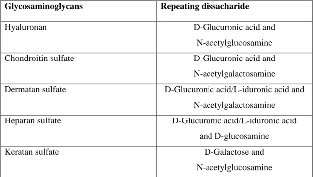
GAGs can be grouped into four categories according to their sugars, the type of linkage between the sugars, and the number and location of sulfate groups. GAG types are hyaluronic acid, chondroitin sulfate and dermatan sulfate, heparan sulfate, and keratin sulfate.47 Repeating dissacharide monomers of GAGs can slightly vary in terms of type and also position of the sulfate groups. Most common forms of dissacharide units of GAGs are given in Table 1.1.
Table 1.1 GAGs and their repeating dissacharide units
Glycosaminoglycans Repeating dissacharide
Hyaluronan D-Glucuronic acid and
N-acetylglucosamine
Chondroitin sulfate D-Glucuronic acid and
N-acetylgalactosamine
Dermatan sulfate D-Glucuronic acid/L-iduronic acid and N-acetylgalactosamine
Heparan sulfate D-Glucuronic acid/L-iduronic acid
and D-glucosamine
Keratan sulfate D-Galactose and
11
1.3.1 Chemical and functional properties of glycosaminoglycans
Apart from HA, GAG chains are usually covalently linked to a protein which is called core protein and form proteoglycans. Generally the properties of the GAGs tend to dominate the biophysical and biochemical characters of the entire molecule. GAGs are highly polyanionic molecules, therefore, in aqueous solution they attract bivalent cations such as Ca2+ and Na2+.48 This creates a high hydrodynamic volume combined with low compressibility.48 GAGs with these properties take a role as a size-selective barrier in which only small molecules can freely diffuse, restricting the bioavailability of larger molecules.48 In this way GAGs modulate water and extracellular cation homeostasis. GAGs are the major organic components of the extracellular matrix.48 As an ECM component, GAGs modulate the attraction and migration of precursor cells, and their subsequent differentiation.44,49 They also attract and regulate the action of proteins that are essential for tissue regeneration. GAGs also act as scaffolds that can modulate the function of diverse proteins, including cytokines, chemokines, growth factors, enzymes, and adhesion molecules.43,50 Due to these interactions, GAGs regulate cellular processes such as adhesion, migration, proliferation, and differentiation of various cell types.51 When processed into collagen scaffolds, they also modulate the structure of the collagen matrix. Their composition changes during the remodeling process such as bone formation, wound healing or scaring.48 It is known that GAGs in the skin and bone undergo quantitative and qualitative changes upon aging and UV radiation.48
The degree of sulfation and polymer length is important to determine the precise action of glycosaminoglycans.52 It has been shown that sulfation degree of GAGs changes their functional properties. In a recent study, native GAGs (HA and CS),
12
highly sulfated HA and CS and GAG free control were compared in different aspects.53 This study revealed that sulfation causes the loss of the proliferative activity of MSCs by about 40%, while it almost completely suppresses necrosis and apoptosis.53 Increased sulfation also modulates MSC differentiation. MSCs cultured in osteogenic medium show increased osteogenic marker gene expression when treated with sulfated GAGs compared to native GAGs. Mineral deposition of MSCs that are treated with sulfated GAGs also increased compared to native GAGs, which is an important marker of osteogenic differentiation53. In another study, it is shown that desulfation and/or shortening of heparin results in loss of its binding ability to heparin binding proteins.48
1.3.2 GAG types: a more detailed explanation
GAGs are classified into six different groups: chondroitin sulfate (CS), dermatan sulfate (DS), heparan sulfate (HS), heparin, keratin sulfate (KS), and hyaluronic acid.54 This classification is done based on their uronic acid composition, amino sugar composition, linkage between amino sugar and uronic acid, chain length of the dissacharide polymer, presence or absence of sulfate groups and their position of attachment to the sugar, nature of the core protein to which they are attached and their tissue distribution.55 CS disaccharide units are arranged in alternating unbranched sequences that can bear sulfate ester substituents in a variety of positions. Variations in molecular weight, chain length and the position of sulfate substitution can be observed depending on the species, which renders sequence heterogeneity.56 CS chains have important functions in central nervous system development, wound repair, infection, growth factor signaling, morphogenesis and
13
cell division, in addition to their conventional structural roles.56 It is also the most abundant GAG in the body with its presence in tendons, ligament, aorta and cartilage.54 It binds to proteins, like aggrecan, to form large aggregates of proteoglycans.54
Dermatan sulfate (DS) is also known as chondroitin sulfate B. DS is first isolated from the skin.57 DS is defined as a chondroitin sulfate if GalNAc is present in the structure. The presence of iduronic acid (IdoA) in DS distinguishes it from chondroitin sulfates-A and -C and bridges it to heparin and heparin sulfate.58 DS is a key biological response modifier. It is a stabilizer of cofactor and/or co-receptor for growth factors, cytokines and chemokines. It regulates enzyme activity.59 DS acts as signaling molecules in response to cellular damage, such as wounding, infection, and tumorigenesis.58 Besides, it is a target for bacterial, viral and parasitic virulence factors for attachment, invasion and immune system evasion.56
Heparin and heparan sulfate (HS) terms can sometimes be confusing. There is not a certain definition or criteria that distinguish between heparan sulfate and heparin. Both molecules are sulfated but heparin is more extensively so, and that provides higher electronegativity.60 Another structural difference between these two molecules is that HS includes a greater proportion of GlcA, whereas heparin contains more IdoA.60 While heparin is only synthesized by connective tissue mast cells, HS is found on cell surfaces or in the extracellular matrix of all mammalian organs and tissues in the form of proteoglycans.61 The initial product in their biosynthesis is a non-sulfated polymer and this polymer then enzymatically transformed into complex sulfated derivatives.62 While heparin is used as an anticoagulant in the clinic, HS exists at the cell surface and ECM.63 As HS is found at the cell surface, it binds to a
14
variety of protein ligands and regulates a wide variety of biological activities, including developmental processes, angiogenesis, blood coagulation and tumor metastasis.63–65
Keratan sulfate (KS) is an atypical GAG type. It is the most heterogeneous GAG type,54 is the only one that does not contain uronic acid.62 The family that it represents does not have acidic residues alternating in the basic unit structure with an N-acetylated amino-sugar.56 It is highly expressed in cornea, bones and brain.59 Corneal transparency is dependent on the amount of KS.66
HA, also called hyaluronan, is the sole GAG that does not form proteoglycan and is not sulfated.54 It is synthesized in the cytoplasm at the plasma membrane and is delivered into the extracellular space without post-modification.67 HA is a major component of the ECM of the skin, joints, eye, and cartilage.68 Due to high degree of polymerization capability, HA molecules can compose large molecules that reach up to several million daltons in weight which exceeds other GAGs in length.67 There are different types of HA-binding proteins that are called hyaladherins.69 Among them, CD44 and CD168 are distinct HA cell surface receptors. CD168 triggers motility by controlling focal adhesion kinase, and activation of mitogen-activated protein kinases.48 CD44 is the main receptor for HA and it modulates macrophage fusion, migration, and polarization of cells, and serves as co-receptor and organizer of the actin skeleton.69,70
HA has extraordinarily wide-ranging biological functions depending on the size despite its uniform and simple primary structure. For example in cartilage, HA is an important structural element of the matrix.71,72 It creates an aggregation centre for aggrecan which is a large chondroitin sulfate proteoglycan that retains its
15
macromolecular assembly in the matrix due to specific HA–protein interactions.73,56 The presence of negatively charged HA in cartilage ECM helps retain high water content and this creates a lubricant ECM that reduce friction between bones which also protects bones by absorbing dumping pressure. They also take role as space-filling, antiangiogenic and immunosuppressive materials.56
1.3.3 Roles of GAGs in biology
Glycosaminoglycans are seen in ECM of all the connective tissues. They modulate cell adhesion and motility. Many cell adhesion promoting components in ECM or on cell surfaces bind GAGs and support adhesion. GAGs and proteoglycans act as co-receptors for integrins and are involved in transducing signals.74 Bioactivity of GAGs is related to the interaction of their negatively charged groups to the positively charged amino groups of proteins. It is hypothesized that these charged functional groups have vital role in the formation of proteoglycans, thus, they are important for biochemical processing/signaling related to cell functionality and survival.45
GAGs also take role in immunity. It is reported that CS creates an anti-inflammatory environment by downregulating the expression of MMPs, IL-1β, TNFα, COX-2, and NOS2 as a result of blocking the NF-κB signaling pathway.48 HS has been shown to regulate leukocyte development and migration, immune activation, and inflammatory processes.75 HA takes a role in the recruitment processes of leukocytes to the inflammation site through its interaction with CD44.76 HA protects the healthy form of skin acting as scavenger for free radicals, that can be form upon exposure to UV light, which may result in oxidative stress on cell.56 HA regulates osteoclast precursor mobility and may act as a diffusion barrier for enzymes from the resorption
16
area. It is suggested that HA may take a role in osteoclast attachment and resorption via CD44.48 The main GAG type in bone tissue is chondroitin-4-sulfate while HA, DS and chondroitin-6-sulfate can be seen in less amounts in bone. Expression of these molecules enhances osteogenic differentiation of mesenchymal stem cells (MSCs).77 Glycosaminoglycans modulate the attraction of bone precursor cells including the actions of proteins essential for bone regeneration.45
The epidermis and dermis, which are different layers of skin, contain various types of GAGs. Although they make up only 0.1–0.3% of the total skin weight, GAGs define skin volume and elasticity due to their large water-retaining capability.78 When it comes to wound healing, the quantity and qualitative composition of GAGs differ in healthy skin and scar tissue.48 In fact, this difference is one of the reasons of scar formation rather than complete healing. The fibroblasts in scar tissue, produce more HA, and more C4S (chondroitin-4-sulfate), but less DS than fibroblasts in healthy skin.48 It is also known that de novo synthesis of GAGs is increased temporarily during the proliferation phase of wound healing. In addition to that persisting high HA levels in granulation tissue are related to scarless healing.48
GAGs are also studied in cancer biology due to their functional properties related to adhesion, motility and signal transduction. Tumor cells must adhere to extracellular matrix (ECM) proteins and molecules on other cells in order to invade and metastasize. Integrins, the receptors that are responsible for cell-to-ECM interactions are therefore important in tumor cell invasion and metastasis.79 GAGs have a modulating role for integrin binding and this is one of the reaction types that makes them important molecules in cancer studies. There are several examples that show GAGs-cancer relation. In one example, α4β1 integrin-mediated melanoma cell
17
adhesion is stopped by removal of cell surface chondroitin sulfate glycosaminoglycan (CSGAG).74
As mentioned above GAGs have diverse biological functions and this makes them a shining candidate for regenerative purposes. Currently, collagen–GAG composites are developed for a wide range of applications in tissue engineering of bone and skin.48 GAGs also can be used as anticoagulation agents. Heparin and heparin like GAGs are used to inactivate coagulation proteases and blood anticoagulation.80 In another study, regeneration potential of GAGs on spinal cord injuries was studied. In this work, chondroitin sulfate with two sulfate groups showed excellent motor recovery in vivo assay, improved axonal growth in vitro, and neuroprotection against the NMDA induced neuronal cell death. Besides, it was observed that GAGs do not inhibit axonal growth.81 The importance of GAGs in neural tissue regeneration is also revealed by studying healthy and crush-injury models of sciatic nerve in vivo. In this study, GAGs are recovered from supernatant and pellets of tissue homogenates and assayed for different GAG types. The study shows that GAG profile in supernatant and pellets differ among injury model and healthy tissue. In addition, GAG amount is two times higher in healthy samples. This indicates the need to modulate the glycosaminoglycan expression pattern in adult neural tissue regeneration after post-traumatic period.82
GAGs have several important roles in tissue healing and remodeling. HA accelerates healing processes of dermal wounds.68 It is shown that hyaluronic acid modulates the inflammation at short term but also at later stages of tendon or ligament healing, also it involves in collagen fibril aggregation and development.83 It improves the healing quality of the injured tissues by increasing cell migration and proliferation in the
18
internal parts of the injured tendon and ligaments and by reducing peritendinous adhesion over the tendon and ligament surface as it decreases cell proliferation. HA is also used as chondroprotective and its effects are shown in vitro and in vivo.68 In addition, HA is used as space-filling matrix of the eye in ophthalmology applications.68
GAGs can also be used in vascular tissue regeneration. Vascular endothelial cells are the cells found in vascular tissue and they synthesize collagen IV and elastin during angiogenesis. In one study, it was shown that collagen IV and elastin deposition in matrix is increased and their mRNA expression is upregulated when endothelial cells are cultured in matrix that incorporated HA and HA+HS combination.84
In another study, GAGs were shown to modulate differentiation of MSCs. It was shown that hMSCs (mesenchymal stem cells isolated from human bone marrow) selectively differentiate into bone when they are cultured on GAG containing tissue culture plates. Except from chondroitin-4-sulfate, all tested GAG types which were DS, chondroitin-6-sulfate, heparin and HA, increase osteoblast differentiation related genes expression.77 To sum up, GAGs of extracellular matrix played a significant role in regulating osteoblast differentiation and could be exploited in the biomimetic approach of fabricating or functionalizing scaffolds.77 Depending on the type of GAG that is used, MSCs were also directed into chondrogenic differentiation by changing the mechanical and morphological properties of the scaffold that they were incorporated in. In the study that compares effects of CS and HA incorporated collagen based scaffolds on MSC differentiation, it was shown that HA scaffold promotes cartilage differentiation. While expression of chondrogenic related gene markers such as Sox-9 and collagen II increased significantly, HA scaffold also
19
provided greater levels of MSC infiltration in comparison to the CS scaffolds which is characteristic for cartilage tissue.85 Conversely, gene expression studies, scaffold morphology and mechanical characterizations revealed that scaffold incorporated with CS induce osteogenic differentiation. Therefore, this study also points out that GAG type determines differentiation fate of the MSCs.
1.3.4 GAG-protein interactions
GAGs are generally believed to exert their biological activities through the localization, stabilization, activation or inactivation of interacting proteins.80 GAG-receptor and GAG-protein interactions are important as these interactions modulate various biological signals. High molecular weight HA (HMW-HA) inhibits osteoclast differentiation via Toll-like receptor (TLR)-4 by interfering with colony stimulating factor (M-CSF) signaling. In the inflammatory phase, GAG binding of chemo-attractants such as IL-8 and TGF-β provides the ECM with cues directing the migration of neutrophils and macrophages, towards the wounded tissue. HA runs most of its reaction upon binding CD44, which is the main receptor for HA.48 The most studied GAG-protein interactions involves HS and growth factors, especially fibroblastic growth factor.61 DS is also shown to bind to hepatocyte growth factor.58 Heparin forms complexes with thrombin and protease inhibitors, and inhibits coagulation.61 There are different factors that affect GAG-protein binding. One of them is presence of internal ion pairing in place of counter-ions. For example Na+ on GAGs and Cl- on the proteins influence GAG-protein interactions. High energy binding sites and the entropy of the environment are other factors that affect interaction80. Amino acid sequence also affects the binding behavior. There are
20
certain amino acids that particularly bind to GAGs when they are found in peptide sequence. Peptides including high amounts of arginine and lysine bind to GAGs with greatest affinity.54 Peptides with high affinity for heparan sulfate are also enriched in other polar amino acids including serine. Asparagine residues are commonly found in heparin binding site.80 For heparan sulfate binding, peptides are enriched in glutamine residues. Tyrosine residues are also enriched in known heparin binding regions.80 These specific preferences are believed to stem from differences in abundance of chemical interactions like hydrophobic and hydrophilic interactions.
1.4 Peptide Amphiphiles
Peptide amphiphiles are molecules that combine structural features of amphiphiles and with functions of peptides that can be biologically active. They consist of two main blocks: an alkyl tail connected to a peptide block. Alkyl tail is the part that gives the structure lipophilicity. Therefore, PAs can also be called as lipopeptides, or amphiphilic peptides.86,87
A typical PA design includes four key regions.88 First region is the hydrophobic domain, which is generally composed of an alkyl tail and gives lipophilicity to the molecule. Second region is located adjacent to the alkyl tail and is composed of a short peptide sequence capable of forming intermolecular hydrogen bonding and β-sheet secondary structure. Third region is designed to enhance water solubility of the PA molecules by providing charges to the molecule. This part is composed of either basic or acidic amino acids. Another function of this region is to make PA molecule assembly controllable by charge, pH, salts and ions. The last part is used to present
21
bioactive signals which may be an epitope to interact with cell receptors, a segment that binds proteins or biomolecules, or a pharmacological agent. This typical design of PAs can change depending on the application purpose. For example, PAs that are designed to functionalize liposomes include proline residues instead of β-sheet forming amino acids in order to trigger micelle structure formation.89
PAs are assembling molecules in aqueous conditions. Forces that drive self-assembly include hydrophobic interactions of the alkyl tails, hydrogen bonding among the middle peptide segments, and electrostatic repulsions between the charged amino acids.88 Amphiphilicity is the main triggering factor for the self-assembly of PA molecules.86 Assembly of PAs can also be triggered by ions and pH change. Upon self-assembly, PAs can form diverse nanostructures. These structures can have α-helical, and parallel or anti-parallel β-sheet secondary structures. Nanofibers, nanotubes, tape-like structure and cylindrical micelles are among the more sophisticated structures that can be obtained by self-assembly PAs.90–93
PAs are biocompatible, biodegradable, and can be easily controlled to form different shapes. Another most important feature of PAs is their high density epitope presenting properties. As they intrinsically self assemble, they present bioactive epitope groups to the environment with a much higher density compared to single bioactive peptide molecules. These features make PAs versatile tools as self-assembled building blocks. PAs can be used as delivery agents for drugs and miRNAs, as MRI contrast agents, antimicrobial agents, incorporated into energy applications, used in vitro to control cell fate and in vivo to induce or suppress (depending on the purpose) angiogenesis and to trigger tissue regeneration.10,11,89,94–97
22
CHAPTER 2
23
2.1 Materials
[4-[-(2’,4’-dimethoxyphenyl) Fmoc-aminomethyl]phenoxy] acetamidonorleucyl-MBHA resin (Rink amide acetamidonorleucyl-MBHA resin), Fmoc-Glu(OtBu)-Wang resin, 2-(1H Benzotriazol-1-yl)-1,1,3,3-tetramethyluronium hexafluoro-phosphate (HBTU), 9-Fluorenylmethoxycarbonyl (Fmoc) and tert-butoxycarbonyl (Boc) protected amino acids were purchased from NovaBiochem, ABCR and Sigma-Aldrich. Fmoc-Ser[-Glc(OAc)4]-OH was purchased from AAPPTec. N,N-diisopropylethylamine (DIEA)
and lauric acid were purchased from Merck. Piperidine, acetic anhydride, dichloromethane (DCM), dimethylformamide (DMF), trifluoroacetic acid (TFA) and triisoproplysilane (TIS) were obtained from Sigma-Aldrich. All other chemicals and materials were purchased from Invitrogen, Fisher, Merck, Alfa Aesar, and SigmaAldrich. Deionized water (ddH2O) used in experiments had a resistance of
18.2 MΩ.cm (Millipore Milli-Q). All chemicals and materials were used as provided. For cell culture experiments, Dulbeccos Modified Eagle Medium (DMEM), Penicillin/Streptomycin (PS) antibiotic mix and Fetal Bovine Serum (FBS) were purchased from Gibco, Life Technologies. LIVE/DEAD Viability/Cytotoxicity Kit was purchased from Invitrogen. Cell Proliferation ELISA, BrdU (colorimetric) kit was purchased from Roche. Safranin-O and Oil Red-O were obtained from Sigma-Aldrich.
24
2.2 α-N-Acetyglucosamine Carboxylic Acid Synthesis
Figure 2.1 Synthesis of 2-Acetamido 2-deoxy-a-D-glucopyranose 3,4,6 triacetate
carboxylic acid derivative. Carboxylic acid functionality appended to
N-acetyl-glucosamine.
2.2.1 Synthesis of Allyl-N-Acetyl-α-D-Glucopyronoside-3,4,6 Triacetate
0.2 mL of BF3.Et2O was added to a solution of 2 g N-acetyl-glucosamine in 50 mL
allyl alcohol and reaction mixture was refluxed at 130 °C for 2 h. After complete conversion allyl alcohol was removed on rotary evaporator to get crude allyl-N-acetyl-α-D-glucopyranoside. The crude product was purified by dissolving in 20 mL ethanol and then adding diethyl ether. Desired compound separates as a white solid. White solid compound was dissolved in 20 mL dry pyridine, acetic anhydride at 0 °C was added and then stirred at room temperature for 12 h. After the completion of reaction, pyridine was removed on rotary evaporator. Then crude material was dissolved in ethyl acetate and washed with dil. HCl, brine and water. Organic layer was dried on anhydrous Na2SO4 and column purified to get desired compound after
evaporating organic solvent. 1H NMR 5.88 (1H, m), 5.72 (1H, J=9.54Hz, d), 5.27 (3H, m), 5.12 (1H, J=9.54, t), 4.88 (1H, J=3.51Hz, d), 4.35 (1H, m), 4.23 (1H, J=12.30 & 4.52Hz, q), 4.18 (1H, m), 4.09 (1H, m), 4.02 (1H,m), 3.96 (1H,m), 2.09 (3H,s), 2.02 (3H,s), 2.01 (3H,s), 1.95 (3H,s).
25
2.2.2 Synthesis of N-Acetyl-α-D-Glucopyronoside-3,4,6 Triacetate Carboxylic
Acid
Into the stirred solution of allyl-N-acetyl-α-D-glucopyronoside-3,4,6 triacetate, dissolved in 23 mL CCl4, 23 mL CH3CN and 30 mL H2O at 0°C, NaIO4 and
RuCl3.H2O was added. The reaction mixture was stirred for 12-16 h and after
completion was checked by thin layer chromatography (TLC), reaction mixture was evaporated on rotary evaporator to get crude carboxylic acid derivative which was directly used for coupling on solid phase. 8Hz; 4.12-4.27 (m, 6H); 3.98 (d, J=15.0Hz, 1H), 3.54-3.79 (m, 9H), 3.20-3.50 (m, 26H), 3.10-3.17 (m, 5H), 2.10-2.25 (m, 5H), 1.93-2.01 (m, 4H), 1.86 (s, 3H), 1.55-1.81 (m,3H), 1.40-1.49 (m, 5H), 1.20-1.30 (m, 25H), 0.8-0.9 (m, 18H).
2.3 Synthesis, Identification and Purification of Peptide Amphiphile
Molecules
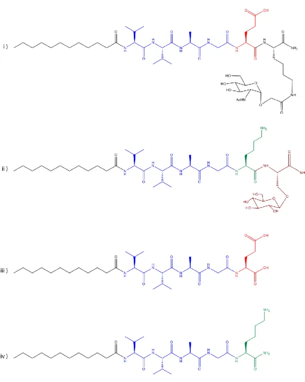
2.3.1 Synthesis of Peptide Amphiphile Molecules
Peptide amphiphile molecules were synthesized by standard SPPS (solid phase peptide synthesis) method. Peptide amphiphiles, except E-PA, were constructed on MBHA Rink Amide resin, and E-PA [Lauryl-VVAGE] was constructed on Wang resin pre-loaded with Fmoc-Glu(OtBu). The resins were swelled in DCM for 30 min. Following resin swelling, DCM solvent was exchanged to DMF, in which all remaining reactions were carried out. All amino acid couplings, except Fmoc-Ser[-Glc(OAc)4]-OH, were performed with 2 equivalents of Fmoc protected amino acid,
26
in DMF. Coupling duration was at least 3 h but varied depending on the type of amino acid that is coupled. Fmoc-Ser[Glc(OAc)4]-OH coupling was performed with
1.2 equivalents of amino acid, 1.1 equivalents of HBTU and 1.8 equivalents of N,N-diisopropylethylamine (DIEA) in DMF. Equivalences are based on the resin that was used for construction.
Fmoc deprotections were performed with 20% piperidine/dimethylformamide (DMF) solution for 20 min. After each coupling reaction, resin was treated with 10% acetic anhydride in DMF for 30 min to block any remaining free amino groups. Before each succeeding event, washing was performed by DMF, DCM, and DMF three times each, respectively. Cleavage of the peptides from resin was carried out with a mixture of triuoroacetic acid (TFA) : triisoproplysilane (TIS) : water in the ratio of 95 : 2.5 : 2.5 for 2 h. Excess TFA was removed by rotary evaporation. The remaining viscous peptide solution was treated with ice-cold diethyl ether overnight at -20 °C. Ether decantation was performed after centrifugation at 4 °C, 8000 rpm for 15 min. After complete evaporation of diethyl ether via air drying, the resulting pellet was dissolved in ddH2O, sonicated for 30 min, freeze-dried at -80 °C, and lyophilized.
Deacetylation of Glc-PA [Lauryl-VVAGKS[β-D-Glc(OAc)4)-Am] was carried out in
solution phase. Acetyl protecting groups on glucose of Glc-PA [Lauryl-VVAGKS(β-D-Glc)-Am] were removed in NaOMe in methanol solution. The reaction was carried out at room temperature, in argon atmosphere, for 3-4 h by stirring. The reaction was quenched at acidic condition which is created by a few drops of acetic acid. Solution was removed by rotary evaporator and obtained product was dissolved in ddH2O and freeze-dried. The PAs were stored at -20 °C.
27
Figure 2.2 Schematic representation of the sequential synthesis of amphiphilic
glycopeptide. N-acetyl-α-D-glucopyronoside-3,4,6 triacetate carboxylic acid was
28
2.3.2 Characterization of Peptide Amphiphile Molecules
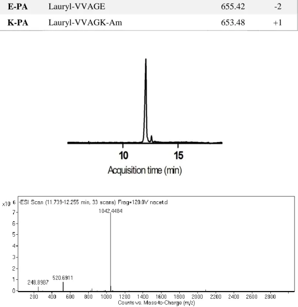
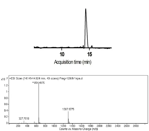
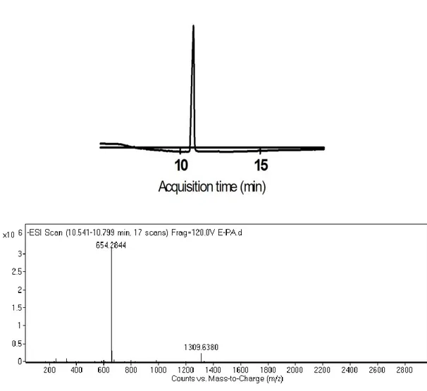
The synthesized peptide was characterized by liquid chromatography mass spectrometry (LC-MS) on an Agilent 6530 Q-TOF mass spectrometer equipped with ESI source and reverse phase analytical high performance liquid chromatography. Basic conditions and acidic conditions were used to identify negatively charged and positively charged PA molecules, respectively. For basic conditions, Zorbax Extend-C18 (4.6 x 50 mm) column and water/acetonitrile gradient with 0.1% volume of NH4OH were used. For acidic conditions, Zorbax 300 SB-C8 (4.6 x100 mm) column
and water/acetonitrile gradient with 0.1% volume of formic acid were used.
2.3.3 Purification of Peptide Amphiphile Molecules
An Agilent 1200 preparative reverse-phase HPLC system was used for purification of PA molecules. As stationary phase, a Luna 5u C8 (2) (21.20 x 100 mm) column for acidic conditions and a Gemni 5u C18 (21.20 x 100 mm) column for basic conditions were used to purify positively charged and negatively charged PA molecules, respectively. As mobile phase, water/acetonitrile gradient with 0.1% volume of NH4OH for basic conditions and water/acetonitrile gradient with 0.1%