THE CHARACTERIZATION AND POTENTIAL
FUNCTIONAL ROLE OF WDR81,
A NOVEL ZEBRAFISH GENE, ASSOCIATED WITH
CEREBELLAR ATAXIA, MENTAL RETARDATION AND
DYSEQUILIBRIUM SYNDROME (CAMRQ) IN HUMANS
A DISSERTATION SUBMITTED TO
THE GRADUATE SCHOOL OF ENGINEERING AND SCIENCE OF BILKENT UNIVERSITY
IN PARTIAL FULFILLMENT OF THE REQUIREMENTS FOR THE DEGREE OF
DOCTOR OF PHILOSOPHY IN
MOLECULAR BIOLOGY AND GENETICS
By
Füsun Doldur-Ballı
April 2016
THE CHARACTERIZATION AND POTENTIAL FUNCTIONAL ROLE OF WDR81, A NOVEL ZEBRAFISH GENE, ASSOCIATED WITH CEREBELLAR ATAXIA, MENTAL RETARDATION AND DYSEQUILIBRIUM SYNDROME (CAMRQ) IN HUMANS
By Füsun Doldur-Ballı April, 2016
We certify that we have read this dissertation and that in our opinion it is fully adequate, in scope and in quality, as a thesis for the degree of Doctor of Philosophy.
________________________ ________________________ Tayfun Özçelik Michelle Adams (Advisor) (Co-Advisor)
________________________ Ayşe Begüm Tekinay
________________________ Hilal Özdağ ________________________ Özlen Konu ________________________ Stefan Fuss
Approved for the Graduate School of Engineering and Science
__________ Levent Onural
Abstract
THE CHARACTERIZATION AND POTENTIAL FUNCTIONAL
ROLE OF WDR81, A NOVEL ZEBRAFISH GENE, ASSOCIATED
WITH CEREBELLAR ATAXIA, MENTAL RETARDATION AND
DYSEQUILIBRIUM SYNDROME (CAMRQ) IN HUMANS
Füsun Doldur-Ballı
PhD in Molecular Biology and Genetics Advisor: Tayfun Özçelik
Co-Advisor: Michelle Adams April, 2016
Cerebellar ataxia, mental retardation and dysequilibrium syndrome (CAMRQ) is a neurodevelopmental disorder. The gene encoding WD repeat containing protein 81 (WDR81) was reported to be associated with CAMRQ2 [MIM 610185]. Human and mouse studies indicated the potential importance of WDR81 in neurodevelopment. The first aim in this study was to characterize the transcript and to reveal the expression profile of wdr81 in zebrafish. The second aim was to perform the initial characterization of wdr81 morphants. In silico analysis indicated that the conserved domains are shared in human, mouse and zebrafish orthologous proteins, implying a conserved function of WDR81 in three species. The characterization of the transcript revealed that wdr81 possessed one ORF and one 5’UTR structure. The predicted sequence for 3’UTR was confirmed along with detection of some variants and an insertion site in samples from ten developmental timepoints and in several adult tissues. This region was not detected in kidney, intestine and gills, which might be pointing out an alternative polyadenylation event. wdr81 appeared to be maternally supplied. 5 hpf and 18 hpf were detected as crucial timepoints regarding wdr81 expression. Expression of wdr81 was found to be increased in the eye and brain regions at 18 hpf and 48 hpf. wdr81 was found to be ubiquitously expressed in the
adult zebrafish. The expression of wdr81 in the adult brain and eye was detected in several regions including retinal layers, presumptive Purkinje cells and some proliferative zones. The splice blocking morpholino which targets the exon 2-intron 2 junction of wdr81 worked at 3 tested doses; 2 ng, 4 ng and 8 ng. The effect of the
wdr81 morpholino was detected to add the intron, which is downstream of the target
exon, to the transcript and introduce a stop codon. Preliminary results indicated a significant reduction in the head sizes at a ratio of 3.88% (p:0.027) in the wdr81 morphant group compared to uninjected group and gbx2 expression was observed to be higher in wdr81 morphants compared to the control groups.
In short, findings of this study emphasize the significance of wdr81 in neurodevelopment and suggest a potential role in neuronal proliferation. This study also serves as a basis for future functional studies.
Özet
İNSANLARDAKİ SEREBELLAR ATAKSİ, ZEKA GERİLİĞİ VE
DENGESİZLİK SENDROMU İLE İLİŞKİLENDİRİLEN VE
ZEBRABALIĞINDA YENİ BİR GEN OLAN WDR81’İN
KARAKTERİZASYONU VE MUHTEMEL İŞLEVSEL ROLÜ
Füsun Doldur-Ballı
Moleküler Biyoloji ve Genetik, Doktora Tez Danışmanı: Tayfun Özçelik Eş Danışmanı: Michelle Adams
Nisan, 2016
Serebellar ataksi, zeka geriliği ve dengesizlik sendromu nörogelişimsel bir hastalıktır. WD tekrarı içeren protein 81’i kodlayan genin bu sendrom ile ilişkilendirildiği daha önce rapor edilmiştir. WDR81 geninin sinir sisteminin gelişim sürecindeki potansiyel önemi insan ve fare çalışmalarıyla ortaya konmuştur. Bu çalışmada ilk amaç, wdr81’in zebrabalığında karakterize edilmesi ve ifade profilinin gösterilmesiydi. İkinci amaç
wdr81 morfantlarının öncül karakterizasyonunun yapılmasıydı. In siliko analizler insan,
fare ve zebrabalığına ait ortolog proteinlerin korunmuş domeynleri paylaştığını gösterdi, bu durum da WDR81’ın işlevinin bu üç türde korunmuş olabileceğine işaret etmektedir. Transkriptin karakterizasyonu, wdr81’in bir açık okuma çerçevesine ve bir 5’UTR yapısına sahip olduğunu gösterdi. 3’UTR’nin öngörülen dizisi, bazı varyantların ve bir içine yerleştirme (insertion) bölgesinin varlığı tespit edilmekle birlikte, teyit edildi. Bu içine yerleştirme (insertion) bölgesi on gelişimsel evreye ait örnekte ve çeşitli yetişkin dokularında tespit edilmiş olup böbrek, bağırsak ve solungaçlarda bulunmadı. Bu durumun da alternatif poliadenilasyon işlemine işaret ettiği öngörülmektedir. wdr81’in yumurta hücresi tarafından sağlanan transkriptler arasında olduğu tespit edildi. wdr81’in ifadesi açısından döllenme sonrası 5. ve 18. saatler kritik evreler olarak saptandı. Döllenme sonrası 18. ve 48. saat evrelerinde wdr81 ifadesinin göz ve beyin bölgelerinde
artmış olduğu gözlendi. wdr81’in test edilen yetişkin dokularının tümünde ifade edildiği, yetişkin göz ve beyin dokularında ise retina katmanları, muhtemel Purkinje hücreleri ve bazı proliferatif alanların da bulunduğu çeşitli bölgelerde ifade edildiği belirlendi. Uçbirleştirmeyi (splicing) etkileyen morfolino wdr81 genine ait ekzon 2-intron 2 bölgesini hedefleyecek şekilde tasarlandı ve denenen üç dozun (2 ng, 4 ng ve 8 ng) da çalıştığı tespit edildi. wdr81 morfolinonun etkisinin, hedef ekzonun aşağı yönündeki intronu transkripte dahil etmek olduğu ve bu dizinin de durdurma kodonu içerdiği tespit edildi. Öncül sonuçlar, wdr81 morfant grubuna ait kafa bölgelerinin ölçümünde, enjeksiyon yapılmayan gruba kıyasla %3,88 oranında anlamlı (p: 0,027) bir düşüş olduğu sonucunu verdi. Ayrıca, gbx2 ifadesi wdr81 morfant grupta kontrol gruplarına kıyasla daha yüksek gözlendi.
Özetle, bu çalışmaya ait bulgular wdr81’in nörogelişimsel önemini anlamakta katkıda bulunmuştur ve bu gen için nöronların çoğalmasına (proliferasyonu) ilişkin olası bir rol önermektedir. Elde edilen veriler aynı zamanda ileride yapılacak fonksiyonel çalışmalar için de temel teşkil edecektir.
Acknowledgements
Having a PhD degree was one of my life goals and I started the program in Department of Molecular Biology and Genetics at Bilkent University with the motivation that having a PhD degree was going to improve me in the progress of becoming the person I wanted to be. Prof. Dr. Tayfun Özçelik accepted to work with me, which was also my only plan when I registered to the program. Prof. Özçelik always guided me to try my best in science and encouraged me to develop critical thinking. I admire his way of synthesizing scientific information and concepts. Another thing I learned from him is to set the career goals at a level of quality and significance accepted by the scientific community in the world. My PhD thesis research continued under co-supervision of Prof. Dr. Tayfun Özçelik and Assoc. Prof. Dr. Michelle Adams. Dr. Adams was always caring, supportive, guiding and encouraging to me. I believe that all the feedback she gave during my thesis research will also help me in my future career. I always appreciated her attitude to the people who work with her. She provided an environment for us to feel comfortable, productive and to be creative. I am grateful to both of my supervisors because of providing me the opportunity to become an independent researcher and of trusting me in the project. I am very thankful to Assoc. Prof. Dr. Ayşe Begüm Tekinay for her patience with my questions, her feedback on the project, being a thesis progress committee and a jury member and most importantly for encouraging me and her belief in me. Her support is always invaluable. I would like to thank to Assoc. Prof. Dr. Özlen Konu for the feedback and perspective she provided during my research and for being a thesis progress committee and a jury member. I am thankful to Prof. Dr. Hilal Özdağ for evaluating my thesis and being a jury member. I thank to Assoc. Prof. Dr. Stefan Fuss for evaluating my thesis and hosting me in his lab in the beginning of my thesis to train me on morpholino microinjection. I also thank to Xalid Bayramlı from his lab for sparing time to teach me how to perform microinjection experiments. I would like to express my gratitude to Assoc. Prof. Dr. Güneş Özhan for her contribution to the study by suggesting to characterize the morphants with marker genes and providing the plasmid constructs. She is always helpful and open to questions. I deeply appreciate her approach to science which is pure and sharing. I would like to express special thanks to Prof. Dr. Hamdullah Aydın who has been a mentor to me since my Master’s degree studies. He broadened my perspective with outstanding ideas and comments.
I have always felt lucky by means of colleagues. One of the best things in my PhD education was to meet Dr. Gülşah Merve Kılınç. I am thankful to her for moral support, encouragement
and suggestions in my research. I also thank to Dr. Emre Onat and Dr. Süleyman Gülsüner as senior researchers of Özçelik Group for their contributions and help in various forms. I would like to record my sincere appreciation to Dr. Ayça Arslan-Ergül for her contributions and support during my research. Her perspective has been always inspiring to me. I would like to thank Dilan Çelebi-Birand for performing the head size and body measurements of injection groups and to Melek Umay Tüz for her help with the whole mount in situ hybridization experiment using gbx2 probe. I also thank other members of Adams Group; Elif Karoğlu, Göksemin Şengül, Ayşegül Dede, Begün Erbaba, Özge Pelin Burhan and zebrafish laboratory technician Tülay Arayıcı for their contributions and help in various forms.
I am pleased to meet and to have known my friends from MBG, who are Dr. Verda Bitirim, Dr. Pelin Telkoparan, Dr. Nilüfer Sayar, Dr. Gurbet Karahan, Dr. Ender Avcı, Umur Keleş, Begüm Kocatürk, Andaç Kipalev Neşelioğlu, Buket Gültekin, Merve Mutlu, Begüm Han Horuluoğlu, Begüm Yıldız, Şahika Cıngır Köker, Ayşegül Örs, Gözde Güçlüler, Özlem Tufanlı, İnci Şimşek Onat, Zeynep Ayyıldız, Emre Yurdusev, Dr. Gökhan Yıldız, Defne Bayık, Merve Aydın and Dr. Çiğdem Özen. The peaceful work environment they created and their help was important, the conversation with them and their feedback was always supportive.
I dedicate this thesis to my spouse Mevlüt Ballı and our future daughter Yaren Rüya Ballı. I would like to express my heartfelt appreciation and eternal love to Mevlüt. He has always been understanding, supportive, encouraging and patient during my graduate studies. His help to my efforts for balancing my career goals with our family life is precious. I hope dedicating this thesis also to her will be a future surprise to our daughter, who will join us in a few months. I am deeply grateful to my parents Elif and Kemal Doldur and my brother Utku Doldur for their endless love, unhesitant support and encouragements during my whole education. My father has been the first and the most strongest inspiration for devising a career plan in science. I would like to also thank to Ayşe Mergenci, Ayşe Can, Hatice Yiğit and Canan Şişman Korkmaz for their support and belief in me.
I was supported by TÜBİTAK 1001 grants, 111S199 and 114S548, in part by a European Molecular Biology Organization Installation Grant to Michelle Adams and Department of Molecular Biology and Genetics during my PhD. Finally, I will share the quotation from Albert Einstein “Play is the highest form of research”, which also partially explains why I preferred a career in science over routine work. I am determined to put every effort to continue playing/practicing science in the future as well.
Table of Contents
ABSTRACT………...III ÖZET………...V ACKNOWLEDGEMENTS………...VIII TABLE OF CONTENTS………...X LIST OF FIGURES………...XIII LIST OF TABLES………...XVI ABBREVIATIONS………...XVII
CHAPTER 1.INTRODUCTION ………1
1.1. CEREBELLAR ATAXIA, MENTAL RETARDATION AND DYSEQUILIBRIUM SYNDROME……….…..1
1.1.1.CAMRQASSOCIATED GENES……….……….2
1.1.1.1.VERY LOW DENSITY LIPOPROTEIN RECEPTOR (VLDLR)………2
1.1.1.2.WDREPEAT CONTAINING PROTEIN 81(WDR81)………..…3
1.1.1.3.CARBONIC ANHYDRASE VIII(CA8)………..………..8
1.1.1.4.ATPASE,AMINOPHOSPHOLIPID TRANSPORTER,CLASS I,TYPE 8A, MEMBER 2(ATP8A2)……….……10
1.2.ZEBRAFISH AS A MODEL ORGANISM……….11
1.2.1.ZEBRAFISH CENTRAL NERVOUS SYSTEM………..15
1.2.1.1.ZEBRAFISH EMBRYO………..15
1.2.1.2.ADULT ZEBRAFISH………...…….16
1.3.CELL PROLIFERATION IN ZEBRAFISH NERVOUS SYSTEM ……….……17
1.3.1.NEUROGENESIS IN ZEBRAFISH………18 1.3.1.1.ZEBRAFISH EMBRYO………..18 1.3.1.2.ADULT ZEBRAFISH………...….19 1.3.2.GLIOGENESIS IN ZEBRAFISH………..…….19 1.3.2.1.ZEBRAFISH EMBRYO………..20 1.3.2.2.ADULT ZEBRAFISH………...….20 1.4.SYNAPTOGENESIS IN ZEBRAFISH………....21
1.5.AIM AND SCOPE OF THIS STUDY………...………...23
CHAPTER 2.MATERIALS AND METHODS……….………25
2.1. METHODS………...………...25
2.1.1. ZEBRAFISH AND EMBRYOS………...…….25
2.1.3. CHARACTERIZATION OF THE WDR81TRANSCRIPT………...……26
2.1.3.1.AMPLIFICATION OF THE OPEN READING FRAME OF WDR81………27
2.1.3.2.RAPID AMPLIFICATION OF CDNAENDS (RACE)..………..28
2.1.3.2.1.CHARACTERIZATION OF THE 5’RACEPRODUCT………28
2.1.3.2.2.CHARACTERIZATION OF THE 3’RACEPRODUCT………30
2.1.4.ANALYSIS OF THE EXPRESSION OF WDR81 IN WILD TYPE ZEBRAFISH…………..32
2.1.4.1.QUANTITATIVE REAL-TIME PCR(QRT-PCR)……….32
2.1.4.2. IN SITU HYBRIDIZATION EXPERIMENTS………...….34
2.1.4.2.1.WHOLE MOUNT IN SITU HYBRIDIZATION (WMISH) ON EMBRYOS………..34
2.1.4.2.2. IN SITU HYBRIDIZATION (ISH) ON BRAIN AND EYE TISSUES……….35
2.1.5.MORPHOLINO MICROINJECTIONS TO KNOCKDOWN WDR81………..36
2.1.5.1. WDR81 MORPHOLINO DOSE CURVE……….37
2.1.5.2.INITIAL CHARACTERIZATION STUDIES……….38
2.1.5.2.1.HEAD SIZE MEASUREMENT………..38
2.1.5.2.2.PHENOTYPE CHARACTERIZATION WITH WMISH METHOD………...39
2.2. MATERIALS………..40
2.2.1. GENERAL CHEMICALS,REAGENTS AND ENZYMES………...40
2.2.2.GENERAL MATERIALS AND EQUIPMENTS………..41
2.2.3.BUFFERS AND SOLUTIONS………..42
2.2.4.MOLECULAR SIZE MARKERS AND PLASMID VECTORS………..…42
CHAPTER 3. RESULTS………..43
3.1. ZEBRAFISH ORTHOLOGUE OF WDR81………..43
3.2. ZEBRAFISH WDR81TRANSCRIPT POSSESSES ONE ORF………..45
3.3. CHARACTERIZATION OF 5’ END OF WDR81 VERIFIED THE PREDICTION AND AN INSERTION SITE WAS DETECTED IN THE 3’END OF SOME TISSUES ………46
3.4. EXPRESSION OF WDR81 IS INCREASED AT 5 HPF AND 18 HPF DURING DEVELOPMENT AND IT IS ENRICHED IN THE EYE AND BRAIN OF EMBRYOS…………..53
3.5. SPATIAL EXPRESSION OF WDR81 AND ITS DETECTION IN PLOLIFERATION ZONES……….61
3.6.INITIAL CHARACTERIZATION OF MORPHANTS………...68
3.6.1.DOSE CURVE OF WDR81 MORPHOLINO……….………68
3.6.2.INITIAL RESULTS FROM PHENOTYPE CHARACTERIZATION OF WDR81 MORPHANTS………..…..67
CHAPTER 4. DISCUSSION………..79
CHAPTER 5. FUTURE PERSPECTIVES………88
REFERENCES ……….…….……90
APPENDIX A–BUFFERS AND SOLUTIONS………..111
APPENDIX B–MOLECULAR SIZE MARKERS……….117
APPENDIX C-CLONING VECTORS………...………..……..119
APPENDIX D–MULTIPLE SEQUENCE ALIGNMENT OF DANIO RERIO (ZEBRAFISH) WDR81 AMINO ACID SEQUENCE WITH WDR81 SEQUENCES FROM VARIOUS SPECIES ……….…..121
List of Figures
FIGURE 1: QUADRUPEDAL GAIT IN A MALE IN 1914 (LEFT) AND IN A FEMALE PATIENT
NOWADAYS (RIGHT)……….1
FIGURE 2: BRAIN MORPHOLOGY OF A CONTROL INDIVIDUAL AND A PATIENT WERE ANALYZED VIA MAGNETIC RESONANCE IMAGING (MRI)……….5
FIGURE 3:PHYLOGENETIC TREE OF PROTEIN SEQUENCES OF WDR81 IN VERTEBRATES……7 FIGURE 4:LATERAL VIEWS OF A FEMALE (F) AND A MALE (M) ADULT ZEBRAFISH………..12 FIGURE 5:FIVE DPF WILD TYPE ZEBRAFISH EMBRYO………16 FIGURE 6: ILLUSTRATION WHICH COMPARES THE BRAIN SIZES OF HUMAN, MOUSE AND
ZEBRAFISH………...17
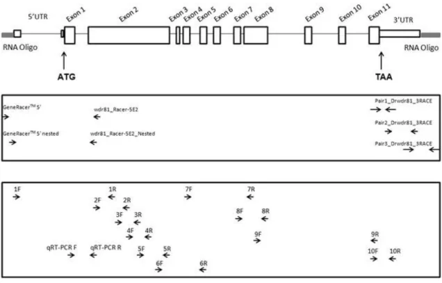
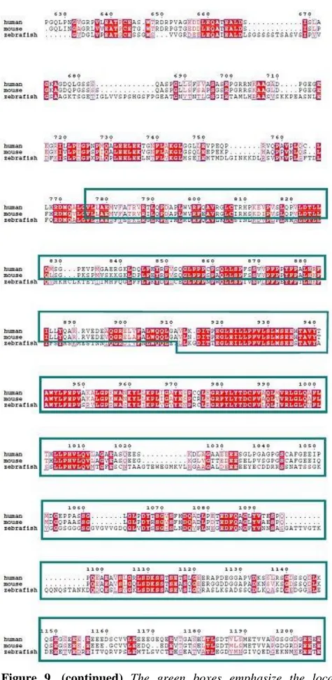
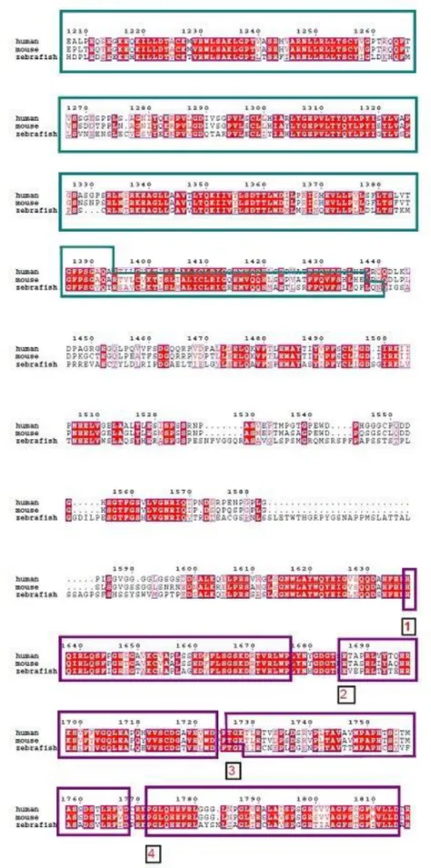
FIGURE 7:.ZEBRAFISH WDR81 GENE, ILLUSTRATION OF ITS GENOMIC STRUCTURE AND BINDING REGIONS OF PRIMERS………44 FIGURE 8:THE CONSERVED DOMAINS AND THE STRUCTURAL ORGANIZATION OF ZEBRAFISH WDR81, PREDICTED IN SILICO………..45 FIGURE 9: ALIGNMENT OF THE PROTEIN SEQUENCES OF HUMAN, MOUSE AND ZEBRAFISH
WDR81………47
FIGURE 10:DIAGRAM SHOWING THE SPLICE VARIANTS OF HUMAN WDR81, DERIVED FROM
ENSEMBL……….51
FIGURE 11: DIAGRAM SHOWING THE SPLICE VARIANTS OF MOUSE WDR81, DERIVED FROM
ENSEMBL……….52
FIGURE 12: DIAGRAM SHOWING THE ZEBRAFISH WDR81 TRANSCRIPT, DERIVED FROM
ENSEMBL……….52
FIGURE 13: AGAROSE GEL ELECTROPHORESIS IMAGE OF THE PCR PRODUCTS OF THE REACTIONS, WHICH ZEBRAFISH WDR81 OPEN READING FRAME WAS CHARACTERIZED……..53 FIGURE 14: THE ILLUSTRATION OF THE STRATEGY IN THIS STUDY TO OBTAIN 5’RACE PRODUCT OF WDR81 BY PERFORMING A NESTED PCR FOLLOWING A TOUCH DOWN PCR…..54
FIGURE 15:AGAROSE GEL ELECTROPHORESIS IMAGE OF THE 5’RACE PRODUCT OF WDR81
TRANSCRIPT……….54
FIGURE 16: THE SCHEMATIC ILLUSTRATION OF THE ORDER OF SEQUENCES IN PLASMIDS EXPECTED TO BE OBSERVED IN THE SANGER SEQUENCING OF 5’RACE PRODUCT OF
FIGURE 17: A CLCBIO MAIN WORKBENCH SOFTWARE ANALYSIS DIAGRAM SHOWING A REPRESENTATIVE SANGER SEQUENCING RESULT OF 5’RACE PRODUCT WITH A 6 BP SHORTER
5’UTR THAN THE PREDICTION………...55 FIGURE 18: THE ILLUSTRATION OF THE STRATEGY IN THIS STUDY TO OBTAIN 3’RACE PRODUCT OF WDR81 BY AMPLIFYING RACE-READY CDNAS AS THREE OVERLAPPING PCR
PRODUCTS………...56
FIGURE 19: AGAROSE GEL ELECTROPHORESIS IMAGE OF THE OVERLAPPING AMPLICONS OBTAINED FROM THE EXPREIMENT IN WHICH 3’END OF THE ZEBRAFISH WDR81 WAS
CHARACTERIZED………...58
FIGURE 20:ALIGNMENT OF THE CLONES OF THE INSERTION SITE DETECTED IN 3’UTR OF ZEBRAFISH WDR81 TRANSCRIPT REVEALED ITS LENGTH AS 266 BP………60 FIGURE 21: AGAROSE GEL ELECTROPHORESIS IMAGE OF THE AMPLICONS OBTAINED FROM THE REACTIONS IN WHICH PRESENCE OF THE INSERTION SITE WAS TESTED IN SAMPLES FROM DEVELOPMENTAL STAGES AND IN ADULT TISSUES……….61 FIGURE 22:RELATIVE EXPRESSION OF WDR81 AT TEN DEVELOPMENTAL STAGES…………63 FIGURE 23: SPATIO-TEMPORAL EXPRESSION OF WDR81 DURING EARLY DEVELOPMENT
REEVALED VIA WMISH………...………...64
FIGURE 24: TRANSVERSE SECTIONS OBTAINED FROM THE HEAD REGIONS OF WMISH
SPECIMENS FROM 3 DEVELOPMENTAL STAGES………...65 FIGURE 25:RELATIVE EXPRESSION OF WDR81 IN TEN ADULT TISSUES………..66 FIGURE 26: DISTRIBUTION OF THE SIGNAL OF RIBOPROBE REVEALED THE EXPRESSION OF
WDR81 IN THE ADULT BRAIN AREAS AND EYE TISSUE……….67 FIGURE 27: AGAROSE GEL ELECTROPHORESIS OF THE PCR PRODUCTS FROM A REACTION SHOWING WDR81 EXPRESSION BETWEEN 1-48 HPF TIMEPOINTS……….70 FIGURE 28: ILLUSTRATION OF THE LOCATION OF THE TARGET OF THE MORPHOLINO SEQUENCE (SHOWN IN RED) WHICH WAS DESIGNED TO KNOCK DOWN WDR81………...71 FIGURE 29: ILLUSTRATION SHOWING THE LOCATION OF THE PRIMER PAIR, WHICH WAS DESIGNED TO REVEAL THE EFFECT OF WDR81 MORPHOLINO ON SPLICING……….73 FIGURE 30: AGAROSE GEL ELECTROPHORESIS OF THE PCR PRODUCTS FROM A REACTION SHOWING THE EFFECT OF THREE DOSES OF WDR81 MORPHOLINO ON SPLICING……….74 FIGURE 31: A REPRESENTATIVE PICTURE (TAKEN WITH A LEICA MZ10F MICROSCOPE) OF AN EMBRYO, WHOSE HEAD SIZE AND BODY LENGTH WAS MEASURED VIA LEICA
APPLICATION SUITE 4.3(LASV 4.3) SOFTWARE……….75 FIGURE 32: COMPARISON OF GBX2 EXPRESSION AMONG 3 EXPERIMENT GROUPS, WHICH WERE 2 NG WDR81 MORPHOLINO INJECTED (A-C), 2 NG STANDARD NEGATIVE CONTROL MORPHOLINO INJECTED (D, E) AND UNINJECTED (F) EMBRYOS………78
FIGURE 33: A TWENTY FOUR HOURS POST FERTILIZATION ZEBRAFISH EMBRYO WHICH WAS INCUBATED WITH BRDU FOR 24 HOURS ……….89 FIGURE B.1: PUC MIX MARKER, 8, SEPERATED ON A 1.7% AGAROSE GEL (SM0303,
FERMENTAS)………..117
FIGURE B.2: MASSRULER DNA LADDER, SEPERATED ON A 1% AGAROSE GEL (SM0403, THERMO SCIENTIFIC)……….118
FIGURE C.1: PCR4-TOPO CLONING VECTOR (INVITROGEN)………...119
FIGURE C.2: PGEM-TEASY CLONING VECTOR (PROMEGA)………..120
FIGURE D.1: MULTIPLE SEQUENCE ALIGNMENT OF DANIO RERIO (ZEBRAFISH) WDR81 AMINO ACID SEQUENCE WITH WDR81 SEQUENCES FROM VARIOUS SPECIES.
List of Tables
TABLE 1:SUMMARY OF THE REPORTED CASES WITH CAMRQ1……….………4
TABLE 2:SUMMARY OF THE REPORTED CASES WITH CAMRQ3………10
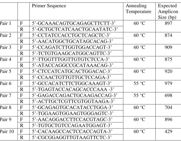
TABLE 3: PRIMER PAIRS DESIGNED TO CHARACTERIZE THE OPEN READING FRAME OF
WDR81...27
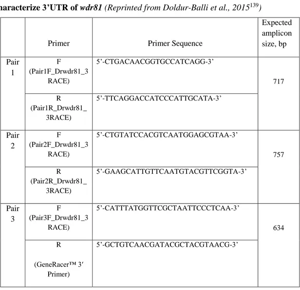
TABLE 4:PRIMER SETS DESIGNED TO OBTAIN THREE OVERLAPPING AMPLICONS IN ORDER TO CHARACTERIZE 3’UTR OF WDR81...31 TABLE 5:PRIMER PAIRS AND PROBES TO BE USED IN QRT-PCR………33
TABLE 6:VARIANTS DETECTED IN THE 3’RACE PRODUCTS OF 24 HPF EMBRYO AND ADULT BRAIN WDR81 EXCEPT THE INSERTION SITE WHICH WAS FOUND TO BE 266 BP LONG……….59 TABLE 7:INTENSITIES OF THE BANDS OF PCR PRODUCTS FROM FIGURE 21……….62
TABLE 8: SURVIVAL RATES OF THE EMBRYOS FROM EXPERIMENT GROUPS IN THE WDR81 MORPHOLINO DOSE CURVE STUDY………..72 TABLE 9:SURVIVAL RATES OF THE EMBRYOS FROM EXPERIMENT GROUPS IN THE HEAD SIZE
MEASUREMENT STUDY………76
TABLE E.1: TBLASTN SCORES OF ZEBRAFISH WDR81……….…..131
TABLE E.2: LIST OF THE HITS OBTAINED AS A RESULT OF TBLASTN SEARCH OF
Abbreviations
ANOVA, Analysis of variance ISH, In situ hybridization ATP8A2, ATPase, aminophospholipid
transporter, class I, type 8A, member 2
MFS, Major facilitator superfamily
BA6, Brodmann area 6 MLF, Medial longitudinal fascicle BEACH, Beige and Chediak-Higashi ORF, Open reading frame
BrdU, 5-bromo-2’-deoxyuridine P15, Post natal day 15
CA8, Carbonic anhydrase VIII qRT-PCR, Quantitative real-time PCR CAMRQ, Cerebellar Ataxia, Mental
Retardation and Dysequilibrium Syndrome
RACE, Rapid amplification of cDNA ends
CNS, Central nervous system RoP, Rostral primary motor neuron
DIG, Digoxigenin SGZ, Subgranular zone
Dpf, Days post fertilization SVZ, Subventricular zone ENU, N-ethyl-N-nitrosourea UTR, Untranslated region
GABA, Gamma-aminobutyric acid VLDLR, Very low density lipoprotein receptor
gbx2, gastrulation brain homeobox 2 WD40, ~40 amino acid structural motifs, often ending with a Tryptophan-Aspartic acid (W-D) dipeptide
GFAP, glial fibrillary acidic protein WDR81, WD repeat containing protein 81
Hpf, Hours post fertilization ZFIN, The Zebrafish model organism database
Chapter 1. Introduction
1.1.
Cerebellar Ataxia, Mental Retardation and Dysequilibrium
Syndrome
Neurodevelopmental disorders are associated with mutations which might affect numerous cellular mechanisms leading to impairment of growth and development of the nervous system. These type of impairments during the neurodevelopmental processes give rise to neurological or psychiatric diseases1. Schizophrenia, autism spectrum
disorder, fragile-X and Rett syndrome are a few examples of such neurodevelopmental disorders2–5. Cerebellar ataxia, mental retardation and dysequilibrium syndrome
(CAMRQ) is also a neurodevelopmental disorder. The inheritance pattern is autosomal recessive and it is a rare condition, which is characterized by mental retardation, cerebellar ataxia and dysarthric speech with or without quadrupedal gait6–14. Quadrupedal gait, ie. walking on all fours, in humans is also referred to as Unertan Syndrome6,15 and was first observed in 1914 in the Black Sea Region16 (Figure 1). CAMRQ is associated with specific mutations and these mutations have been shown in consanguineous families with one exception17. Homozygosity mapping, linkage analysis and targeted next generation sequencing of homozygous regions revealed genetic heterogeneity in this syndrome.
Figure 1. Quadrupedal gait in a male in 1914 (left)16 (by courtesy of Prof. Dr. Üner Tan) and in a female patient nowadays (right)18 (Reprinted with permission from
1.1.1.
CAMRQ Associated Genes
VLDLR (very low-density lipoprotein receptor), WDR81 (WD repeat containing protein
81), CA8 (carbonic anhydrase VIII) and ATP8A2 (ATPase, aminophospholipid transporter, class I, type 8A, member 2) have been reported to be associated with CAMRQ1 (MIM: 224050)8–10,13,17,19–21, CAMRQ2 (MIM: 610185)6–8,12, CAMRQ3 (MIM: 613227)11,22,23 and CAMRQ4 (MIM: 615268)14, respectively. The CAMRQ associated genes are mapped to chromosomes 8, 9, 13 and 17; CA8 to chromosome 8q12, VLDLR to chromosome 9p24, ATP8A2 to chromosome 13q12 and WDR81 to chromosome 17p13.
1.1.1.1. Very Low Density Lipoprotein Receptor (VLDLR)
VLDLR is a gene made up of 19 exons24 and encodes a receptor protein which is a member of the low-density lipoprotein receptor family25. It is an integrative element in the reelin pathway and functions in guiding neuroblast migration in the developing cerebral cortex and cerebellum26,27. ApoER2 (Apolipoprotein E Receptor 2) and VLDLR, which are reelin receptors, are both required for the coordination of alignment of Purkinje cell subsets in the developing cerebellum28,29. Reelin mediates cortical layer formation and dendrite development in hippocampus via VLDLR/ApoER2-Dab1 (Disabled 1) pathway30. Recent studies showed that VLDLR is actively involved in regulation of formation and development of dendritic spines31,32. In addition, VLDLR is
expressed in synapses, both presynaptically and postsynaptically. Knockdown of
VLDLR is reported to significantly decrease the synaptophysin puncta number and also
to decrease glutamate receptor subunits such as GluN1 levels and levels of GluA1 at the cell surface32. Besides its role in the reelin signaling pathway, VLDLR fulfills critical
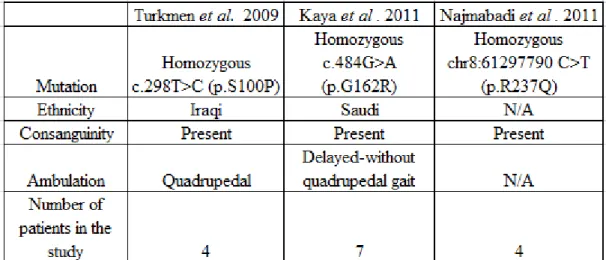
functions in triglyceride metabolism. It has an effect on the uptake of VLDL triglycerides in peripheral tissues33. It is also shown that the expression of VLDLR is essential in promoting adipocyte differentiation34. The summary of the reported cases with VLDLR-associated CAMRQ (CAMRQ1) and the information about the VLDLR mutations are given in Table 1.
3
The common morphological abnormalities in the brains of the patients with CAMRQ1 are the hypoplasia of cerebellum8,10,13,17,19–21 and moderate gyrial simplification of the
cerebral cortex8,10,13,17,20. The pons was observed particulary small in some patients8,13,17,21 while mild hypoplasia of the corpus callosum was observed by Turkmen
et al. (2008)19. Moheb et al. (2008) was unable to obtain any MRI (magnetic resonance imaging) or CT (computerized tomography) scans of the brains from any patients, and for this reason the brain abnormalities of the patients in this study could not be evaluated9.
1.1.1.2. WD Repeat Containing Protein 81 (WDR81)
The gene encoding WD repeat containing protein 81 (WDR81) was found to be mutated in some CAMRQ patients. It is a missense mutation and it lies in exon 1 of the WDR81 isoform 1, WDR81 p.P856L. The most significant morphological changes in the CAMRQ patients carrying this mutation were significant decline in the volumes of cerebellum and corpus callosum. Additional analysis showed that morphological differences in the precentral gyrus and Brodmann areas BA6, BA44 and BA45 took place in the patients’ brains12 (Figure 2). When researchers investigated the
neuro-ophthalmic aspect of CAMRQ, they observed that four patients who carry the missense mutation in WDR81 gene had downbeat nystagmus and two male patients among four patients had also bilateral temporal disc pallor and ring-shaped macular atrophy35. A similar phenotype to CAMRQ was obtained with a mutant mouse line nur5. The N-ethyl-N-nitrosourea (ENU) induced mutation in this model was a missense mutation, L1349P, and it was also located in the predicted major facilitator superfamily domain of the Wdr81 protein as in patients. This homozygous missense mutation caused loss of Purkinje cells, which was detected at P28 (young adult) and loss of photoreceptor cells, which was detected on P15 (infant) the earliest tested timepoints in this mouse model36.
The mitochondrial defects in the dendrites of Purkinje cells was detected in mutant mice at P21 (juvenile) and this finding led the conclusion that the mitochondrial abnormalities are followed up by death of Purkinje cells during development36.
Table 1. Summary of the reported cases with CAMRQ1 (Adapted from Ali et al. 201220)
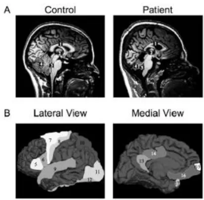
Figure 2. Brain morphology of a control individual and a patient were analyzed via magnetic resonance imaging (MRI). The patient was reported to be affected from
WDR81 associated CAMRQ (CAMRQ2, MIM: 610185). (A) A healthy control individual (left) and a patient (right) were scanned. The areas, which were assigned with numbers indicate regions where differences in volumes are distinguishable: corpus callosum (1), third ventricle (2), fourth ventricle (3), and cerebellum (4). (B) Lateral and medial views of a reference cortex on which the significantly affected morphometric parameters are numbered: BA45 (5), BA44 (6), BA6 (7), precentral (8), superior temporal (9), superior parietal (10), lateral occipital (11), fusiform (12), isthmus cingulated (13), posterior cingulated (14), frontal pole (15), medial orbitofrontal (16), and temporal pole (17)12. Reprinted with permission from Cold Spring Harbor
Laboratory Press.
WDR81 gene was shown to be mutated in cancer and it appears that it locates in a
metabolically important loci. WDR81 gene was reported to be mutated and expressed in >10% of the evaluated 23 colorectal cancer cell lines37. Transethnic meta-analysis of European ancestry and Japanese genome-wide association studies revealed that
SERPINF2-WDR81 loci as one of the six significant loci for serum albumin38. Serum
albumin level is metabolically important because it is inversely associated with cardiovascular risk and mortality risk39,40.
WDR81 was shown to be expressed in all of the tested human tissues and its level was
the highest in cerebellum and corpus callosum among brain regions12. Expression of
Wdr81 was detected in Purkinje cell neurons, photoreceptor cells, deep cerebellar nuclei
neurons and neurons of brainstem among the central nervous system neurons in wild-type adult mouse. Localization of Wdr81 was observed in the mitochondria of Purkinje cells using electron microscopy. Wdr81 expression was also detected in all of the evaluated adult tissues36. Gulsuner et al. also revealed that Wdr81 expression was higher in the Purkinje cell neurons and molecular layer of cerebellum in mouse embryonic brain12.
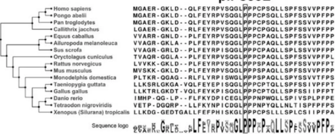
The function of WDR81 is not currently known, however it can be predicted in silico with information about the domains of the protein. The presence of conserved domains in proteins may lead to the indication of their function as well. Moreover, conservation of the identical domains in homologous proteins from different species sheds light on the critical importance of the protein of interest at several evolutionary levels. WDR81 protein is conserved among vertebrates, the mutation also hits a conserved domain of the protein of interest12 (Figure 3). The putative domains of human and mouse WDR81 proteins are Beige and Chediak-Higashi (BEACH) domain, a major facilitator superfamily (MFS) domain and six WD40 repeat domains. Both proteins are predicted to be transmembrane proteins12,36. A BEACH-domain containing proteins are proposed to perform as scaffold proteins. They are mostly large proteins and they take place in membrane-related events. These events include vesicle fusion and fission, so that they might function in vesicular transport, apoptosis, receptor signaling, formation of synapses, autophagy and membrane dynamics41. WD40 domain is given this name because of the length of the repeats of 40 aminoacids and the repeats often end with the
Figure 3. Phylogenetic tree of protein sequences of WDR81 in vertebrates. The box
implies the amino acid, mutated in CAMRQ patients12. Reprinted with permission from
Cold Spring Harbor Laboratory Press.
WD40 domain-containing proteins take place in numerous cellular functions such as cell cycle control, signal transduction, regulation of transcription, apoptosis, chromatin dynamics, vesicular trafficking and cytoskeletal assembly. The WD40 domains of BEACH domain-containing proteins serve as a scaffold for protein or protein-DNA interactions41,42. The MFS transporters function in response to chemiosmotic ion gradients and they transport small molecules. They are single-polypeptide secondary carriers and act as uniporters, symporters or antiporters43. Taken together, the WDR81 protein might have an importance related to both the central nervous system and the metabolism. The findings from mouse and human studies indicate that WDR81 might be a critical gene in neurodevelopment, but the exact function of the gene is still not fully understood.
The family of proteins named as WDR and followed by a number, is composed of almost 100 proteins in human genome. Some of them were reported to be associated with diseases. The association of WDR10 with cranioectodermal dysplasia 1 [MIM 218330]44, WDR11 with hypogonadotropic hypogonadism 14 with or without anosmia
with short-rib thoracic dysplasia 11 with or without polydactyly [MIM 615633]48,49,
WDR35 with cranioectodermal dysplasia 2 [MIM 613610]50 and short-rib thoracic
dysplasia 7 with or without polydactyly [MIM 614091]51, WDR36 with primary open angle glaucoma [MIM 609887]52, WDR56 with short-rib thoracic dysplasia 2 with or without polydactyly [MIM 611263]53,54, WDR60 with short-rib thoracic dysplasia 8 with or without polydactyly [MIM 615503]55, WDR62 with microcephaly 2, primary, autosomal recessive, with or without cortical malformations [MIM 604317]56–60 were reported. Dolichocephaly and high forehead were observed in cranioectodermal dysplasia as a result of abnormal development of the cranium and skeleton44. Anosmia was associated with the absence or underdevelopment of olfactory bulbs and their nerve fibers61. Hypogonadism was found to be related with the failure in migration of neurons which synthesize gonadotrophin releasing hormone during development62. Senior-Loken syndrome 8 is a progressive disorder characterized by retinal and renal failure63. Impairment in the organs such as brain, eye, heart, kidneys, liver, pancreas, intestines, and genitalia as wells as cleft lip/palate might accompany the short-rib thoracic dysplasia 11 with or without polydactyly condition64. Primary open angle glaucoma was characterized by the defect in the vision, optic nerve damage and increased intraocular pressure65. Primary microcephaly was characterized by mental retardation and brain
size, which is smaller than normal56. The brain malformations found to be related with
WDR62 mutations were pachygria56,57, underdevelopment of corpus callosum57,59,
polymicrogyria58,59, schizencephaly, subcortical heterotropia59, microlissencephaly, band heterotropias and dysplastic cortex56. Thus, mutations in several genes encoding WD40 domain containing proteins, similar to WDR81, were shown to be associated with brain and eye malformations during development.
1.1.1.3. Carbonic Anhydrase VIII (CA8)
Another CAMRQ associated gene CA8 is comprised of 9 exons24 and encodes a protein which has a critical function in motor control66. It has been classified as a member of the carbonic anhydrase gene family because of the sequence similarity, however the gene product of CA8 does not possess the enzymatic activity to catalyze the reaction of
hydration of carbon dioxide reversibly67. CA8 binds to IP
3R1 (inositol 1,4,5 triphospate
receptor type 1) and binding of CA8 to IP3R inhibits binding of IP3 to the receptor by
decreasing its affinity. Expression of both CA8 and IP3R1 is abundant in Purkinje cells
and they co-localize in the cytoplasm, dendrites and axons. This might explain why the sensitivity of IP3R1 to IP3 for IP3-induced Ca+2 release in this layer is low68. Expression
of CA8 in human fetal brain was detected in neuroprogenitor cells in the subventricular zone and in the neurons, which migrate to the cortex. In the adult human, CA8 expression was found out in the neural cell bodies in most regions of the central nervous system, such as cerebrum, diencephalon, cerebellum, pons and medulla69. The “waddles” (wdl) mouse is an animal model of Car8 null mutation66. Car8 is the
homologous protein of CA8 in mouse which shows a 98% identity70. Ataxia and appendicular dystonia comprised the phenotype of the wdl mouse and the gait disorder continued througout life-time66. CA8 overexpression in neuronal cell lines under staurosporine induced apoptotic stress reduced the cell death and overexpression of CA8 in neuronal cell lines and mouse cerebellar granule neurons increased neuronal migration and invasion. CA8 downregulation decreased cell migration and invasion ability and resulted in abnormal Ca+2 release in cerebellar granule neurons. Knockdown
of ca8 in zebrafish was achieved by using morpholino injection and ca8 morphants at 3 days post fertilization exhibited a decrease in motility71. Knockdown of ca8 via
microinjection of morpholino oligonucleotides in another study resulted in neuronal death and malformations in cerebellum and muscle. Body axis was curved and motor functions and coordination were defective, showing a similar phenotype to human condition72. Also, CA8 expression has been found to be increased in non-small lung cancer and colorectal cancer73,74.
The summary of the reported cases with CA8-associated CAMRQ (CAMRQ3) and the information about the CA8 mutations are given in Table 2. Among the three publications summarized in Table 2, only Kaya et al. (2011) provided results of brain imaging studies on patients. The MRI study showed variable volume loss in cerebellum and vermis in patients in this study22.
Table 2. Summary of the reported cases with CAMRQ3
N/A: not applicable, information not provided in the article.
1.1.1.4. ATPase, Aminophospholipid Transporter, Class I, Type 8A,
Member 2 (ATP8A2)
ATP8A2 (ATPase, aminophospholipid transporter, class I, type 8A, member 2) is made
up of 37 exons24 and encodes a protein, which is a member of P4 ATPase protein family75. This protein family facilitates transport of phospholipids from exoplasmic leaflet to cytosolic leaflet and thereby are involved in formation of the asymmetry of the cell membrane. They are also called lipid flippases and they take roles directly or indirectly in dynamics of cytoskeleton, signaling and metabolism of lipids, cell division and membrane trafficking76. ATP8A2 is expressed in brain, retina and testis among evaluated human tissues. Expression of ATP8A2 in the brain was detected in both fetal and adult tissues77. In adult human brain, all the tested regions showed expression of the
gene and the highest level of expression was found to be in cerebellum14. In mouse,
Atp8a2 expression was detected in brain, spinal cord, retina and testis78,79. The function
of Atp8a2 was revealed as to transfer aminophospholipids (phosphatidylserine and phosphatidyethanolamine) from exoplasmic to cytosolic leaflet in an ATP-dependent manner78. Overexpression and downregulation studies on Atp8a2, in conjuction with the results from the overexpression and downregulation of Cdc50a, demonstrated that
indicates a critical role in rat hippocampal neuronal differentiation80. A mutant mouse
model, wabbler lethal, which has ataxia and neurodegeneration, was shown to carry loss of function mutations in Atp8a2 gene. It was concluded Atp8a2 is associated with axon degeneration and neurodegenerative disease79. Disruption of the ATP8A2 gene was also reported to be associated with severe mental retardation and hypotonia in a patient77.
ATP8A2-associated CAMRQ (CAMRQ4) was reported in a consangineous family from Turkey. Four CAMRQ patients were detected in the family, however DNA from one patient could not be included in the study. Patients showed delayed ambulation and have quadrupedal gait8. The index patient was unable to walk at the time of the study. A missense mutation in ATP8A2 (c. 1128 C>G, p.I376M) was found out to be associated with the condition. Moderate hypoplasia of inferior cerebellum, corpus callosum and cerebral cortex were observed in the patients14.
1.2.
Zebrafish as a Model Organism
The model organism used in the current work is the zebrafish (Danio rerio) (Figure 4). George Streisinger and his colleagues established zebrafish as a genetic model system 35 years ago81. Zebrafish have become a promising model organism for scientists in developmental biology, neurophysiology, biomedical research and ethology fields82. Comparison of human reference genome and zebrafish reference genome revealed that one clear zebrafish orthologue gene exists for approximately 70% of human genes83. It is a vertebrate with an integrated nervous system and possesses common organs and tissues like brain and spinal cord84. Zebrafish produce large batches of externally fertilized and transparent embryos that enable observation of development with microscopy techniques. Moreover, embryos develop at a fast rate and key developmental events take place earlier and faster compared to mammalians. For example neurogenesis begins around 10 hours post-fertilization (hpf), synaptogenesis and the first behaviors of embryos begin around 18 hpf and hatching is observed around 52 hpf82,84. After approximately 2 days of development, zebrafish embryo has its internal organs, eyes, ears and a brain, which has been already divided into
compartments. Thus the embryo has formed all of the common main characteristics of a vertebrate body by this timepoint85.
Figure 4. Lateral views of a female (F) and a male (M) adult zebrafish. wt: wild
type, L: left side of the bodies. Scale bar indicates 2 mm86. Reprinted with permission from Elsevier.
Various molecular genetic techniques have been devised and used in zebrafish research and new techniques are continuing to emerge over time. For example, transgenic zebrafish lines, which express a fluorescent protein in specific types of neurons, are useful in studying the development of nervous system and neural circuits87. Bacterial artifical chromosome (BAC) mediated transgenesis is managed by using BAC which is engineered with homologous recombination to express fluorescent reporter in target cells88. Although the efficiency of BAC mediated transgenesis is low, this technique is
advantageous in obtaining the reporter genes and regulatory sequences together in the construct instead of putting effort in laborious subcloning work87,89. The efficiency of integration of the DNA injected to the zebrafish embryos increases based on the activity of the transposases, such as sleeping beauty90 and tol-291 transposases. Employing transgenes under control of heat-inducible promoters provides conditional control of gene expression92,93. Using the adapted form of the yeast galactose inducible94 and Cre-loxP systems95, which efficiently works in mouse models, also provide conditional control of gene expression and permanent labeling of cell lineages in zebrafish models.
Targeted mutation is managed by zinc finger nucleases (ZFNs)96 and TAL effector nucleases (TALENs)97,98 in zebrafish. A double strand break induced by these nucleases
is repaired by non-homologous end joining. This often results in insertion or deletions and ultimately a loss-of-function phenotype is obtained96–98. A recent method, Clustered
regularly-interspaced short palindromic repeats (CRISPR)/Cas9 is reported to be more efficient than ZFN and TALEN methods in terms of germline transmission. This system is a genome editing tool, already used in various model organisms and has advantages over other mutagenesis methods by its simplicity in design, being practical to use and enabling to target more than one genes at the same time99.
Morpholino antisense technology is used in loss-of-function experiments. Morpholino sequences knock down the gene of interest either by targeting the RNA splicing or the translation initiation. These sequences are stable and introduced to the embryos preferably at one-cell stage however their effect lasting for up to seven days and the concentration is diluted as cell division continues. Possible off-target effects needs to be evaluated as well. If apoptosis is triggered as an off-target effect of the morpholino, co-injection of p53 morpholino might be considered89,100. ENU mutagenesis is utilized to provide random mutagenesis also in zebrafish101 and Targeted Induced Local Lesions in Genomes (TILLING) method can reveal the mutations caused by ENU102.
Applying morpholino antisense technology was preferred in the present study since establishment of this technique would be faster compared to other genetic manipulation tools. The data obtained with the transient knockdown of the gene of interest might be investigated further by employing stable knockout systems. Besides, the effects of knockdown of wdr81 gene in zebrafish is possible to be searched starting from early developmental stages up to seven days post fertilization, which encompasses the critical timepoints of neurodevelopment. This provides an important advantage when it is considered that CAMRQ is a neurodevelopmental disease.
Zebrafish was used as a model organism to investigate cerebellar disorders. Knockdown of ataxin-7 via morpholino microinjection was applied by producing mild and severe phenotypes in order to study spinocerebellar ataxia 7 (SCA7). SCA7 was characterized by loss of Purkinje cells and granule cells of cerebellum and rod-cone photoreceptors.
While severe phenotype ended up with increased lethality of embryos and developmental defects, moderate phenotype led the observations close to the human condition. The differentiation of photoreceptors, Purkinje cells and granule cells was prevented via partial depletion of ataxin-7. Moreover, the phenotypes could be rescued by using human transcript, which might be evaluated as an evidence of conserved function103.Another study in which involvement of a gene to a condition was searched by utilizing morpholino technique demonstrated cerebellar and cerebral atrophy. Knockdown of clpb, which encodes caseinolytic peptidase B protein homolog, led to this phenotype and proved involvement of the gene of interest to 3-methylglutaconic aciduria, progressive brain atrophy, intellectual disability, congenital neutropenia, cataracts and movement disorder. This phenotype was also rescued via injection of human transcript104. Microinjection of sorting nexin 14 (snx14) translation blocking morpholino to embryos of a zebrafish line, which expresses GFP in the hindbrain, showed decrease in the intensity of the signal from GFP. Microinjection of the morpholino to embryos of a wild type strain showed decreased number of Purkinje cells. Data from both experiments propose that snx14 was necessary for hindbrain and formation and survival of Purkinje cells. Rescue was achieved by using the human transcript105. Epistatic interaction of two genes was investigated in association with
ataxia, dementia and hypogonadotropism. rnf216, encoding a ubiquitin E3 ligase and
otud4, encoding a deubiquitinase were silenced separately in zebrafish and impairments
in the cerebellum, optic tectum and eye were obtained. Coinjection of morpholinos targeting both genes showed a more severe phenotype. All the phenotypes were rescued with human transcript106. Homozygous mutant zebrafish, carrying loss of function mutation in qars, which encodes glutaminyl tRNA synthetase, demonstrated a phenotype similar to human patients. The phenotype was composed of small brain, neurodegeneration and small eye size107. A transgenic zebrafish line was generated in order to examine whether excitability of motor neurons were affected by spinocerebellar ataxia type 13 (SCA13) associated mutations. Human dominant mutation in the Kv3.3 voltage-gated K+ channel was expressed in zebrafish and this mutation was found to be associated with the loss in locomotion and decreased excitability108. Another research group utilized from several transgenic lines of zebrafish in order to screen mutations, which affect the development of cerebellum109.Hence, zebrafish became an attractive model organism for studying human diseases. Rescuing morphants with human
transcripts helps proving that the function of the gene of interest was conserved through evolution as well. Zebrafish community is also capable of generating stable models such as transgenic lines or mutants. These models might be used to examine the cellular changes underlying the diseases further.
1.2.1.
Zebrafish Central Nervous System
Zebrafish central nervous system (CNS) will be discussed under two sections: the first “1.2.1.1. Central Nervous System in Zebrafish Embryos” and the second “1.2.1.2. Central Nervous System in Adult Zebrafish” in order to review information about both the development of the CNS and the developed CNS, respectively.
1.2.1.1 Central Nervous System in Zebrafish Embryos
The neuroectoderm needs to be specified on the dorsal part of the embryo via neural induction in order to form neural plate. The neural plate is transformed into a neural tube following a combination of signaling and morphogenetic movements. The anterior region of the neural tube forms the brain and the posterior region forms the spinal cord110,111.
Emergence of some critical structures related with zebrafish CNS are earlier than that of mammalians. The brain rudiment appears at sphere stage (blastula, 4-4.33 hpf), primary motor neurons appear at 1-4 somites stage (segmentation, 10.33-11.66 hpf), neural tube appears at 10-13 somites stage (segmentation, 14-16 hpf), cerebellum appears at 26+ somites stage (segmentation, 22-24 hpf), spinal cord appears at prim-5 (pharyngula, 24-30 hpf) and immature eye appears at 5-9 somites stage (segmentation, 11.66-14 hpf)112.
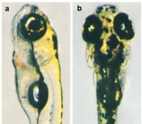
All of the main components of the brain have formed by 5 days post fertilization (dpf)85.
The size of the larval brain at 5 dpf facilitates microscopic research in vivo. Its thickness is around 500 micrometers and the length is 1.5 milimeters (mm), makes all neurons possible to be studied with this technique82 (Figure 5).
Figure 5. Five dpf wild type zebrafish embryo. a) Lateral view b) dorsal view113. Reprinted with permission from John Wiley and Sons.
1.2.1.2 Central Nervous System in Adult Zebrafish
The regions of the zebrafish adult brain are telencephalon (olfactory bulbs, ventralis
telencephali, area dorsalis telencephali, telencephalic tracts and commissures),
diencephalon (area praeoptica, epithalamus, dorsal thalamus, ventral thalamus, posterior tuberculum, hypothalamus, synencephalon, pretectum, diencephalic tracts and commissures), mesencephalon (tectum opticum, torus semicircularis, tegmentum), rhombencephalon (cerebellum, medulla oblongata), medulla spinalis and brain stem114. One of the most striking features of adult zebrafish brain is the dominance of the optic tectum in the dorsal midbrain, as also observed in other teleosts. One disadvantage of studying with zebrafish telencephalon is eversion of this region dorsally, which is not present in other vertebrates (Figure 6). This feature, partly causes difficulties in finding homologous regions between fish and mammalian forebrain regions85.
The length of the adult zebrafish brain is around 4.5 mm and its thickness is between 0.4 -2 mm. The zebrafish brain size and number of neurons enable research with advanced microscopy techniques such as multiphoton microscopy and 3D electron microscopy (Figure 6). Since activity analysis and connectivity pattern studies possess size restrictions from a circuit neuroscience point of view82.
1.3.
Cell Proliferation in Zebrafish Nervous System
The differentiated cells of the central nervous system originate from multipotent neuroepithelial stem cells. These stem cells give rise to mature and functional neurons or glia after the processes called neurogenesis and gliogenesis, respectively115. Because
of this phenomenon, cell proliferation in zebrafish central nervous system will be discussed under two titles of “1.3.1. Neurogenesis in Zebrafish” and “1.3.2. Gliogenesis in Zebrafish”.
Figure 6. Illustration which compares the brain sizes of human, mouse and zebrafish. (A) Comparison of the lateral views of the adult brains from three species to
scale. (B) Major brain areas of zebrafish in a linear plane. OB, olfactory bulb; Tel, Telencephalon; Di, Diencephalon; Mes, Mesencephalon; Rh, Rhombencephalon; sc, Spinal cord116 (By courtesy of Associate Professor Philippe Mourrain). The small size of the zebrafish brain enables observation of the whole brain under microscopy, especially in neuroscience research.
1.3.1.
Neurogenesis in Zebrafish
Neurogenesis is defined as the process which first starts with neural induction and ends up with differentiated functional neurons117. Neurogenesis in zebrafish will be discussed under two sections: the first “1.3.1.1. Neurogenesis in Zebrafish Embryos” and the second “1.3.1.2. Neurogenesis in Adult Zebrafish”.
1.3.1.1. Neurogenesis in Zebrafish Embryos
Neural induction, which is defined as specification of the neuroectoderm, is required before neurogenesis. Neural induction is directed by extrinsic and intrinsic factors. Indeed, the interplay of extrinsic bone morphogenetic protein (BMP), wingless-integrated (Wnt) and fibroblast growth factor (Fgf) family members and intrinsic factors (SRY-box containing genes B1 (SoxB1) family members) decides the vertebrate neural induction. After the neuroectoderm is specified, it forms the neural plate111. The neural plate is converted into the neural tube as a result of the process called as neurulation. In contrast with most vertebrates, zebrafish generates the neural keel. The neural plate is firstly transformed into a solid structure in zebrafish instead of folding the lateral edges of the neural plate and forming a tube with a lumen by fusing the edges in the dorsal midline. However the typical neural tube is obtained after the cells in the center detach. Despite the differences, fish and mammals produce very similar neural tubes111,118.
Neurogenesis take place in two stages in zebrafish: primary neurogenesis and secondary neurogenesis. Primary neurogenesis starts at late gastrulation and proceeds during embryogenesis. This process yields early-born, big neurons which possess long axons. The brain epiphyseal and post-optic clusters, Rohon-Beard sensory neurons, Mauthner cells, and the three types of primary spinal motoneurons (the rostral (RoP), middle (MiP) and and caudal (CaP) are produced as a result of primary neurogenesis. The primary neurons form the first functional neuronal scaffold at embryonic and early larval development. Axonogenesis starts to appear between 14-24 hpf119. Secondary
neurogenesis, which is also called postembryonic neurogenesis starts at around 2 dpf in zebrafish. This second stage of neurogenesis turns the primary system into a network which gets more fine-tuned and complex120. Neuronal migration, differentiation and survival are the critical steps of producing functional neurons after the cells leave mitosis and multipotent progenitors determine particular cell types119.
1.3.1.2. Neurogenesis in Adult Zebrafish
Although adult mammals have a restricted neurogenic capacity, teleosts including zebrafish possess a larger potential for neurogenesis. Two neurogenic areas in adult mammalian brain are the subventricular zone (SVZ) of the lateral ventricle and the subgranular zone (SGZ) of the dentate gyrus in the hippocampus121. Neurogenic regions in the adult zebrafish brain are more widespread and there are regions homologous to mammalian SVZ and SGZ. The number of newly born cells in the adult zebrafish brain is approximately 6000 cells during every 30 minutes111.
The zebrafish has become an attractive model for adult neurogenesis studies in recent years both because of the extensive capacity of neurogenesis and its advantages over mammalian models. In contrast to mammalian neural tissue, zebrafish brain does not exhibit scar formation after an injury. The zebrafish is a promising model organism to ultimately devise a theurapeutic approach for humans by providing the mechanisms of the plasticity and of regeneration by suppressing the scar formation111.
1.3.2.
Gliogenesis in Zebrafish
Gliogenesis, which is defined as the generation of mature glia cell types, in zebrafish will be discussed under two sections: the first is “1.3.2.1. Gliogenesis in Zebrafish Embryos” and the second is “1.3.2.2. Gliogenesis in Adult Zebrafish”.
1.3.2.1. Gliogenesis in Zebrafish Embryos
Glial cell types in central nervous system of mammals are macroglia (astrocytes, oligodendrocytes and ependymocytes) and microglia. Schwann cells, satellite glial cells, and enteric glia constitute the glial cell types in the peripheral nervous system of mammalian organisms. The glial cell types in zebrafish have not been described profoundly yet, however there is an accumulating data about the similarities between the glial cells of fish and mammals89.
Glial cells appear at 26+ somites stage (segmentation, 22-24 hpf) in zebrafish112.
Oligodendrocytes, in both mammals and zebrafish, appear to originate from the ventral regions of the neural tube. Oligodendrocyte precursor cells migrate to the appropriate regions and divide in the central nervous system. They, then leave the cell cycle and cover axons with myelin sheath89,115. Schwann cells of both mammalian organisms and zebrafish generate from neural crest tissues and the developmental process they pass through is similar. During the process, they migrate to and eventually cover peripheral axons with myelin sheath89.
Radial glia cells and a subpopulation of astrocytes serve as neural stem cells in the adult mammalian brain. Neurons, ependymal cells, astrocytes, intermediate progenitor cells that give rise to neurons (nIPCs), and intermediate progenitor cells that give rise to oligodendrocytes (oIPCs) originate from the neuroepithelial-radial glia-astrocyte line122.
1.3.2.2. Gliogenesis in Adult Zebrafish
As previously mentioned under “1.3.2.1. Zebrafish Embryo” section, zebrafish possesses mammalian counterparts of oligodendrocytes and Schwann cells. Expression analysis showed that satellite and enteric glia of zebrafish are also similar to their
mammalian counterparts123,124. Ependymal cells have not been detected in zebrafish and
low number of cells are detected with the known star shaped morphology of astrocytes. Radial glia cells appear to carry the characteristics of ependymal cells and astrocytes and to fulfill their functions. A large number of radial glia cells in zebrafish express glial fibrillary acidic protein (GFAP), gluthamine synthase and aquaporin-4. GFAP and gluthamine synthase are markers often used to detect astrocytes in mammalian model organisms and aquaporin-4 is a water channel found on astrocytes89. Microglia are present in zebrafish. The origin of microglia cells is mesoderm125 and they are mononuclear phagocytes, which function in the defense of the central nervous system126,127.In both mammals and zebrafish this glial cell type takes place in the parenchyma of the central nervous system126.
Gliogenesis continues in the adult zebrafish as neurogenesis does; radial glia cells and a subpopulation of astrocytes can re-enter the cell cycle and can give rise to macroglia cell types as well as to neurons128. The radial glia cell population reduces in the adult mammalian brain, but still in the neurogenic areas of the brain, which are the subventricular zone (SVZ) of the lateral ventricle in telencephalon and the subgranular zone (SGZ) of the dentate gyrus in the hippocampus, some radial glia cells serve as neural stem cells. In contrast to mammalian adult brain, zebrafish holds the large numbers of radial glia cells both in the late embryonic development and during adulthood89.
1.4.
Synaptogenesis in Zebrafish
Synaptogenesis is a multi-step process to form synapses. This process is critical in neurodevelopment and has a continuing significance in learning, memory, plasticity, cognition and adaptation throughout adulthood129,130. Synapses are the contact sites of neurons where one transfers signals to another. These signals might be either electrical or chemical. Chemical synapses are more common in the vertebrate nervous system. Electrical signals, in the form of action potential, start at the axon hillock and then move
through the axon of the neuron, arrive at the presynaptic terminal and this triggers a release of neurotransmitters into the synaptic cleft. This is where electrical signal is converted into a chemical one. The neurotransmitters bind to their receptors at the membrane of the postsynaptic neuron. The signal is converted back to electrical one within the postsynaptic dendrite. Excitatory chemical synapses are asymmetric structures, and inhibitory chemical synapses are symmetric structures and occur between neurons and other neurons, muscles or glands130.
Synaptic vesicle fusion does not take place randomly, active zones are specialized sites for synaptic vesicles to dock and fuse with the plasma membrane in the presynaptic terminal. Another specialized region in the postsynaptic membrane which is occupied with the receptors, secondary messenger molecules and voltage-gated ion channels is called postsynaptic density. The size, organization and thickness of the active zone and postsynaptic density of the synapses in the central nervous system depend on three parameters: type of the synapse, function of the synapse and efficacy of the synapse. Type of the synapse is defined by the type of the released neurotransmitter. The synapse might be classified into three groups based on their functions: excitatory, inhibitory or modulatory. Efficacy of the synapses is determined by its reliable or unreliable, continous or sporadic activity130.
Synaptogenesis in neurodevelopment starts in embryonic stages and continues through early postnatal stage. It is closely related with neuronal differentiation and formation of the neural circuits. The genes which encode synaptic proteins are activated a short time after the neurons differentiate and form their axons and dendrites. The axons and dendrites make some contacts and form synapses. These initial synapses are frequently transient. A set of molecules and proteins (such as receptors and cell surface adhesion molecules) and signaling process (specialization of active zone and postsynaptic density) are required for synapse formation. Eventually, the decision on keeping or eliminating a synapse is determined based on its activity both during neurodevelopment and processes going on in mature brain130.