Mutations in the very low-density lipoprotein
receptor
VLDLR cause cerebellar hypoplasia
and quadrupedal locomotion in humans
Tayfun Ozcelik*†‡, Nurten Akarsu§¶, Elif Uz*, Safak Caglayan*, Suleyman Gulsuner*, Onur Emre Onat*, Meliha Tan储,
and Uner Tan**
*Department of Molecular Biology and Genetics, Faculty of Science and‡Institute of Materials Science and Nanotechnology, Bilkent University, Ankara 06800, Turkey;§Department of Medical Genetics and¶Gene Mapping Laboratory, Department of Pediatrics, Pediatric Hematology Unit, Ihsan Dogramaci Children’s Hospital, Hacettepe University Faculty of Medicine, Ankara 06100, Turkey;储Department of Neurology,
Baskent University Medical School, Ankara 06490, Turkey; and **Faculty of Sciences, Cukurova University, Adana 01330, Turkey
Edited by Mary-Claire King, University of Washington, Seattle, WA, and approved January 16, 2008 (received for review October 22, 2007) Quadrupedal gait in humans, also known as Unertan syndrome, is
a rare phenotype associated with dysarthric speech, mental retar-dation, and varying degrees of cerebrocerebellar hypoplasia. Four large consanguineous kindreds from Turkey manifest this pheno-type. In two families (A and D), shared homozygosity among affected relatives mapped the trait to a 1.3-Mb region of chromo-some 9p24. This genomic region includes the VLDLR gene, which encodes the very low-density lipoprotein receptor, a component of the reelin signaling pathway involved in neuroblast migration in the cerebral cortex and cerebellum. Sequence analysis of VLDLR revealed nonsense mutation R257X in family A and single-nucleotide deletion c2339delT in family D. Both these mutations are predicted to lead to truncated proteins lacking transmembrane and signaling domains. In two other families (B and C), the phenotype is not linked to chromosome 9p. Our data indicate that mutations in VLDLR impair cerebrocerebellar function, conferring in these families a dramatic influence on gait, and that hereditary disorders associated with quadrupedal gait in humans are genet-ically heterogeneous.
genetics兩 Unertan syndrome
O
bligatory bipedal locomotion and upright posture of mod-ern humans are unique among living primates. Studies of fossil hominids have contributed significantly to modern under-standing of the evolution of posture and locomotion (1–5), but little is known about the underlying molecular pathways for development of these traits. Evaluation of changes in brain activity during voluntary walking in normal subjects suggests that the cerebral cortices controlling motor functions, visual cortex, basal ganglia, and the cerebellum might be involved in bipedal locomotor activities (6). The cerebellum is particularly impor-tant for movement control and plays a critical role in balance and locomotion (7).Neurodevelopmental disorders associated with cerebellar hy-poplasias are rare and often accompanied by additional neuro-pathology. These clinical phenotypes vary from predominantly cerebellar syndromes to sensorimotor neuropathology, ophthal-mological disturbances, involuntary movements, seizures, cog-nitive dysfunction, skeletal abnormalities, and cutaneous disor-ders, among others (8). Quadrupedal locomotion was first reported when Tan (9, 10) described a large consanguineous family exhibiting Unertan syndrome, an autosomal recessive neurodevelopmental condition with cerebellar and cortical hy-poplasia accompanied by mental retardation, primitive and dysarthric speech, and, most notably, quadrupedal locomotion. Subsequent homozygosity mapping indicated that the phenotype of this family was linked to chromosome 17p (11). Thereafter, three additional families from Turkey (12–14) and another from Brazil (15) with similar phenotypes have been described, and video recordings illustrating the quadrupedal gait have been
made (10–12). Here, we report that VLDLR is the gene respon-sible for the syndrome in two of these four Turkish families and report additional gene mapping studies that indicate the disorder to be highly genetically heterogeneous.
Author contributions: T.O., N.A., and U.T. designed research; T.O., N.A., E.U., S.C., S.G., and O.E.O. performed research; T.O., N.A., E.U., S.C., S.G., and M.T. analyzed data; and T.O., N.A., and U.T. wrote the paper.
The authors declare no conflict of interest. This article is a PNAS Direct Submission.
†To whom correspondence should be addressed. E-mail: [email protected]. This article contains supporting information online atwww.pnas.org/cgi/content/full/ 0710010105/DC1.
© 2008 by The National Academy of Sciences of the USA
Fig. 1. Phenotypic (A) and cranial radiologic (B) presentation of quadrupe-dal gait in families A and D. (A) Affected brothers VI:20 and VI:18 and cousin VI:25 in family A (Upper) and the proband II:2 in family D (Lower) display palmigrate walking. This is different from quadrupedal knuckle-walking of the great apes (2). The hands make contact with the ground at the ulnar palm, and consequently this area is heavily callused as exemplified by VI:20. Stra-bismus was observed in all affected individuals. (B) Coronal and midsagittal MRI sections of VI:20, demonstrating vermial hypoplasia, with the inferior vermial portion being completely absent. Inferior cerebellar hypoplasia and a moderate simplification of the cerebral cortical gyri are noted. The brainstem and the pons are particularly small (Left and Center). Similar findings are observed for II:2 (Right).
Results
The proband of Family A (12) is a 37-year-old male with habitual quadrupedal gait (Fig. 1A Upper Left and Fig. 2A, VI:20). He did not make the transition to bipedality during his childhood despite the efforts of his healthy parents. He has dysarthric speech with a limited vocabulary, truncal ataxia, and profound mental retardation. He was not aware of place or of the year,
month, or day. His MRI brain scan revealed inferior cerebellar and vermial hypoplasia, with the inferior vermial portion being completely absent. Whereas corpus callosum appeared normal, a moderate simplification of the cerebral cortical gyri accom-panied by a particularly small brainstem and the pons was observed (Fig. 1 B Left and Center). Subsequently, we studied the proband’s affected brother and cousin (Fig. 1 A Upper Center and
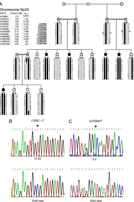
Fig. 2. Homozygosity mapping of cerebellar hypoplasia and quadrupedal locomotion to chromosome 9p24 (A) and identification of the VLDLR c769C 3 T mutation in family A (B) and of the VLDLR c2339delT mutation in family D (C). (A) Pedigree of family A; filled symbols represent the affected individuals. Squares indicate males, and circles indicate females. Black bars represent the haplotype coinherited with the quadrupedal phenotype in the family. Recombination events in individuals VI:16 (obligate carrier) and VII:4 (normal sibling) positioned the disease gene between markers rs7847373 and rs10968723. Physical positions and pairwise lod scores for each marker are shown on the upper left. Zmaxrepresents the maximum lod score obtained at ⫽ 0.00 cM. (B and C) Sequences of critical regions of VLDLR for wild-type and homozygous mutant genotypes.
Upper Right and Fig. 2 A, VI:18 and VI:25) and other branches of the family living in nearby villages in southeastern Turkey. All affected individuals were offspring of consanguineous marriages (Fig. 2 A). With the exception of one female (VII:1), who was an occasional biped with ataxic gait, all affected persons in family A had quadrupedal locomotion.
The proband of family D (14) is a 38-year-old male (Fig. 1 A Lower Left and Center). Like all other quadrupedal individuals in these families, he did not make the transition to bipedality during his early childhood. He is profoundly retarded and exhibits dysarthric speech along with truncal ataxia. His MRI brain scan images are consistent with moderate cerebral cortical simplification and inferior cerebellar and vermial hypoplasia (Fig. 1B Right). The 65-year-old aunt and 63-year-old uncle of the proband are both mentally retarded and continue to walk on their wrists and feet despite their advanced ages. The family is consanguineous; all relatives were raised in neighboring villages on the western tip of the Anatolian peninsula.
All patients in these four families had significant developmen-tal delay noted in infancy (Table 1). They sat unsupported between 9 and 18 months, and began to crawl on hands and knees or feet. Whereas normal infants make the transition to bipedal walking in a short period, the affected individuals continued to move on their palms and feet and never walked upright. All patients had severe truncal ataxia affecting their walking pat-terns. They can stand from a sitting position and maintain the upright position with flexed hips and knees. However, they virtually never initiate bipedal walking on their own and instead ambulate efficiently in a quadrupedal fashion. All patients had hyperactive lower leg and vivid upper extremity reflexes. Normal tone and power were observed in motor examination. All affected persons were mentally retarded to the degree that consciousness of place, time, or other experience appeared to be absent. However, no autistic features were expressed. The affected individuals all had good interpersonal skills, were friendly and curious to visitors, and followed very simple ques-tions and commands. Additional clinical information on families A and D is provided insupporting information (SI) Table 2.
To identify the chromosomal locale of the gene or genes responsible for this phenotype, we carried out genome-wide linkage analysis and homozygosity mapping in families A–C (see
Materials and Methods below). Although the families lived in isolated villages 200–300 km apart and reported no ancestral relationship, the rarity of the quadrupedal gait in humans led us to expect a single locus shared by affected individuals in all families. Instead, the trait mapped to three different chromo-somal locales. In family A, linkage analysis and homozygosity mapping positioned the critical gene on chromosome 9p24 between rs7847373 and rs10968723 in a 1.032-Mb region (Fig. 2 A andSI Fig. 4). In family B, the trait mapped to chromosome 17p13, confirming a previous study (11). In family C, highly negative logarithm of odds (lod) scores were obtained for both
chromosomes 9p24 and 17p13 (SI Figs. 5 and 6); gene mapping
in this family is ongoing. In family D, polymorphic markers from the critical intervals of chromosomes 9p24 and 17p13 were genotyped, and homozygosity was detected with markers on 9p24. Together, these results indicate that the syndrome includ-ing quadrupedal gait, dysarthric speech, mental retardation, and cerebrocerebellar hypoplasia is genetically heterogeneous.
The chromosome 9p24 region linked to the trait in families A and D includes VLDLR, the very low-density lipoprotein recep-tor. We hypothesized that a gene involved in neural develop-ment, cell positioning in brain, and cerebellar maturation could be involved in the pathogenesis of quadrupedal gait. In addition, cerebellar hypoplasia with cerebral gyral simplification was shown to be associated with a genomic deletion that includes VLDLR (16). We therefore considered VLDLR (17) to be a prime positional candidate for our phenotype and sequenced the
gene in genomic DNA from probands of the four families (SI
Table 3). The VLDLR sequence of affected members of family
A was homozygous for a nonsense mutation in exon 5 (c769C 3 T; R257X) (Fig. 2B). The VLDLR sequence of the proband of family D was homozygous for a single-nucleotide deletion in exon 17 resulting in a stop codon (c2339delT; I780TfsX3) (Fig. 2C). VLDLR sequences excluded the possibility of compound heterozygosity in families B and C (SI Fig. 7). In families A and D, homozygosity for the VLDLR mutations was perfectly coin-herited with quadrupedal gait (SI Figs. 8 and 9). Both mutations were absent from 100 unaffected individuals who live in the same local areas of southeastern and western Turkey as families A and
D (SI Fig. 10).
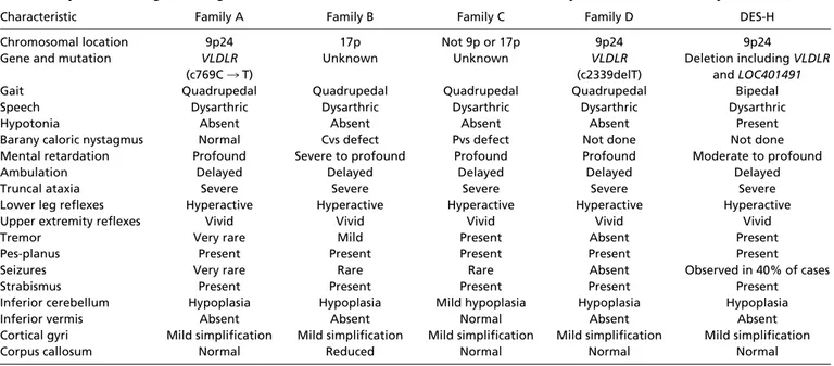
Table 1. Physical, radiological, and genetic characteristics of the Turkish families in this study and of Hutterite family DES-H (27)
Characteristic Family A Family B Family C Family D DES-H
Chromosomal location 9p24 17p Not 9p or 17p 9p24 9p24
Gene and mutation VLDLR (c769C 3 T)
Unknown Unknown VLDLR
(c2339delT)
Deletion including VLDLR and LOC401491
Gait Quadrupedal Quadrupedal Quadrupedal Quadrupedal Bipedal
Speech Dysarthric Dysarthric Dysarthric Dysarthric Dysarthric
Hypotonia Absent Absent Absent Absent Present
Barany caloric nystagmus Normal Cvs defect Pvs defect Not done Not done Mental retardation Profound Severe to profound Profound Profound Moderate to profound
Ambulation Delayed Delayed Delayed Delayed Delayed
Truncal ataxia Severe Severe Severe Severe Severe
Lower leg reflexes Hyperactive Hyperactive Hyperactive Hyperactive Hyperactive
Upper extremity reflexes Vivid Vivid Vivid Vivid Vivid
Tremor Very rare Mild Present Absent Present
Pes-planus Present Present Present Present Present
Seizures Very rare Rare Rare Absent Observed in 40% of cases
Strabismus Present Present Present Present Present
Inferior cerebellum Hypoplasia Hypoplasia Mild hypoplasia Hypoplasia Hypoplasia
Inferior vermis Absent Absent Normal Absent Absent
Cortical gyri Mild simplification Mild simplification Mild simplification Mild simplification Mild simplification
Corpus callosum Normal Reduced Normal Normal Normal
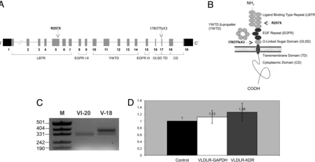
VLDLR㛭R257X is in the ligand-binding domain, and VLDLR㛭I780TfsX3 is in the O-linked sugar domain of the VLDLR protein (Fig. 3 A and B). Mutant VLDLR transcripts were expressed in endothelial cells from blood of affected individuals (Fig. 3C), and in these cells, levels of mutant and wild-type transcript expression appeared approximately equal (Fig. 3D; please also seeSI Text). Because the stop codons of both mutations are located in the extracellular domain of VLDLR (Fig. 3B), the encoded mutant proteins could not be inserted into the membrane and could not function as receptors for reelin.
We propose VLDLR-associated Quadrupedal Locomotion (VLDLR-QL) or Unertan Syndrome Type 1 to describe the phenotype of families A and D.
Discussion
The identification of these VLDLR mutations provides molec-ular insight into the pathogenesis of neurodevelopmental move-ment disorders and expands the scope of diseases caused by mutations in components of the reelin pathway (18). Reelin is a secreted glycoprotein that regulates neuronal positioning in cortical brain structures and the migration of neurons along the radial glial fiber network by binding to lipoprotein receptors VLDLR and APOER2 and the adapter protein DAB1 (19). In the cerebellum, reelin regulates Purkinje cell alignment (20), which is necessary for the formation of a well defined cortical plate through which postmitotic granual cells migrate to form the internal granular layer (21). Homozygous mutations in the reelin gene (RELN) cause the Norman–Roberts type lissencephaly syndrome, associated with severe abnormalities of the cerebel-lum, hippocampus, and brainstem (OMIM 257320) (22). Muta-tion of Reln in the mouse results in the reeler phenotype and disrupts neuronal migration in several brain regions and gives rise to functional deficits such as ataxic gait and trembling (23). In contrast, mice deficient for Vldlr appear neurologically normal
(24), but the cerebellae of these mice are small, with reduced foliation and heterotopic Purkinje cells (17).
In humans, homozygosity for either of two VLDLR truncating mutations leads to cerebrocerebellar hypoplasia, specifically vermial hypoplasia, accompanied by mental retardation, dysar-thric speech, and quadrupedal gait. In the Hutterite population of North America, homozygosity for a 199-kb deletion encom-passing the VLDLR gene leads to a form of Disequilibrium Syndrome (DES-H, OMIM 224050), characterized by nonpro-gressive cerebellar hypoplasia with moderate-to-profound men-tal retardation, cerebral gyral simplification, truncal ataxia, and delayed ambulation (16). The designation Disequilibrium Syn-drome was originally given to cerebral palsy characterized by a variety of congenital abnormalities, including mental retarda-tion, disturbed equilibrium, severely retarded motor develop-ment, muscular hypotonia, and perceptual abnormalities (25, 26). Neither DES-H nor other disequilibrium syndromes have been reported to include quadrupedal gait. The movement of most DES-H patients was so severely affected that independent walking was not possible. Those who could walk had a wide-based, nontandem gait (27).
The neurological phenotypes in the Turkish families and in the Hutterite families appear similar, with the most striking differ-ence being the consistent adoption of efficient quadrupedal locomotion by the affected Turkish individuals (Table 1). In our view, the movement disorder described for the Hutterite patients may be a more profound deficit, with the patients perhaps lacking the motor skills for quadrupedal locomotion. The 199-kb deletion in DES-H encompasses the entire VLDLR gene and part of a hypothetical gene. LOC401491, the hypothetical gene, is an apparently noncoding RNA that shares a CpG island and likely promoter with VLDLR, and is represented by multiple alternative transcripts expressed in brain. It has been suggested that the DES-H phenotype could be the result of deletion of VLDLR or both VLDLR and the neighboring gene (16).
Fig. 3. Functional domains of VLDLR with positions of the mutations relative to the exons (A), domains (B), and the analysis of VLDLR transcript (C and D). (A) The gene consists of 19 exons. Arrows indicate the locations of the mutations. (B) VLDLR consists of ligand-binding type repeat (LBTR), epidermal growth factor repeat (EGFR) I–III, YWTD-propeller (YWTD), O-linked sugar domain (OLSD), transmembrane domain (TD), and cytoplasmic domain (CD) (34) (www.expasy.org/uniprot/P98155). (C) Restriction-based analysis with HphI revealed the presence of only the mutant (347 bp) and both the mutant and wild type (396 and 347 bp; please note that the 49-bp fragment is not visible) VLDLR transcripts in patient VI:20 and carrier V:18 (both from family A), respectively. M is a DNA size marker. (D) Quantitative RT-PCR analysis of VLDLR transcript from peripheral blood samples of all probands in families A and D and controls was performed. Relative expression ratios were normalized according to the housekeeping gene GAPDH (glyceraldehyde-3-phosphate dehydrogenase) and the endothelial marker KDR (kinase insert domain receptor).⌬Ct values were calculated from duplicate samples and were converted to linear scale (35). Control denotes ‘‘VLDLR expression in controls,’’ VLDLR-GAPDH denotes ‘‘VLDLR expression in patients normalized to GAPDH,’’ and finally VLDLR-KDR denotes ‘‘VLDLR expression in patients normalized to KDR.’’
It has been suggested that in the Turkish families, lack of access to proper medical care exacerbated the effects of cere-bellar hypoplasia, leading to quadrupedality. Although it may be true that family B lacked proper medical care, families A and D had consistent access to medical attention, and both families actively sought a correction of quadrupedal locomotion in their affected children. An unaffected individual in family A is a physician who was actively involved in the medical interventions. In family D, the proband’s mother sought a definitive diagnosis and correction of the proband’s quadrupedal locomotion from private medical practices and from two major academic medical centers. The parents in family A discouraged quadrupedal walking of their affected children, but without success. We conclude that social factors were highly unlikely to contribute to the quadrupedal locomotion of the affected individuals.
In conclusion, we suggest that VLDLR-deficiency in the brain at a key stage of development leads to abnormal formation of the neural structures that are critical for gait. Given the heteroge-neity of causes of quadrupedal gait, identification of the genes in families B and C promises to offer insights into neurodevelop-mental mechanisms mediating gait in humans.
Materials and Methods
Study Subjects. Parents of patients and other unaffected individuals gave
consent to the study by signing the informed consent forms prepared accord-ing to the guidelines of the Ministry of Health in Turkey. The Ethics Commit-tees of Baskent and Cukurova Universities approved the study (decision KA07/47, 02.04.2007 and 21/3, 08.11.2005, respectively).
Genome-Wide Linkage Analysis. Linkage analysis was performed by SNP
geno-typing with the commercial release of the GeneChip 250K (NspI digest) or 10K
Affymetrix arrays as described (28). In addition, genotype data were analyzed by hand to identify regions of homozygosity. The parametric component of the Merlin package v1.01 was used for the multipoint linkage analysis assum-ing autosomal recessive mode of inheritance with full penetrance (29, 30). The analysis was carried out along a grid of locations equally spaced at 1 cM. Haplotype analysis was performed on chromosomal regions with positive lod scores (Fig. 2 A andSI Figs. 4 – 6). Pairwise linkage was analyzed by using the MLINK component of the LINKAGE program (FASTLINK, version 3) (31–33). Markers D17S1298 (3.51 Mb) and D9S1779 (0.4 Mb), D9S1871 (3.7 Mb) were used to test for homozygosity to chromosomes 17p13 and 9p24, respectively. Mutation Search. Sequencing primers were designed for each VLDLR exon by using Primer3, BLAST, and the sequence of NC㛭000009. DNA from all of the probands was sequenced in both directions by using standard methods. The mutations in exons 5 (c769C 3 T) and 17 (c2339delT) were detected in all affected (homozygous) and carrier (heterozygous) individuals of families A and D, respectively. The c769C 3 T mutation creates a restriction site for the enzyme HphI (5⬘-GGTGA(N)8 2 3⬘), and the c2339delT mutation abolishes a restriction site for the enzyme MboI (5⬘-G2 ATC-3⬘). Assays using these restriction enzymes were developed to test for the mutations in all four families and in 200 healthy controls from the Turkish population. Restriction based mutation and quantitative RT-PCR analyses of VLDLR transcript in patients and controls was also performed (please seeSI Textrelating to Fig. 3
C and D).
ACKNOWLEDGMENTS. We thank the patients and family members for their participation in this study, E. Tuncbilek and M. Alikasifoglu for providing the microarray facility at Hacettepe University, Iclal Ozcelik for help in writing the manuscript, and Mehmet Ozturk for support. This work was supported by the Scientific and Technological Research Council of Turkey Grant TUBITAK-SBAG 3334, International Centre for Genetic Engineering and Biotechnology Grant ICGEB-CRP/TUR04-01 (to T.O.), and by Baskent University Research Fund KA 07/47 and TUBITAK-SBAG-HD-230 (to M.T.).
1. Spoor F, Wood B, Zonneveld F (1994) Nature 369:645– 648. 2. Richmond BG, Strait DS (2000) Nature 404:382–385. 3. Bramble DM, Lieberman DE (2004) Nature 432:345–352.
4. Alemseged Z, Spoor F, Kimbel WH, Bobe R, Geraads D, Reed D, Wynn JG (2006) Nature 443:296 –301.
5. Wood B (2006) Nature 443:278 –281.
6. Fukuyama H, Ouchi Y, Matsuzaki S, Nagahama Y, Yamauchi H, Ogawa M, Kimura J, Shibasaki H (1997) Neurosci Lett 228:183–186.
7. Morton SM, Bastian AJ (2007) Cerebellum 6:79 – 86. 8. Fogel BL, Perlman S (2007) Lancet Neurol 6:245–257. 9. Tan U (2005) Neuroquantology 4:250 –255. 10. Tan U (2006) Int J Neurosci 116:361–369.
11. Turkmen S, Demirhan O, Hoffmann K, Diers A, Zimmer C, Sperling K, Mundlos S (2006)
J Med Genet 43:461– 464.
12. Tan U, Karaca S, Tan M, Yilmaz B, Bagci NK, Ozkur A, Pence S (2008) Int J Neurosci 118:1–25.
13. Tan U (2006) Int J Neurosci 116:763–774. 14. Tan U (2008) In J Neurosci 118:211–225.
15. Garcias GL, Roth MG (2007) Int J Neurosci 117:927–933.
16. Boycott KM, Flavelle S, Bureau A, Glass HC, Fujiwara TM, Wirrell E, Davey K, Chudley AE, Scott JN, McLeod DR, Parboosingh JS (2005) Am J Hum Genet 77:477– 483. 17. Trommsdorff M, Gotthardt M, Hiesberger T, Shelton J, Stockinger W, Nimpf J, Hammer
RE, Richardson JA, Herz J (1999) Cell 97:689 –701. 18. Tissir F, Goffinet AM (2003) Nat Rev Neurosci 4:496 –505.
19. Hiesberger T, Trommsdorff M, Howell BW, Goffinet A, Mumby MC, Cooper JA, Herz J (1999) Neuron 24:481– 489.
20. Miyata T, Nakajima K, Mikoshiba K, Ogawa M (1997) J Neurosci 17:3599 –3609. 21. Wechsler-Reya RJ, Scott MP (1999) Neuron 22:103–114.
22. Hong SE, Shugart YY, Huang DT, Shahwan SA, Grant PE, Hourihane JO, Martin ND, Walsh CA (2000) Nat Genet 26:93–96.
23. D’Arcangelo G, Miao GG, Chen SC, Soares HD, Morgan JI, Curran T (1995) Nature 374:719 –723.
24. Frykman PK, Brown MS, Yamamoto T, Goldstein JL, Herz J (1995) Proc Natl Acad Sci 92:8453– 8457.
25. Hagberg B, Sanner G, Steen M (1972) Acta Paediat Scand 61(Suppl. 226):1– 63. 26. Sanner G (1973) Neuropaediatrie 4:403– 413.
27. Glass HC, Boycott KM, Adams C, Barlow K, Scott JN, Chudley AE, Fujiwara TM, Morgan K, Wirrell E, McLeod DR (2005) Dev Med Child Neurol 47:691– 695.
28. Matsuzaki H, Dong S, Loi H, Di X, Liu G, Hubbell E, Law J, Berntsen T, Chadha M, Hui H,
et al. (2004) Nat Methods 1:109 –111.
29. Abecasis GR, Cherny SS, Cookson WO, Cardon LR (2002) Nat Genet 30:97–101. 30. Abecasis GR, Wigginton JE (2005) Am J Hum Genet 77:754 –767.
31. Lathrop GM, Lalouel JM (1984) Am J Hum Genet 36:460 – 465.
32. Cottingham RW, Jr, Idury RM, Schaffer AA (1993) Am J Hum Genet 53:252–263. 33. Schaffer AA, Gupta SK, Shriram K, Cottingham RW, Jr (1994) Hum Hered 44:225–237. 34. Herz J, Bock HH (2002) Annu Rev Biochem 71:405– 434.
35. Pfaffi MW (2004) in A-Z of Quantitative PCR, ed Bustin S (International University Line, La Jolla, CA), pp 89 –120.