Contents lists available atScienceDirect
Results in Physics
journal homepage:www.elsevier.com/locate/rinp
Solid substrates decorated with Ag nanostructures for the catalytic
degradation of methyl orange
Menekse Sakir
a, M. Serdar Onses
a,b,⁎aDepartment of Materials Science and Engineering, Nanotechnology Research Center (ERNAM) Erciyes University, Kayseri 38039, Turkey bUNAM, National Nanotechnology Research Center, Institute of Materials Science and Nanotechnology, Bilkent University, Ankara 06800, Turkey
A R T I C L E I N F O Keywords:
Catalytically active surfaces Ag nanostructures Polymer thin films Catalytic degradation Methyl orange
A B S T R A C T
There is a strong demand for development of catalytically active solid substrates for heterogeneous catalysis applications. This study reports fully solution-processable and scalable fabrication of solid substrates decorated with Ag nanostructures for the degradation of organic dyes. Ag nanostructures were prepared by direct surface-growth from Pt nanoparticles that were immobilized on Si substrates modified with a layer of end-grafted poly (2-vinylpyridine). The proper choice of the growth conditions and seed-selective growth from Pt nanoparticles were critically important in fabricating Ag nanostructures with high catalytic activity and large surface coverage. The catalytic performance of the presented platform was studied by the reduction of methyl orange by bor-ohydride ions and monitored using UV–visible spectrometry. The substrates exhibited high catalytic activity enabling degradation of 10−5M methyl orange solution in less than an hour with an apparent reaction rate
constant of 33.5 × 10⁻³min−1. The substrates can be easily removed from the degradation medium and used
multiple times. Our approach presents an effective strategy for waste water management applications avoiding the agglomeration and separation issues of colloidal catalysts and overcoming the need for tedious and costly fabrication of thin films.
Introduction
The growth of the population together with industrialization have resulted in a strong demand for clean water. A source of water pollution consists of organic dye molecules used in textile, paper, leather, plastic, cosmetic, food, and pharmaceutical industries [1–3]. The presence of these hazardous dye molecules in natural water resources poses serious risks to the ecological system and human health. Besides their toxicity and carcinogenicity, these dyes limit penetration of sunlight and reduce the amount of the dissolved oxygen in water [4]. The removal of or-ganic dyes from water is challenging, due to their stable and complex aromatic molecular structure[5]. These dyes, such as methyl orange (MO), and Congo red, are characterized by nitrogen to nitrogen double bounds (eN]Ne), named as azo linkage. The cleavage of azo bond causes to the decoloration of dyes. These dyes can be highly hazardous. MO, for example, is highly toxic, may be fatal if inhaled, swallowed or absorbed though skin [6]. It is known that the reduction of MO is thermodynamically favorable but kinetically unfavorable [7]. This challenge strongly motivates research into classes of materials that can transform these organic dye molecules into environmentally benign
forms.
The use of nanomaterials for catalytic degradation of organic dyes presents an effective solution to this challenge. The large surface to volume ratio of nanoparticles (NPs) and high activity of atoms with low coordination number make nanomaterials appealing for heterogenous catalysis applications[8,9]. Pt NPs are one of the most efficient cata-lysts due to their stability, the abundance of active edges, and high specific surface area[10]. The limited reserves and high cost of Pt, together with opportunities to synergistically improve their catalytic properties through the charge transfer, lattice strain and tailored sur-face compositions resulted in investigation of bimetallic nanostructured catalysts prepared by alloying Pt with various noble metals (Au, Ag, Pd) [11–13]and transition metals (Ni, Co, Fe)[14–16]. With its relatively low-cost, unique plasmonic and catalytic properties, Ag is a logical choice[17]. In addition, both Pt and Ag have a face-centered cubic (fcc) crystal structure and their lattice parameters are close to each other, favoring preparation of bimetallic catalysts [18]. The catalytic effi-ciency of bimetallic nanostructures depends on their morphology, size, composition and chemical structure. As a result, a range of different nanostructured catalysts consisting of Ag and Pt has been reported for
https://doi.org/10.1016/j.rinp.2018.12.084
Received 19 December 2018; Accepted 25 December 2018
⁎Corresponding author at: Department of Materials Science and Engineering, Nanotechnology Research Center (ERNAM) Erciyes University, Kayseri 38039,
Turkey.
E-mail address:[email protected](M.S. Onses).
Available online 30 December 2018
2211-3797/ © 2018 The Authors. Published by Elsevier B.V. This is an open access article under the CC BY-NC-ND license (http://creativecommons.org/licenses/BY-NC-ND/4.0/).
catalytic degradation of nitrophenol, Congo red, rhodamine B, and methyl violet dyes in water using Ag@Pt core-shell[19], Pt@Ag core-shell [20], hollow Pt/Ag nanocomposite [21], and Ag/Pt bimetallic catalysts[22]. The size, structure and shape of the nanostructures were critically important in the catalytic activity[23,24]. To provide flex-ibility, reusability and enhanced catalytic activity, these heterogeneous catalysts are often used in colloidal forms either by doping metal oxide crystals such as Al2O3and SiO2 [25,26] or by attachment on other
nanomaterials including graphene oxide layers[27], Si nanowires[28], and nanofibers [29]. Despite the high catalytic activity of colloidal nanomaterials, a number of processing steps, such as centrifugation, filtration, and washing, is necessary for their separation and reuse[30]. Recycled catalysts have significant technological value in the energy, environmental and biomedical fields [31,32]. Additionally, colloidal nanomaterials can easily aggregate resulting in loss of the catalytic activity associated with the decrease in the active surface area.
A promising approach to overcome these problems in colloidal catalysts involves the use of solid substrates as supports for the catalytic nanostructures. The direct growth of nanostructures on the surface of solid substrates via vapor-phase deposition methods, such as magnetron sputtering and atomic layer deposition, is a commonly adopted strategy. Miotella et al. synthesized Co NPs embedded B matrix films by pulsed laser deposition (PLD) for hydrogen production by hydrolysis of NaBH4and NH3BH3. They further assembled Co oxide NPs on thin films
fabricated by reactive PLD for the degradation of methylene blue[33]. Henkel et al. prepared TiO2thin films on quartz and phosphor doped Si
substrates by magnetron sputtering for the degradation of methylene blue[34]. Zeng et al. reported substantial improvements in the pho-tocatalytic yields for CO2reduction to CO through the surface
passi-vation of InP with TiO2 deposited by ALD [35]. These vapor-phase
deposition methods allow for fabrication of uniform films of catalytic nanostructures on the surface of the substrates with precise control over the structure and composition of the NPs; however, the need for ex-pensive and complex infrastructure together with limitations in the surface coverage and, throughput challenge their use in practical ap-plications. In a complementary strategy, catalytically active solid sub-strates can be fabricated by immobilization of colloidal nanostructures or wet-chemistry based surface growth of nanostructures. Catalytically active nanocomposite surfaces were fabricated by in situ synthesis of Pd and Pt NPs inside poly(2-vinylpyridine) (P2VP) brushes[36]. In a re-cent study, polymer ionic liquids grafted to a glassy carbon electrode, was presented as an efficient electrocatalyst for oxygen reduction re-action[37]. Constantini et al. reported surface-initiated polymerization followed by in-situ reduction of metal ions to functionalize the inner walls of glass microreactors for the reduction of 4-nitrophenol (4-NP) [38]. Mussel inspired polydopamine is another approach to prepare catalytically active solid substrates through aqueous functionalization followed by the immobilization or growth of metallic NPs[39,40]. A challenge associated with these previous demonstrations is that the surface coverage of NPs was low and there was only one type of na-nostructure limiting the processes of electron transfer for development of synergetic catalytically active solid substrates.
Herein we report a scalable, solution-processing based and versatile strategy for fabrication of catalytically active solid substrates. Our strategy relies on surface-growth of Ag nanostructures from Pt NPs that were immobilized on solid substrates functionalized with a layer of end-grafted P2VP. The use of a seed selective reducing agent, hydroquinone, enabled direct surface growth of the Ag nanostructures with high sur-face coverage on the solid substrates. The catalytic degradation of MO dye by borohydride ions was studied as a model reaction. The catalytic activity of the fabricated substrates was monitored using UV–visible spectrometry. The impact of the growth conditions on the size and structure of the nanostructures and their catalytic activity were in-vestigated. The fabricated solid substrates enabled complete degrada-tion of 10−5M MO solution in less than an hour, showing the promise
of the presented platform for catalytic degradation of organic dyes. Our
approach presents a simple yet effective strategy avoiding the ag-glomeration and separation issues of colloidal catalysts and overcoming the need for tedious and costly fabrication of thin films.
Experimental section
Materials
Si wafers (〈1 0 0〉) were purchased from Wafer World Inc. Silver nitrate (AgNO3, ≥99.5%), hydroquinone (C6H6O2, abbreviated as HQ,
≥99%), N, N-dimethylformamide (HCON(CH3)2, abbreviated as DMF,
≥99%), methyl orange (C14H14N3NaO3S, abbreviated as MO), sodium
borohydride (NaBH4, ≥98%) were purchased from Sigma-Aldrich.
Hydroxyl-terminated poly(2-vinyl pyridine) (Mn= 20.5 kg/mol,
poly-dispersity index = 1.04, abbreviated as P2VP-OH) was purchased from Polymer Source Inc. Citrate-stabilized Pt NPs with an average diameter of 30 nm were purchased from NanoComposix. All aqueous solutions were prepared with purified water.
Fabrication of solid substrates decorated with Ag nanostructures Immobilization of Pt seeds
Si substrates (1 × 1 cm2) were cleaned in a UV-ozone chamber for
20 min. A film of P2VP-OH was then deposited on the freshly cleaned Si substrate by spin-coating from a 3% solution in DMF at 3000 rpm for 30 s. The substrates were then thermally annealed at 180 °C for 5 min on a hot-plate in a glove box filled with argon. The annealing step was followed by washing with DMF under sonication for three times, 3 min each, and drying with nitrogen. Citrate-stabilized Pt NPs were then immobilized on the P2VP-grafted substrates by spotting a suspension (50 µL/cm2) of the particles for 3 h. The substrates were then washed
with water under sonication for 2 min and dried with nitrogen.
Growth of Ag nanostructures
The substrates with the immobilized seed particles were immersed into the growth solution including 0.2 mM AgNO3and 0.4 mM HQ in
50 mL water for varying times (1 h, 4 h, 24 h, 48 h) under constant agitation in a dark environment. Following the growth of the Ag na-nostructure on Pt seeds, the substrates were then rinsed with water and dried with nitrogen.
Characterization
The morphology and chemical composition of the substrates were studied using scanning electron microscopy (SEM, Zeiss EVO LS10) at 25 keV and energy dispersive ray spectroscopy (EDX, Bruker). The X-ray thin film diffraction pattern was recorded with a Rigaku SmartLab diffractometer operating at 40 kV and 30 mA by using Cu Kα radiation source and a scanning rate of 5°/min in the range of 35–70°. UV–visible spectroscopy (Perkin Elmer Lambda 25) was recorded in the wave-length range of 355–575 nm to monitor the catalytic degradation of MO. The thickness of the grafted P2VP layer was measured via an el-lipsometer (Gaertner LSE Stokes). The refractive index of P2VP was assumed as 1.595. The compositions of Pt seeds and Ag nanostructures were examined with X-ray photoelectron spectroscopy (XPS, Specs-Flex) using XRm50 M (UXC1000) source exciting radiation (1486.71 eV). All binding energies were referenced with respect to the C 1s peak at 284.8 eV.
Catalytic degradation of methyl orange
The catalytic reduction of MO was selected as a model reaction. The catalytic reduction of MO was carried out in a glass beaker with a vo-lume of 80 mL. In a typical experiment, an aqueous solution of MO with a concentration of 10−5M and a volume of 35 mL was prepared. The Si
was immersed in the MO solution (Fig. S1). The solution was magne-tically stirred at room temperature for 30 min to facilitate interaction between the dye molecules and catalytic substrate. The reduction pro-cess was initiated by adding 25 mL of freshly prepared 0.033 M NaBH4
solution. The reaction solution was pipetted into a quartz cell and its absorption spectrum was measured for different reaction times using an UV–visible spectrophotometer. The color of the MO solution gradually changed from yellow to colorless, indicating that the reduction of MO catalyzed by the substrate. The catalytic degradation of MO is sche-matically illustrated inFig. S2. The degradation % was calculated ac-cording to the following expression:
= ×
Degradation A A A
% 0 t 100
0 (1)
where Aois the initial absorbance of MO at 464 nm and Atis the
ab-sorbance of MO at 464 nm after “t” min. The effect of NaBH4was
in-vestigated with a control experiment where the degradation was per-formed in the absence of the catalytic substrate. To test the stability and reusability, the substrates were subjected to 4 consecutive cycles of the catalytic degradation reaction. The catalytic substrates were sonicated in water after each cycle for 15 min to remove the weakly bound dye molecules from the substrate.
Results and discussion
Fabrication and structural characterization of the catalytically active substrates
Our process to prepare catalytically active solid substrates begun with grafting P2VP-OH onto the freshly cleaned Si substrates (Fig. 1a). The grafting was achieved through a condensation reaction between the hydroxyl-terminus of the polymer and surface silanol groups. The washing of the substrates in DMF under sonication was important to remove excess and unreacted polymers from the surface. The thickness of the grafted P2VP layer was 5.6 nm with a grafting density of ∼0.16 chains per square nanometer. The grafted P2VP layer provided a covalently bonded, robust interface for the uniform immobilization of seed NPs[41]. The pyridine group is capable of binding to citrate-sta-bilized metallic NPs through electrostatic interactions[42,43]. We used citrate-stabilized Pt NPs with an average diameter of 30 nm as seeds. The immobilization was performed by drop-casting the suspension of Pt NPs followed by washing in water under sonication. SEM imaging (Fig. 1b) verified the uniform immobilization of Pt NPs with an average surface density of 136 particles/µm2. The P2VP grafted substrates with
the immobilized Pt NPs were then immersed in the growth solution containing the metal salt and reducing agent. At proper concentrations
and ratios of AgNO3and HQ, the growth resulted in high density Ag
nanostructures over the entire substrate area (Fig. 1c). The surface growth of the Ag nanostructures was achieved via the use of a specific reducing agent, HQ, that only functions in the presence of the seed particles[44]. Control experiments with the P2VP-grafted substrates in the absence of Pt NPs showed that the seed particles are necessary for the growth of Ag nanostructures (Fig. S3).
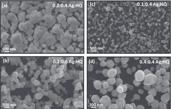
The concentration of the reducing agent and its ratio to the metal salt was important for the successful growth of the Ag nanostructures. We first fixed the concentration of AgNO3at 0.2 mM and varied the
concentration of the reducing agent. The growth was performed for 24 h on the P2VP-grafted substrate with the immobilized Pt NPs. At a HQ concentration of 0.4 mM, the entire substrate was covered with the Ag nanostructures with an average size of 511 nm (Figs.2a,S4a). The further increase of the HQ concentration resulted in the decrease of the size (320 nm) of the Ag nanostructures together with a slight reduction of the surface coverage and increased stacking of the nanostructures (Figs.2b,S4b). At a fixed HQ concentration of 0.4 mM, the average sizes (Fig. S4c, d) of the Ag nanostructures were 110 nm, 511 nm and 433 nm for AgNO3concentrations of 0.1 mM, 0.2 mM and 0.4 mM,
re-spectively. The reduction in the size of the nanostructures at 0.1 mM AgNO3could be related to the lack of sufficient metal ions in the
so-lution. HQ has two hydroxyl groups, which get oxidized to reduce silver ions. Our results suggest that a slight excess of HQ is necessary for the high surface coverage of Ag nanostructures that are grown from im-mobile Pt NPs. These results are in agreement with our previous study on the growth of the Ag nanostructures on solid substrates decorated with Au NPs[44]. The further increase of the HQ concentration (e.g. Fig. 2b) adversely affected the growth process, as reported in the case of colloidal synthesis of gold NPs[45].
The size of the Ag nanostructures was highly dependent on the growth time.Fig. 3presents SEM images of the Ag nanostructures that were prepared by incubating the substrates with the immobilized Pt NPs in 0.2 mM AgNO3and 0.4 mM HQ for different times. The size of
Ag nanostructures increased with the growth time. At a short growth time of 1 h, the average size of Ag nanostructures was 208 nm with a low surface coverage (Figs.3a,S5a). Increasing the growth time to 4 h resulted in a high surface coverage of the Ag nanostructures with a slight increase in their sizes (230 nm) (Figs.3b,S5b). For a growth time of 24 h, the entire substrate was covered with the Ag nanostructures with an average size of 511 nm (Figs.3c,S5c). The further increase of the growth time resulted in significantly larger (average size of 1.7 µm) structures, which likely resulted from coalescence of the smaller na-nostructures (Figs.3d,S5d). At such long growth times, detachment of these large particles from the substrate resulted in low surface density of Ag structures. Accordingly, the growth of catalytically active
Fig. 1. Fabrication of catalytically active solid substrates. (a) The surface growth of Ag nanostructures on top of the Pt NPs that were immobilized on P2VP grafted
substrates. (b) SEM image of the immobilized Pt NPs with an average diameter of 30 nm. (c) SEM image of the surface following the growth of Ag nanostructures. The growth was performed for 24 h in a solution containing 0.2 mM AgNO3and 0.4 mM HQ.
substrates was performed in 0.2 mM AgNO3and 0.4 mM HQ for 24 h, in
the rest of the study.
The chemical composition and crystal structure of the nanos-tructures were characterized by EDX and XRD analysis. The EDX spectrum given inFig. S6clearly shows the presence of the character-istic peak (25 keV) of Ag, further verifying the growth of the Ag
nanostructures. The peak at ∼2.0 keV corresponds to Pt, which con-firms the presence of the seed NPs after the growth process. The ratio of Ag:Pt, as calculated from EDX spectrum, is approximately 4:1. The crystal structure of the substrate decorated with Ag nanostructures was probed with XRD analysis (Fig. 4a). The strong peaks at 38.10°, 44.31° and 64.45° were attributed to diffraction from the (1 1 1), (2 0 0), and
Fig. 2. SEM images of the Ag nanostructures grown at varying concentrations of the reducing agent and metal precursor. The growth solutions consisted of (a)
0.2 mM AgNO3and 0.4 mM HQ, (b) 0.2 mM AgNO3and 0.6 mM HQ, (c) 0.1 mM AgNO3and 0.4 mM HQ, and (d) 0.4 mM AgNO3and 0.4 mM HQ. The substrate
consisted of Pt NPs immobilized on P2VP grafted Si substrates. All growth processes were terminated after 24 h by rinsing with water.
Fig. 3. Influence of the growth time on the size and morphology of Ag nanostructures. SEM images of the Ag nanostructures grown for (a) 1 h, (b) 4 h, (c) 24 h, and
(2 2 0) planes of fcc structured Ag (JCPDS card # 089-3722), respec-tively[46]. There is an induced strain in the Pt-Ag system[47,48]due to the lattice incompatibility between Pt (3.92 Å) and Ag (4.08 Å). Therefore, Ag ions did not interfere with Pt and grew on the Pt seeds in the crystal structure. The crystal growth is dominated in the (1 1 1) plane, since this plane has a low energy[49]. The standard diffraction peaks at 38.69°, 44.97° and 65.49° from the (1 1 1), (2 0 0), and (2 2 0) planes of Pt (JCPDS card # 087-0644) were absent in the XRD pattern of the substrate [46]. The absence of Pt is likely a result of the in-sufficiently low intensity signals from the Pt NPs underneath the Ag nanostructures[19].
The chemical composition of the nanostructures was further char-acterized via XPS analysis. The XPS survey scan of the substrates given in Fig. 4b showed the presence of Ag, Pt, C, O and N elements. The characteristic peaks associated with C, O and N likely originated from the growth byproducts and grafted P2VP layer. The predominant peaks centered around 368 eV were referred to the Ag 3d electrons[50]. The Ag 3d peaks confirmed that Ag ions were successfully reduced and deposited onto the Pt seeds. The Pt 4f peaks located around 71.6 eV were also observed in the spectra indicating the presence of Pt seeds [51]. The intensities of the Pt peaks were significantly lower than the peaks of Ag. This result together with the surface-sensitive nature of XPS collecting data from a depth of ∼5 nm, further supports that the Ag nanostructures grew on the Pt seeds over the entire substrates with a high surface coverage. To further probe the interaction between Ag and Pt, we analyzed the Pt 4f and Ag 3d regions of the XPS spectra. The Pt 4f XPS spectrum (Fig. 4c) of the Ag nanostructures exhibited two peaks that correspond to Pt 4f7/2and Pt 4f5/2electrons at 71.4 eV and 74.9 eV,
respectively. In the absence of the growth of Ag nanostructures, Pt seed
NPs immobilized on the P2VP grafted substrates displayed peaks cen-tered at 71.8 eV (Pt 4f7/2) and 75.2 eV (Pt 4f5/2). The growth of Ag
nanostructures from the Pt seed NPs resulted in a negative shift in the binding energies of Pt. The high-resolution XPS spectrum of the Ag nanostructures over Ag 3d region (Fig. 4d) exhibited the characteristic peaks, which were split into 5/2 and 3/2 states positioned at 368.3 and 374.3 eV, respectively. To compare the electronic structure of Ag na-nostructures that were grown on the Pt seeds, we prepared a P2VP grafted substrate and immobilized citrate-stabilized Ag NPs. The Ag 3d5/2 and 3d3/2 peaks were located at 369.0 eV and 375.0 eV for the
immobilized Ag NPs. The binding energies of the 3d electrons for the Ag nanostructures have a negative shift of 0.7 eV in comparison with that of Ag NPs. The shifts in the binding energies of both Pt 4f and Ag 3d electrons suggest the modification of the electronic structure of Pt and Ag atoms as a result of the growth of Ag nanostructures from Pt NPs immobilized on the P2VP grafted substrate. This type of modification in the electronic structure was observed for bimetallic catalysts that con-sist of Au and Pt[52]. The modification of the electronic structure can be related to the transfer of electrons between Ag and Pt. The electro-negativity of Ag is lower than Pt, which implies that electrons can be transferred from Ag to Pt. All the structural and spectroscopic char-acterization confirmed the successful growth of Ag nanostructures on Pt seeds that were immobilized on the grafted P2VP layers.
Catalytic activity
Degradation of methyl orange and kinetics of reaction
The catalytic activity of the substrates decorated with Ag nanos-tructures was investigated using the reduction of MO in the presence of
Fig. 4. Structural and compositional characterization of Ag nanostructures. (a) XRD pattern, (b) XPS survey spectra of the Ag nanostructures. (c, d) High resolution
XPS spectra of (c) Pt 4f and (d) Ag 3d regions. Substrate with Ag nanostructures were prepared by seed-mediated growth of Ag from Pt NPs immobilized on P2VP grafted substrates. Substrates with Pt and Ag NPs were prepared by immobilization of citrate-stabilized, spherical Pt and Ag NPs on P2VP grafted substrates, respectively.
NaBH4. These experiments were performed by immersing the substrates
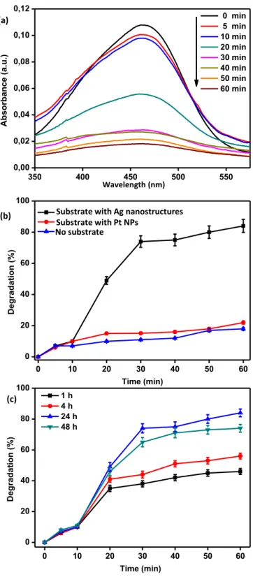
into a solution of MO. The degradation process started with the addition of the reducing agent, NaBH4.Fig. 5a presents UV–visible absorption
spectra of the MO solution taken at different times (0, 5, 10, 20, 30, 40, 50 and 60 min). Before the degradation process, MO exhibited the maximum absorbance in the visible region at a wavelength of 464 nm
associated with the azo linkage[53,54]. This absorbance was taken as the basis for monitoring the degradation process in the rest of the study and the degradation percentage was calculated using Eq.(1). When the catalytically active substrate and the freshly prepared reducing agent were added to the dye solution, the absorption at 464 nm decreased as the time progressed. The degradation rate increased rapidly on the Ag nanostructures following the first 10 min, perhaps related with the ac-tivation of the surface and diffusion of the dye and reducing agent to the catalytically active sites. The large surface area, high surface cov-erage and irregular shape of the Ag nanostructures likely contribute to this behavior. The absence of such an abrupt increase in the degrada-tion rate for the solid substrates decorated solely with the Pt NPs can be related to the low degradation rate together with accessibility of the surface of the NPs due to large particle-to-particle distances. At the end of 60 min, 83% of the initial MO was degraded. We performed control experiments to understand the origin of this catalytic activity (Fig. 5b). In the first set of experiments, the degradation process was monitored in the absence of any substrate (Fig. S7a). The degradation proceeded slowly and only 18% of the initial dye was degraded at the end of 60 min when there was no substrate in the solution. The second control experiment was performed using just the Pt seed NPs immobilized on the P2VP grafted substrate (Fig. S7b). The degradation percentage was as high as 22% at the end of 60 min. Despite a slight increase in the efficiency with the presence of the immobilized Pt NPs, the degradation is much more limited in comparison with the Ag nanostructures grown on the Pt seeds. This result clearly demonstrated the high catalytic activity of the fabricated substrates. Three factors contribute to this high catalytic activity: i) The total catalytically active surface area of the Ag nanostructures is considerable larger than the surface area of the Pt seeds, due to the high coverage of the former structure and the re-duced active area of the Pt NPs associated with the interaction with the end-grafted P2VP layer. ii) The work function of Ag NPs is lower than then Pt NPs[55]. iii) The processes of electron transfer between Pt NPs and Ag nanostructures can synergistically improve the catalytic activity [56].
The catalytic activity of the substrate depended on the extent of Ag growth on the immobilized Pt NPs.Fig. 5c compares the degradation of MO using substrates prepared by growth of Ag for 1 h, 4 h, 24 h and 48 h. The degradation was highest in the case of Ag nanostructures grown for 4 h. After 60 min of degradation, for example, 83%, 74%, 56%, and 46% of the initial MO were degraded for substrates with the Ag growth times of 24 h, 48 h, 4 h and 1 h, respectively (Fig. S8). Considering these results with the SEM images of these structures (Fig. 3) suggest that both the size of Ag nanostructures and their surface coverage are important for the catalytic activity. At long growth times, the large size of Ag structures approaching the micrometer length scale likely decreased the catalytic activity of the substrate. The short growth times, on the other hand, were not sufficient for the growth of Ag na-nostructures over the entire substrate.
Kinetics and mechanism of degradation of methyl orange
We investigated the kinetics of the catalytic degradation reaction on the substrates decorated with Ag nanostructures. In the presence of excess of the reducing agent, the rate of the degradation of organic dye molecules is commonly[57]expressed by pseudo-first order kinetics, which is derived from the Langmuir-Hinshelwood mechanism[58]. The rate of the reaction expression is given by Eq.(2)where Ctrefers to the
concentration of MO at time t. The concentration of MO can be ap-proximated by the absorbance, At, at 464 nm. The linearized form of the
expression is obtained by integrating the expression and given by Eq. (3), where A0 and At are the initial and final absorbance of MO at
464 nm, respectively[7]. = = dC dt dA dt k A t t app t (2)
Fig. 5. Catalytic activity of the substrates. (a) UV–visible spectra of MO as a
function of time after addition of the reducing agent for the Ag nanostructures. (b) The degradation percentage as a function of time for different types of substrates. (c) The degradation percentage as a function of time for different Ag growth times. The growth was performed in a solution containing 0.2 mM AgNO3and 0.4 mM HQ.
= = ln C C ln A A k t t t app 0 0 (3)
Fig. 6a presents the plot of ln(At/A0) against time for different types
of substrates. A reasonably good linear relationship was obtained in the plot, showing that the degradation follows a pseudo first-order kinetics. The apparent reaction rate constant, kapp, was derived from the slope of
the linear fit to the plot. The apparent reaction rate constant for the Ag nanostructures was 33.5 × 10⁻³min−1, which was roughly ten times
higher than the constant in the case of just Pt seeds (kapp= 3.4 × 10⁻³min−1) and degradation in the absence of a
sub-strate (kapp= 2.29 × 10⁻³min−1). This result further highlights the
significant enhancement of the degradation with the use of the sub-strates decorated with Ag nanostructures. The reaction rate constant of our platform is higher than the recent reports based on the green Ag NPs [4], AgCl nanowires decorated with Au NPs [59] and organic semiconductor film@Au[60].
The degradation of MO on the substrates decorated with Ag na-nostructures is likely to proceed through the mechanism depicted in Fig. 6b. The catalytic degradation process relies on the transfer of electrons from the donor (NaBH4) to the acceptor (MO). The
simulta-neous adsorption of both the dye and BH4−ions on the surface of the
nanostructures is the first step for the degradation. The Ag nanos-tructures serve as electron relay systems facilitating the electron transfer between the electrophilic dyes and nucleophilic BH4− ions [61]. The processes of electron transfer results in the gain of electrons by MO, leading to the reduced form of the dye molecule. Previous studies suggested that the electron transfer from BH4−ions to the dye
molecules mediated by the metallic nanostructures in water results in formation of BO33−[62]. Such electron transfer can further proceed
and result in more reduced forms of the dye molecule[63]. The high catalytic activity of our platform can be related to the large surface area to volume ratio, high surface coverage and irregular shape of the Ag nanostructures, which facilitate electron transfer and enable over-coming the kinetic barrier for the degradation reaction. Since the re-action occurs on the surface, increasing the area enhances the rate of the degradation. The processes of electron transfer between Ag and Pt suggested by XPS analysis can further contribute to this catalytic ac-tivity[50]. The interaction of functional groups[64]presented by the end-grafted P2VP with noble metals can also play a role in the catalytic activity of the presented platform.
Reusability of catalytic substrates
Inherent advantages of the catalytically active solid substrates are the ease of separation from the reaction medium and ability for the repeated use of the catalyst. Together with the efficiency of the cata-lytically active surfaces, the stability of the material is highly critical for practical waste-treatment applications. To investigate the stability of the solid substrates decorated with Ag nanostructures, we performed four consecutive cycles of degradation on the same substrate. The substrate was simply removed from the reaction medium without any additional processing. The substrate was washed under sonication to remove the bound dye molecules, prior to the following cycle.Fig. 7 presents the degradation percentage of MO after each cycle. The sub-strate retained most of its catalytic activity with a slight decrease in the degradation efficiency with the increasing number of cycles (Fig. S9). The degradation, for example was reduced by 3.5% and 12% after the second and fourth cycles with respect to the first cycle, respectively. This reduction in the degradation efficiency is perhaps associated with
Fig. 6. The kinetics and proposed mechanism of the degradation reaction. a) The variation of ln(At/A0) as a function of time for different substrates. The catalytic
degradation experiments were performed using 10−5M MO and 0.033 M NaBH
4. (b) Proposed mechanism for the catalytic reduction of MO by NaBH4in the presence
of the Ag nanostructures.
Fig. 7. Reusability of the substrates decorated with Ag nanostructures. (a) Degradation percentage as a function of time for the consecutive cycles. (b) Degradation
the incomplete removal of the bound dye molecules from the surface, resulting in the decay of the catalytically active sites. The possibility of destruction and removal of the nanostructures are excluded because SEM imaging of the substrates following four cycles showed the re-tainment of the Ag nanostructures (Fig. S10). These results further verify the easy separation of the catalyst from the reaction medium without any additional processing steps and ability of reusing the fab-ricated catalytically active solid substrates.
Conclusions
In conclusion, this study demonstrated the promise of solid sub-strates decorated with Ag nanostructures for the catalytic degradation of organic dyes. The versatility and scalability of our strategy emerge from the all-solution processing-based fabrication of catalytically active solid substrates. The conditions for growth of Ag on Pt NPs immobilized on end-grafted P2VP determine the size, surface coverage and catalytic activity of the nanostructures. The seed-mediated growth of Ag na-nostructures on the immobilized Pt NPs plays a key role in the high degradation performance of our platform by presenting large areas of catalytically active surfaces. The easy removal of the solid substrates from the degradation medium together with the ability to reuse the substrates are important advantages for practical applications. The presented results motivate research in other classes of materials for fabrication of catalytically active solid substrates decorated with na-nostructures.
Acknowledgements
This work was supported by Research Fund of the Erciyes University (Project Number: FDK-2017-7270). MS acknowledges support from TUBITAK (Scientific and Technical Research Council of Turkey) priority areas doctoral scholarship program. MSO acknowledges support from the Turkish Academy of Sciences Distinguished Young Scientist Award (TUBA-GEBIP).
Appendix A. Supplementary data
Supplementary data to this article can be found online athttps:// doi.org/10.1016/j.rinp.2018.12.084.
References
[1] Robinson T, McMullan G, Marchant R, Nigam P. Remediation of dyes in textile effluent: a critical review on current treatment technologies with a proposed al-ternative. Bioresour Technol 2001;77:247–55.
[2] Li K, Rong Z, Li Y, Li C, Zheng Z. Preparation of nitrogen-doped cotton stalk mi-croporous activated carbon fiber electrodes with different surface area from hex-amethylenetetramine-modified cotton stalk for electrochemical degradation of methylene blue. Results Phys 2017;7:656–64.
[3] Hao Z, Iqbal A. Some aspects of organic pigments. Chem Soc Rev 1997;26:203–13. [4] Bhakya S, Muthukrishman S, Sukumaran M, Muthukumar M, Senthil Kumar T, Rao MV. Catalytic degradation of organic dyes using synthesized silver nanoparticles: a green approach. J Bioremed Biodegr 2015;6:1–9.
[5] Martínez-Huitle CA, Brillas E. Decontamination of wastewaters containing synthetic organic dyes by electrochemical methods: a general review. Appl Catal B 2009;87:105–45.
[6] Rauf MA, Meetani MA, Hisaindee S. An overview on the photocatalytic degradation of azo dyes in the presence of TiO2doped with selective transition metals. Desalination 2011;276:13–27.
[7] Ucar A, Findik M, Gubbuk IH, Kocak N, Bingol H. Catalytic degradation of organic dye using reduced graphene oxide-polyoxometalate nanocomposite. Mater Chem Phys 2017;196:21–8.
[8] Shi J. On the synergetic catalytic effect in heterogeneous nanocomposite catalysts. Chem Rev 2013;113:2139–81.
[9] Bell AT. The impact of nanoscience on heterogeneous catalysis. Science 2003;299:1688–91.
[10] Yu W, Porosoff MD, Chen JG. Review of Pt-based bimetallic catalysis: from model surfaces to supported catalysts. Chem Rev 2012;112:5780–817.
[11] Devarajan S, Bera P, Sampath S. Bimetallic nanoparticles: a single step synthesis, stabilization, and characterization of Au-Ag, Au-Pd, and Au-Pt in sol-gel derived silicates. J Colloid Interface Sci 2005;290:117–29.
[12] Pan YT, Yan L, Shao YT, Zuo JM, Yang H. Regioselective atomic rearrangement of Ag-Pt octahedral catalysts by chemical vapor-assisted treatment. Nano Lett 2016;16:7988–92.
[13] Kong F, Du C, Ye J, Chen G, Du L, Yin G. Selective surface engineering of hetero-geneous nanostructures: in-situ unravelling the catalytic mechanism on Pt-Au cat-alyst. ACS Catal 2017;7:7923–9.
[14] Stamenkovic V, Mun BS, Mayrhofen KJJ, Ross PN, Markovic NM, Rossmeisl J, et al. Changing the activity of electocatalysts for oxygen reduction by tuning the surface electronic structure. Angew Chem Int Ed 2006;45:2897–3290.
[15] Swearer DF, Zhao H, Zhou L, Zhang C, Robatjazi H, Martirez JMP, et al. Heterometallic antenna-reactor complexes for photocatalysis. Proc Natl Acad Sci 2016;113:8916–20.
[16] Kibsgaard J, Jackson A, Jaramillo TF. Mesoporous platinum nickel thin films with double gyroid morphology for the oxygen reduction reaction. Nano Energy 2019;29:243–8.
[17] Verma P, Yuan K, Kuwahara Y, Mori K, Yamashita H. Enhancement of plasmonic activity by Pt/Ag bimetallic nanocatalyst supported on mesoporous silica in the hydrogen production from hydrogen storage material. Appl Catal B 2018;223:10–5. [18] Rashid M, Jun TS, Jung Y, Kim YS. Bimetallic core-shell Ag@Pt
nanoparticle-de-corated MWNT electrodes for amperometric H2sensors and direct methanol fuel cells. Sens Actuators B 2015;208:7–13.
[19] Ma Y, Wu X, Zhang G. Core-shell Ag@Pt nanoparticles supported on sepiolite na-nofibers for the catalytic reduction of nitrophenols in water: enhanced catalytic performance and DFT study. Appl Catal B 2017;205:262–70.
[20] Salem MA, Bakr EA, El-Attar HG. Pt@Ag and Pd@Ag core/shell nanoparticles for catalytic degradation of Congo red in aqueous solution. Spectrochim Acta Part A Mol Biomol Spectrosc 2018;188:155–63.
[21] Kim MR, Lee DK, Jang DJ. Facile fabrication of hollow Pt/Ag nanocomposites having enhanced catalytic properties. Appl Catal B 2011;103:253–60. [22] Chandraboss VL, Senthilvelan S, Natanapatham L, Murugavelu M, Loganathan B,
Karthikeyan B. Photocatalytic effect of Ag and Ag/Pt doped silicate non crystalline material on methyl violet-experimental and theoretical studies. J Non-Cryst Solids 2013;368:23–8.
[23] Jiang X, Fu G, Wu X, Liu Y, Zhang M, Sun D, et al. Ultrathin AgPt alloy nanowires as a high-performance electrocatalyst for formic acid oxidation. Nano Res 2018;11:499–510.
[24] Jiang X, Yan X, Ren W, Jia Y, Chen J, Sun D, et al. Porous AgPt@Pt nanooctahedra as an efficient catalyst toward formic acid oxidation with predominant dehy-drogenation pathway. ACS Appl Mater Interfaces 2016;8:31076–82.
[25] Babucci M, Fang CY, Hoffman AS, Bare SR, Gates BC, Uzun A. Tuning the selectivity of single-site supported metal catalysts with ionic liquids. ACS Catal
2017;7:6969–72.
[26] Wu HC, Chen TC, Chen YC, Lee JF, Chen CS. Formaldehyde oxidation on silica-supported Pt catalysts: the influence of thermal pretreatments on particle formation and on oxidation mechanism. J Catal 2017;355:87–100.
[27] Hsieh SH, Hsu MC, Liu WL, Chen WJ. Study of Pt catalyst on graphene and its application to fuel cell. Appl Surf Sci 2013;277:223–30.
[28] Shen W, Wu B, Liao F, Jiang B, Shao M. Optimizing the hydrogen evolution reaction by shrinking Pt amount in Pt-Ag/SiNW nanocomposites. Int J Hydrogen Energy 2017;42:15024–30.
[29] Kayaci F, Vempati S, Akgun CO, Donmez I, Biyikli N, Uyar T. Selective isolation of the electron or hole in photocatalysis: ZnO-TiO2and TiO2-ZnO core-shell structured heterojunction nanofibers via electrospinning and atomic layer deposition. Nanoscale 2014;6:5735–45.
[30] Vidhu VK, Philip D. Catalytic degradation of organic dyes using biosynthesized silver nanoparticles. Micron 2014;56:54–62.
[31] Jones CW. On the stability and recyclability of supported metal-ligand complex catalysts: myths, misconceptions and critical research needs. Top Catal 2010;53:942–52.
[32] Zhang X, Jiang W, Zhou Y, Xuan S, Peng C, Zong L, et al. Magnetic recyclable Ag catalysts with a hierarchical nanostructure. Nanotechnology 2011;22:375701. [33] Miotello A, Patel N. Pulsed laser deposition of cluster-assembled films for catalysis
and the photocatalysis relevant to energy and the environment. Appl Surf Sci 2013;278:19–25.
[34] Henkel B, Vahl A, Aktas OC, Strunskus T, Faupel F. Self-organized nanocrack net-works: a pathway to enlarge catalytic surface area in sputtered ceramic thin films. Showcased for photocatalytic TiO2. Nanotechnology 2018;29:035703.
[35] Zeng G, Qiu J, Hou B, Shi H, Lin Y, Hettick M, et al. Enhanced photocatalytic re-duction of CO2to CO through TiO2passivation of InP in ionic liquids. Chem Eur J 2015;21:13502–7.
[36] Koenig M, Simon F, Formanek P, Müller M, Gupta S, Stamm M, et al. Catalytically active nanocomposites based on palladium and platinum nanoparticles in poly(2-vinylpyridine) brushes. Macromol Chem Phys 2013;214:2301–11.
[37] Truong TNP, Randriamahazaka H, Ghilane J. Polymer brushes ionic liquid as a catalyst for oxygen reduction and oxygen evolution reactions. ACS Catal 2018;8:869–75.
[38] Costantini F, Benetti EM, Tiggelaar RM, Gardeniers HJGE, Reinhoudt DN, Huskens J, et al. A brush-gel/metal-nanoparticle hybrid film as an efficient supported cat-alyst in glass microreactors. Chem Eur J 2010;16:12406–11.
[39] Zhang L, Liu Z, Wang Y, Xie R, Ju XJ, Wang W, et al. Facile immobilization of Ag nanoparticles on microchannel walls in microreactors for catalytic applications. Chem Eng J 2017;309:691–9.
[40] Yilmaz M, Bakirci G, Erdogan H, Tamer U, Demirel G. The fabrication of plasmonic nanoparticle-containing multilayer films via a bio-inspired polydopamine coating. RSC Adv 2016;6:12638–41.
rapid and facile deposition of polymer brushes for immobilization of plasmonic nanoparticles. Appl Surf Sci 2016;385:299–307.
[42] Malynych S, Luzinov I, Chumanov G. Poly(Vinyl Pyridine) as a universal surface modifier for immobilization of nanoparticles. J Phys Chem B 2002;106:1280–5. [43] Onses MS, Wan L, Liu X, Kiremitler NB, Yilmaz H, Nealey PF. Self-assembled
na-noparticle arrays on chemical nanopatterns prepared using block copolymer li-thography. ACS Macro Lett 2015;4:1356–61.
[44] Sakir M, Pekdemir S, Karatay A, Kucukoz B, Ipekci HH, Elmali A, et al. Fabrication of plasmonically active substrates using engineered silver nanostructures for SERS applications. ACS Appl Mater Interfaces 2017;9:39795–803.
[45] Sirajuddin A, Mechler AAJ, Torriero A, Nafady CY, Lee AM, Bond AP, et al. The formation of gold nanoparticles using hydroquinone as a reducing agent through a localized pH change upon addition of NaOH to a solution of HAuCl4. Colloids Surf A 2010;370:35–41.
[46] Lee CL, Tseng CM, Wu CC, Chou TC, Syu CM. High activity of hexagonal Ag/Pt nanoshell catalyst for oxygen electroreduction. Nanoscale Res Lett 2009;4:193–6. [47] Lahiri D, Bunker B, Zhang BMZ, Doudna DMM, Bertino MF, Blum FD, et al.
Bimetallic Pt-Ag and Pd-Ag nanoparticles. J Appl Phys 2005;97:094304. [48] Chen S, Thota S, Singh G, Aímola TJ, Koenigsmann C, Zhao J. Synthesis of hollow
Pt-Ag nanoparticles by oxygen-assisted acid etching as electrocatalysts for the oxygen reduction reaction. RSC Adv 2017;7:46916–24.
[49] Chaudhuri RG, Paria S. Au and Ag/Au double-shells hollow nanoparticles with improved near infrared surface plasmon and photoluminescence properties. J Colloid Interface Sci 2016;461:15–9.
[50] Schaal MT, Hyman MP, Rangan M, Ma S, Williams CT, Monnier JR, et al. Theoretical and experimental studies of Ag-Pt interactions for supported Ag-Pt bi-metallic catalysts. Surf Sci 2009;603:690–6.
[51] Pan S, Cai Z, Duan Y, Yang L, Tang B, Jing B, et al. Tungsten diselenide/porous carbon with sufficient active edge-sites as a co-catalyst/Pt-support favoring ex-cellent tolerance to methanol-crossover for oxygen reduction in acidic medium. Appl Catal B 2017;219:18–29.
[52] Ren F, Zhai C, Zhu M, Wang C, Wang H, Bin D, et al. Electrochim Acta 2015;153:175–83.
[53] Feng W, Nanasheng D, Helin H. Degradation mechanism of azo dye C. I. reactive red
2 by iron powder reduction and photooxidation in aqueous solutions. Chemosphere 2000;41:1233–8.
[54] Li X, Li X, Yang W, Chen X, Li W, Luo B, et al. Preparation of 3D PbO2 nano-spheres@SnO2nanowires/Ti electrode and its application in methyl orange de-gradation. Electrochim Acta 2014;146:15–22.
[55] Gupta N, Singh HP, Sharma RK. Metal nanoparticles with high catalytic activity in degradation of methyl orange: an electron relay effect. J Mol Catal A
2011;335:248–52.
[56] Li J, Rong H, Tong X, Wang P, Chen T, Wang Z. Platinum-silver alloyed octahedral nanocrystals as electrocatalyst for methanol oxidation reaction. J Colloid Interface Sci 2018;513:251–7.
[57] Piella J, Merkoçi F, Genç A, Arbiol J, Bastús NG, Puntes V. Probing the surface reactivity of nanocrystals by the catalytic degradation of organic dyes: the effect of size, surface chemistry and composition. J Mater Chem A 2017;5:11917–29. [58] Kumar KV, Porkodi K, Rocha F. Langmuir-Hinshelwood kinetics- a theoretical
study. Catal Commun 2008;9:82–4.
[59] Sun Y. Conversion of Ag nanowires to AgCl nanowires decorated with au nano-particles and their photocatalytic activity. J Phys Chem C 2010;114:2127–33. [60] Yilmaz M, Erkartal M, Ozdemir M, Sen U, Usta H, Demirel G. Three-dimensional
Au-coated electrosprayed nanostructured BODIPY films on aluminum foil as surface-enhanced raman scattering platforms and their catalytic applications. ACS Appl Mater Interfaces 2017;9:18199–206.
[61] Khan MM, Lee J, Cho MH. Au@TiO2nanocomposites for the catalytic degradation of methyl orange and methylene blue: an electron relay effect. J Ind Eng Chem 2014;20:1584–90.
[62] Jiang ZJ, Liu CY, Sun LW. Catalytic properties of silver nanoparticles supported on silica spheres. J Phys Chem B 2005;209:1730–5.
[63] Yang Y, Liao H, Tong Z, Wang C. Porous Ag/polymer composite microspheres for absorption and catalytic degradation of organic dyes in aqueous solutions. Compos Sci Technol 2015;107:137–44.
[64] Ma Y, Zhang G. Sepiolite nanofiber-supported platinum nanoparticle catalysts to-ward the catalytic oxidation of formaldehyde at ambient temperature: efficient and stable performance and mechanism. Chem Eng J 2016;288:70–8.
![Fig. 2b) adversely affected the growth process, as reported in the case of colloidal synthesis of gold NPs [45].](https://thumb-eu.123doks.com/thumbv2/9libnet/6023776.127257/3.892.149.743.827.1061/fig-adversely-affected-growth-process-reported-colloidal-synthesis.webp)