IDENTIFICATION OF GENES INDUCED BY BRCA1 IN BREAST CANCER CELLS
A THESIS SUBMITTED TO
THE DEPARTMENT OF MOLECULAR BIOLOGY AND GENETICS AND THE INSTITUTE OF ENGINEERING AND SCIENCE OF
BİLKENT UNIVERSITY
IN PARTIAL FULFILLMENT OF THE REQUIREMENTS FOR THE DEGREE OF DOCTOR OF PHILOSOPHY
By
ARZU ATALAY December 2002
I certify that I have read this thesis and that in my opinion it is fully adequate, in scope and in quality, as a thesis for the degree of Doctor of Philosophy.
________________________________
Prof. Dr. Emin Kansu
I certify that I have read this thesis and that in my opinion it is fully adequate, in scope and in quality, as a thesis for the degree of Doctor of Philosophy.
________________________________ Prof. Dr. Wayne Criss
I certify that I have read this thesis and that in my opinion it is fully adequate, in scope and in quality, as a thesis for the degree of Doctor of Philosophy.
________________________________
Prof. Dr. Ay Öğüş
I certify that I have read this thesis and that in my opinion it is fully adequate, in scope and in quality, as a thesis for the degree of Doctor of Philosophy.
________________________________
Prof. Dr. Mehmet Öztürk
I certify that I have read this thesis and that in my opinion it is fully adequate, in scope and in quality, as a thesis for the degree of Doctor of Philosophy.
________________________________ Asst. Prof. Işık G.Yuluğ
Approved for the Institute of Engineering and Science
_______________________________
Prof. Dr. Mehmet Baray
ABSTRACT
IDENTIFICATION OF GENES INDUCED BY BRCA1 IN BREAST CANCER CELLS
ARZU ATALAY
Ph.D. in Molecular Biology and Genetics Supervisor: Assist. Prof. Işık G.Yuluğ
December 2002, 235 Pages
Inherited mutations of the BRCA1 gene predispose to cancer of the breast, ovaries and other organs. The BRCA1 protein product is implicated in the maintenance of chromosomal integrity as BRCA1-deficient cells display gross chromosomal rearrangements. Chromosomal instability in BRCA1-deficient cells is related to inappropriate DNA double-strand break repair. The role of the BRCA1 gene in the maintenance of chromosomal integrity is linked to a number of biological properties of its protein product including transcriptional regulation. The aim of this study is to identify genes that are regulated by BRCA1. Initial attempts to overexpress BRCA1 in breast cancer cells with the tightly-regulated ecdysone inducible system did not result in the desired levels of BRCA1 protein and ectopic BRCA1 expression was therefore performed by using the constitutive expression vector. In this study, we have identified genes whose expression levels are upregulated as a result of BRCA1 overexpression in MCF7 breast carcinoma cells by using the suppression subtractive hybridisation (SSH) method. Differential screening, sequencing and homology search studies showed that BRCA1 overexpression in breast cancer cells leads to transcriptional upregulation of distinct classes of genes encoding proteins involved in cellular processes such as DNA repair, chromosome assembly and segregation, signal transduction, RNA surveillance, ubiquitin-mediated proteolysis, amino acid transport, RNA metabolism and glucose metabolism. This study is the first to report BRCA1-induced genes in breast carcinoma cells with the SSH technique. The identified genes in this study may provide new insights into the tumour suppressor functions of BRCA1.
ÖZET
MEME KANSERİ HÜCRELERİNDE BRCA1 TARAFINDAN İNDÜKLENEN GENLERİN TANIMLANMASI
ARZU ATALAY
Doktora Tezi, Moleküler Biyoloji ve Genetik Bölümü Tez Yöneticisi: Yar. Doç. Dr. Işık G.Yuluğ
Aralık 2002, 235 sayfa
BRCA1 geninin kalıtsal mutasyonları meme, rahim ve diğer organlarda kansere yatkınlığa yol açar. BRCA1-eksik hücrelerde yeni kromozomal düzenlenmeler görüldüğünden BRCA1 protein ürününün kromozomal bütünlüğün devam ettirilmesinde görevli olduğu düşünülmektedir. BRCA1-eksik hücrelerdeki kromozomal dengesizlik DNA çift-ipliğinin yanlış tamiriyle ilişkilidir. BRCA1 geninin kromozomal bütünlüğün sağlanmasındaki rolü protein ürününün transkripsiyonel düzenleme dahil bir kaç biyolojik özelliğine bağlıdır. Bu çalışmanın amacı BRCA1 tarafından düzenlenen genlerin tanımlanmasıdır. BRCA1'i meme kanseri hücrelerinde sıkı-düzenlenen "ecdysone" ile indüklenebilen bir sistemle fazla ifade etmek için yapılan ilk çalışmalar, istenen düzeyde BRCA1 proteini vermemiştir ve bu nedenle ektopik BRCA1 ifadesi konstitütif ekspresyon vektörü kullanılarak yapılmıştır. Bu çalışmada MCF7 meme kanseri hücrelerinde BRCA1’in fazla ifade edilmesi sonucu ifade düzeyleri artan genler "Suppression Subtractive Hybridisation" (SSH) metodu kullanılarak tanımlanmıştır. Ayrımsal tarama, dizileme ve homoloji araştırma çalışmaları meme kanseri hücrelerinde BRCA1’in fazla ifade edilmesinin DNA tamiri, kromozom yapımı ve ayrılması, sinyal iletimi, RNA tarama, ubikitin aracılıklı proteoliz, amino asit taşınımı, RNA metabolizması ve glukoz metabolizması gibi hücresel olaylarda yer alan proteinleri kodlayan farklı gen gruplarının ifadelerinde artışa yol açtığını göstermiştir. Bu çalışma meme karsinom hücrelerinde SSH tekniği kullanılarak BRCA1 tarafından indüklenen genleri gösteren ilk çalışmadır. Bu çalışmada tanımlanan genler BRCA1'in tümör baskılayıcı görevlerine yeni bir bakış açısı sağlayabilir.
ACKNOWLEDGEMENTS
I would like to express my gratitude to my advisor Assoc. Prof. Işık G.Yuluğ for her guidance, supervision and continuous support throughout my studies.
I would like to express my special thanks to Prof. Dr. Mehmet Öztürk for sharing his excellent scientific logic, for his support and instructive comments.
I would like to thank to researchers who provided vectors and cell lines. In particular, I am very grateful to Dr. Tim Crook for supplying the critical materials besides his instructive comments.
Thanks to those at Hacettepe University, Medical Biology department for their help and in particular to Assoc. Prof. Pervin Dincer for supplying the wet transfer facility. Thanks to the Scientific and Technical Research Council of Turkey and the British Council, Academic Links Scheme Program in Turkey for their support.
Many thanks to MBG family, past and present, who have helped me during my studies.
TABLE OF CONTENTS SIGNATURE PAGE ii ABSTRACT iii ÖZET iv ACKNOWLEDGEMENTS v TABLE OF CONTENTS vi
LIST OF FIGURES xiii
LIST OF TABLES xvi
ABBREVIATIONS xvii
CHAPTER 1. INTRODUCTION 1.1 Risk factors for breast and ovarian cancer 1 1.2 Genes implicated in breast cancer 3
1.3 Mechanism of BRCA-mediated breast cancer formation 6 1.4 BRCA1 9
1.4.1 Mutational spectrum of BRCA1 9 1.4.2 BRCA1 gene and protein structure 10 1.4.3 Cellular expression of BRCA1 12
1.4.4 BRCA1-deficient cells and animal models 13 1.4.5 BRCA1 and p53 14
1.5 Functions of BRCA1 15 1.5.1 BRCA1 in damage signaling 20
1.5.3 BRCA1 in DNA repair 23 1.5.4 BRCA1 as a modulator of gene transcription 26
1.5.5 Other potential functions of BRCA1 30
1.6 Identified BRCA1 regulated targets 31
1.7 Strategy 39
CHAPTER 2. MATERIALS AND METHODS
2.1 Materials 42
2.1.1 General reagents 42
2.1.2 Nucleic Acids and Proteins 42
2.1.3 Oligonucleotides 42
2.1.4 Enzymes 43
2.1.5 Bacterial strains 43
2.1.6 Plasmids 43
2.1.7 Nucleic acid and protein transfer materials 44
2.1.8 Radioisotopes 44
2.1.9 Sequencing reagents 44
2.1.10 Tissue culture reagents and cell lines 44
2.1.11 Kits 45
2.1.12 Antibodies 46
2.2 Solutions and Media 46
2.2.1 General solutions 46
2.2.2 Microbiological media and antibiotics 47
2.2.3 Tissue culture solutions 48
2.2.5 Protein extraction and western blotting solutions 49
2.2.6 Immunostaining solutions 50
2.3 General methods 51
2.3.1 Transformation of E.coli 51
2.3.2 Plasmid DNA preparation 52
2.3.3 Restriction enzyme digestion of plasmid DNA 53
2.3.4 Agarose gel electrophoresis of DNA 53
2.3.5 Extraction of DNA fragments from agarose gel 54
2.3.6 Radioactive labelling of DNA 55
2.3.7 DNA isolation from tissue culture cells 55 2.3.8 Amplification of DNA by Polymerase Chain 56 Reaction (PCR)
2.3.9 DNA sequencing 60
2.3.10 Computer analysis of DNA sequences and 61 homology search
2.3.11 Extraction of total RNA from tissue culture cells 61 2.3.12 Extraction of poly (A)+ RNA and removal of 62
DNA contamination
2.3.13 Northern Blotting 62
2.3.14 Protein extraction from tissue culture cells 63
2.3.15 Immunoprecipitation 64
2.3.16 Western Blotting 65
2.3.17 Annexin V and Hoechst staining 67
2.4 Construction of BRCA1 expressing eukaryotic expression vectors 68
2.4.1 Construction of pIND.myc.BRCA1 70
2.4.2 Construction of pIND.BRCA1 71
2.4.3 Construction of pIND.HA.BRCA1 73
2.5 Tissue Culture Techniques 74
2.5.1 Growth conditions of cell lines 74
2.5.2 Determination of cell sensitivity to selective system 75
2.5.3 Cryopreservation of cell lines 75
2.5.4 Transfection of mammalian cells 76
2.5.4.1 Electroporation of mammalian cells 76 2.5.4.2 Calcium phosphate transfection of mammalian cells 76
2.5.4.3 β-galactosidase assay 77
2.6 Subtractive Hybridization 79
2.6.1 Double strand cDNA synthesis 81
2.6.2 RsaI digestion 82
2.6.3 Adaptor ligation 82
2.6.4 First Hybridization 84
2.6.5 Second Hybridization 85
2.6.6 PCR amplification 85
2.6.7 Subtraction Efficiency test 86
2.6.8 Cloning and pre-screening of forward subtracted library 87
2.7 Differential Screening 88
2.7.1 Preparation of cDNA dot blots of forward subtracted library 88
2.7.2 Probe preparation for dot blots 88
2.7.4 Hybridization of the arrays with the subtracted cDNAs 89 2.7.5 Analysis of differential screening results 89
CHAPTER 3. RESULTS
3.1 Generating stable pVgRXR expressing MCF-7 breast carcinoma cell line 91 3.2 Construction and analysis of eukaryotic expression vectors 96
3.2.1 Construction and analysis of eukaryotic expression vector 96 pIND.myc.BRCA1
3.2.1.1 In vitro transcription and translation- 96 Immunoprecipitation assay result of
pcDNA3.myc.BRCA1
3.2.1.2 Construction of eukaryotic expression vector 98 pIND.myc.BRCA1
3.2.1.3 Generating a U2OS cell line that stably expresses 101 pIND.myc.BRCA1
3.2.1.4 Analysing the induction of BRCA1 expression in 102 pIND.myc.BRCA1 clones
3.2.2 Construction and analysis of eukaryotic expression 104 vector pIND.BRCA1
3.2.2.1 In vitro transcription and translation- 104 Immunoprecipitation assay result of pCR3.BRCA1
3.2.2.2 Cloning of BRCA1 from pCR3.BRCA1 into 105 pIND vector
3.2.2.3 Sequencing of eukaryotic expression vector 108 pIND.BRCA1
3.2.2.4 Analysing BRCA1 expression by transient 110 transfection into U2OS cells
3.2.3 Construction and analysis of eukaryotic expression vector 112 pIND.HA.BRCA1
3.2.3.1 Subcloning of BRCA1 from pRc/CMV.BRCA1 112 into pIND
3.2.3.2 Sequencing of pIND.HA.BRCA1 114
3.2.3.3 Analysing the induction of BRCA1 expression 118 in transiently pIND.HA.BRCA1 transfected U2OS cells
3.2.3.4 Analysing the induction of p53 expression in transiently 120 pIND.p53 transfected U2OS cells
3.2.4 Ectopic expression of BRCA1 in MCF7 cells by 121 using pCMVmycBRCA1
3.3 Subtractive hybridization 127 3.3.1 Preparation of DNA-free mRNA for subtractive hybridization 127
3.3.2 ds cDNA synthesis 128
3.3.3 RsaI digestion 129
3.3.4 Adaptor ligation 129
3.3.5 First and second hybridizations for subtraction 131
3.3.6 Primary and secondary PCRs 131
3.3.7 Subtraction efficiency test 133
3.3.8 Cloning and pre-screening of forward subtracted library 133
3.4 Differential Screening of clones 136
3.4.1 Arraying forward subtracted clones 136 3.4.2 Dot blot hybridization with subtracted cDNA probes 137
3.4.3.1 Sequencing, homology search and classification 141 of BRCA1 upregulated genes
3.4.3.2 Confirmation of the induction of BRCA1 147 upregulated candidate genes
CHAPTER 4. DISCUSSION 151
CHAPTER 5. FUTURE PERSPECTIVES 159
REFERENCES 161
APPENDICES
Appendix A Sequence and map of pCMV.myc.BRCA1 177
Appendix B Characteristics of cell lines 195
Appendix C Phosphoimager values of 6 arrays (500 clones) 199 Appendix D Actin normalized average values of 210 clones 209
Appendix E Sequencing results of the clones 214
Appendix F BLAST search results of the sequenced clones 230
LIST OF FIGURES
Figure 1 Relationship between mutation events and formation 7 of breast or ovarian cancers.
Figure 2 Features of the human BRCA Proteins 11
Figure 3 Repair genes as caretakers of the genome. 16 Figure 4 Multiple cellular functions of BRCA1 17
Figure 5 Putative roles of BRCA1 17
Figure 6 Signaling of DSBs. 21
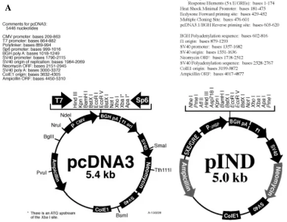
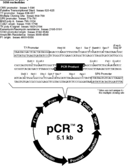
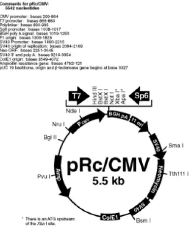
Figure 7 Principle of ecdysone inducible system 69 Figure 8 Multiple cloning sites and properties of vectors 70 Figure 9 Multiple cloning sites and properties of pCR3 vector 72 Figure 10 Multiple cloning sites and properties of pRc/CMV vector 73 Figure 11 Properties of the vectors pVgRXR and pIND.lacZ 78 Figure 12 Principle of suppression subtractive hybridization 80
Figure 13 Map of pGEM-T Easy vector 87
Figure 14 Analysis of pVgRXR transfected MCF7 cells. 93 Figure 15 In vitro transcription-translation and immunoprecipitation 97 assays of pcDNA3.myc.BRCA1 construct
Figure 16 BamHI-XbaI double digestion profiles of pIND and 98
pcDNA3.myc. BRCA1
Figure 17 BamHI-XbaI double digestion profile of pIND+1.6 kb 99 myc colonies.
Figure 18 Determination of DNA concentrations by using the 99 ethidium bromide containing agarose gels.
Figure 19 XbaI-XhoI double digestion profiles of 100 pIND.myc.BRCA1 colonies.
Figure 20 EcF-TC31 amplification results of pIND.myc.BRCA1 102 colonies.
Figure 21 Western blot result of stable U2OS cell clones for 103 pIND.myc.BRCA1 construct analyzed with 5HU.
Figure 22 Western blot result of stable U2OS cell clones for 103 pIND.myc.BRCA1 construct analyzed with MS110.
Figure 23 In vitro transcription and translation-Immunoprecipitation 105 assay with the pCR3.BRCA1 construct
Figure 24 Digestion profiles of pCR3.BRCA1 and pIND. 106 Figure 25 EcoRI digestion profiles of pIND.BRCA1 colonies. 106 Figure 26 AvaI digestion profiles of pIND.BRCA1 colonies. 107 Figure 27 HindIII-NotI digestion profiles of pIND.BRCA1 colonies. 108
Figure 28 Sequencing result of pIND.BRCA1 109
Figure 29 Western blot result of the cells transiently expressing 110 BRCA1 in U2OS cells.
Figure 30 Western blot result of U2OS cells which were transiently 111 transfected with pCMV.myc.BRCA1
Figure 31 Western blot result of MCF7 cells which were transiently 111 transfected with pCMV.myc.BRCA1
Figure 32 Restriction enzyme digestion profiles of pIND and 112 pRc/CMV.BRCA1
Figure 33 HindIII-NotI digestion profiles of pIND.HA.BRCA1 113 colonies.
Figure 34 HindIII-NotI-PvuI digestion profiles of 113 pIND.HA.BRCA1 colonies.
Figure 35 Sequencing data of pIND.HA.BRCA1 sequenced by EcF 115 Figure 36 Sequencing data of pIND.HA.BRCA1 sequenced by EcR 116
Figure 37 Sequencing results of pIND.HA.BRCA1 117
Figure 38 Western blot result of BRCA1 expressing cells that were 118 transiently transfected with pIND.HA.BRCA1
Figure 39 Western blot result of BRCA1 expressing cells that 118 were transiently transfected with pIND.HA.BRCA1 and
pCMVmycBRCA1
Figure 40 Immunoprecipitation results of the U2OS cells transiently 119 transfected with pIND.HA.BRCA1 and pCMV.myc.BRCA1.
Figure 41 Western blot result of the cells transiently transfected 120 with pIND.p53.
Figure 42 Ectopic expression of BRCA1 in MCF7 breast 122 carcinoma cells.
Figure 43 pEGFP transfected MCF7 breast carcinoma cells. 124
Figure 44 Annexin V staining with method I. 125
Figure 45 Annexin V staining with method II. 126 Figure 46 RT-PCR results with sequence specific primers 128
from DnaseI treated mRNA samples
Figure 47 RsaI digestion of ds cDNAs 130
Figure 48 Analysis of ligation efficiency 130
Figure 49 Optimization of primary and secondary PCR conditions 132
Figure 50 Secondary PCR results 132
Figure 51 Analysis of subtraction efficiency test 133 Figure 52 T7-Sp6 amplified cDNA inserts from white colonies 134-136
of the forward subtracted library
Figure 53 PCR products of control cDNAs 137
Figure 54 PCR products of forward and reverse subtracted library 138 Figure 55 RsaI digestion of forward and reverse subtracted library PCR 138 products
Figure 56 Differential screening of SSH-selected cDNA clones with 139 forward and reverse subtracted probes.
Figure 57 Differential screening of 210 selected clones after 140 first screening
Figure 58 Purified PCR products selected for sequencing 141 Figure 59 BRCA1 mediated upregulation of RAD21 and MSH2 149
DNA repair genes.
Figure 60 BRCA1 mediated upregulation of genes involved 150 in cell signalling or tumour suppression.
LIST OF TABLES
Table 1 BRCA1 regulatory proteins 8
Table 2 Mutation types in BRCA1 and BRCA2 10
Table 3 BRCA1 interacting proteins 18
Table 4 BRCA1 modifying proteins 20
Table 5 Summary of methods for gene expression profiling 32
Table 6 List of BRCA1 upregulated genes 33
Table 7 List of BRCA1 downstream genes 34
Table 8 Genes downregulated in Brca1 -/- stem cells 35 Table 9 Genes with altered expression following induction of BRCA1 37
Table 10 List of primers 57
Table 11 Results of the β-gal assay of U2OS cells after 94 transfection with pIND.lacZ reporter plasmid.
Table 12 Results of the β-gal assay of MCF7/RXR clones after 95 transfection with pIND.lacZ.
ABBREVIATIONS
Ad1 Adaptor 1
Ad2R Adaptor 2R
APS Ammonium persulphate
BASC BRCA1 Associated Genome Surveillance Complex
BBS Bes buffered saline
Bp Base pair
BRCA1 BReast CAncer susceptibility gene 1 BRCA2 BReast CAncer susceptibility gene 2
BRCT BRCA1 Carboxyl Terminus
BSA Bovine serum albumin
cAMP Cyclic adenosine mono phosphate
cDNA Complementary DNA
Ci Curie
CIAP Calf intestinal alkaline phosphatase
Cpm Counts per minute
dCTP Cytosine deoxyribonucleotide
ddH2O Double distilled water
DEPC Diethylpyrocarbonate
DMEM Dulbecco’s Modified Eagle’s Medium
DMSO Dimethylsulphoxide
DNA Deoxyribonucleic acid
dNTP Deoxyribonucleotide triphosphate
ds Double strand
DOC Deoxycholate
DSB Double strand break
EDTA Ethylenediaminetetra-acetic acid
ER Estrogen receptor
EtBr Ethidium bromide
FCS Fetal calf serum
G3PDH Glyceraldehyde 3-phosphate dehydrogenase
GFP Green fluorescent protein
HDAC Histone deacetylase
HBS Hepes buffered saline
HR Homologous recombination
h Hour
IP Immunoprecipitation
IPTG Isopropyl β-D-thiogalactopyronoside
IR Ionising radiation
IRIF Ionising radiation induced foci
IVTT In vitro transcription-translation
kb Kilobase
LB Luria-Bertani medium
LOH Loss of heterozygosity
M Molar mA Muristerone A µg Microgram mJ Milijoule min Minute ml Mililiter µl Microliter mm milimeter mM Milimolar
MOPS 3-(N-morpholino)propane-sulphonic acid
mRNA Messenger RNA
N Normal
NaCl Sodium chloride
NaOH Sodium hydroxide
ng Nanogram
NHEJ Nonhomologous end joining
Oligo(dT) Oligodeoxythymidylic acid
pA Ponasterone A
PAGE Polyacrylamide gel electrophoresis
PBS Phosphate buffered saline
PCR Polymerase chain reaction
pmol Picomole
RNA Ribonucleic acid
rpm Revolution per minute
SDS Sodium dodecyl sulphate
Sec Second
ss Single strand
SSH Suppression subtractive hybridization
TBS Tris buffered saline
TCR Transcription coupled repair TEMED N,N,N’,N’-tetramethylenediamine
U unit
UV Ultraviolet
V Volt
v/v volume for volume
xg gravity
X-gal 5-bromo-4-chloro-3-indonyl-β-D-galactosidase
ZBRK1 Zinc binding and BRCA1-interacting protein with a KRAB domain
CHAPTER 1.
INTRODUCTION
Breast cancer, which is the most common female malignancy, is a major cause of death in middle-aged women and its incidence is increasing (Parkin and Coleman, 1990). Breast cancer affects almost one million women worldwide at any given time. One in eight American women will develop breast cancer at some point in their lifetimes, and of these women approximately 30% will develop the metastatic form which is ultimately fatal (Bowcock, 1999).
The breast epithelium undergoes distinct developmental programs during puberty and pregnancy. During puberty, in particular, rapid proliferation of breast tissue occurs. Lobules of the breast are clonal (Kordon and Smith, 1998) and the progeny of this proliferative burst are retained within the breast lobule. In this way, breast epithelial cells have the potential to retain “memory” of genetic alterations that occurred earlier in breast development. In contrast, some other epithelia that are characterized by rapid proliferation, such as the intestinal epithelium, shed cells continuously.
1.1 Risk factors for breast and ovarian cancer
It is well understood that one of the most important risk factors for developing breast cancer is a family history of the disease. However, many other nongenetic risk factors contribute to disease etiology. Besides sex and country of birth, increasing age is an important risk factor. Other suggested risk factors are influences of hormones. Early age at menarche, late menopause, and late first full-term pregnancy, all confer increased risk for breast cancer. Even the prenatal environment might be of importance. It has been shown repeatedly that estrogen exposure is directly associated
with risk for developing breast cancer. High body-mass index after menopause, high alcohol consumption and some dietary factors increase the risk (Martin and Weber, 2000). Exposure to ionising radiation is a well-known risk factor for different kinds of malignancies; young women who received mantle radiation for Hodgkin’s lymphoma have a markedly increased risk for developing breast cancer (Wolden et al., 1998). All these mentioned nongenetic factors increase the risk by about 1.5- to three-fold.
Multiple epidemiological studies have reported that a family history of breast cancer is a reproducible predictor of breast cancer risk (Lynch et al., 1981; Ottman et al., 1986). The risk conferred by having relatives with breast cancer varies with closeness of kinship, numbers of affected relatives and the age of onset. A more than two-fold increase in risk for women with one first-degree relative with early onset breast cancer has been reported in many studies (Arver et al., 2000). Having more than one close relative with breast cancer, or cases of bilateral disease among relatives, seem to confer even higher risks.
Families with three or more close relatives with breast cancer are, in the literature, classified as ‘breast cancer families’. One of the earliest descriptions of a breast cancer family was written in 1866 by the French surgeon P. Broca in the publication ‘Traite des tumeurs’. Numerous pedigrees from families with apparent inherited breast cancer susceptibility have been reported (Lindblom et al., 1993; Lynch et al., 1978) and it was noticed early on that ovarian cancer was frequent in many of these families (Go et al., 1983). Other features found in breast cancer families are early age of onset and bilateral disease (Lynch, 1990; Lynch et al., 1978). In ovarian cancers, about 10% of cases have a hereditary basis. A family history of ovarian cancer is the strongest risk factor for developing the disease. Germline mutations in BRCA genes appear to account for most hereditary ovarian cancers. DNA repair gene mutations (in HNPCC families) account for a much smaller fraction of cases (Lynch et al., 1998).
1.2 Genes implicated in breast cancer
BRCA1, BRCA2, p53, STK11/LKB1, PTEN, MSH2/MLH1, ATM are the genes implicated in formation of hereditary breast cancer. Besides, CYP1A1, GSTs, NAT2 and androgen receptor are some of the low penetrance breast cancer susceptibility genes.
BRCA1 and BRCA2: Familial breast and ovarian cancer predisposition syndromes
have long been recognized. Their genetic bases have become clear with the cloning of two major disease susceptibility genes BRCA1 and BRCA2 (Hall et al., 1990; Miki et al., 1994; Wooster et al., 1995; Wooster et al., 1994). Each has characteristics of a tumour suppressor gene; inheritance within affected families follows an autosomal-dominant pattern of inheritance and loss of heterozygosity (LOH) at the relevant gene locus is seen in familial tumours with retention of the disease-predisposing allele (Collins, 1995; Gudmundsson et al., 1995; Neuhausen and Marshall, 1994). The spectrum of disease-associated mutations includes frequent truncating mutations and less frequent missense mutations. Although LOH is frequently at the BRCA1 or BRCA2 locus in sporadic breast cancer, the retained allele is almost wild-type (Futreal et al., 1994; Lancaster et al., 1996). Thus, in contrast to the casual role of BRCA1 gene mutation in the hereditary syndrome, BRCA1 gene mutation in sporadic breast and ovarian cancer seldom conforms to Knudson’s model for tumour suppressor genes (Knudson, 1971). Cancer risk in BRCA gene mutation carriers may be increased modestly in other organs, but, highly penetrant, early-onset, site specific cancer is restricted to the breast and ovary.
p53: p53 germline mutations are thought to be a rare cause of breast cancer, except in
the setting of Li-Fraumeni syndrome. The first documentation of Li-Fraumeni syndrome was in 1969, in which four families with children with soft tissue sarcomas were found to have an excess of sarcomas in other relatives. In addition these families exhibited an excess of early onset breast cancer and other cancers such as childhood leukemia, adrenocortical carcinoma and brain cancer (Li and Fraumeni, 1969). In 1990, the presence of germline p53 mutations in approximately half of the families with classic Li-Fraumeni syndrome was reported (Malkin et al., 1990). In women
with germline p53 mutations who survive childhood cancers, it is estimated that 50% will have developed breast cancer by the age of 50 (Easton et al., 1993b).
STK11/LKB1: Peutz-Jeghers syndrome is caused by germline mutations in
STK11/LKB1, a serine-threonine kinase located on chromosome 19q13.3 (Jenne et al., 1998). Peutz-Jeghers syndrome is characterized by hamartomatous polyps in the small bowel and pigmented macules of the buccal mucosa, lips, fingers and toes. In spite of the early-onset breast cancer that can be seen in patients with Peutz-Jeghers syndrome, mutations in STK11/LKB1 do not appear to play an important role in sporadic breast cancers, based on the very low prevalence of mutations in the population.
PTEN: Cowden syndrome is a rare autosomal dominant predisposition to both benign
and malignant neoplasms. Breast cancer develops in 20-30% of carrier women. Other tumours seen among patients with Cowden syndrome include adenomas and follicular cell carcinomas of the thyroid gland, polyps and adenocarcinomas of the gastrointestinal tract and ovarian cysts and carcinoma (Hanssen and Fryns, 1995). Cowden syndrome is caused by germline mutations in the PTEN gene (MMAC1/TEP1). PTEN, a tumour suppressor gene on 10q23.3, is a dual-specifity phosphatase (Steck et al., 1997). PTEN does not play a role in familial breast cancer, apart from its role in Cowden syndrome.
MSH2/MLH1: Muir-Torre syndrome is defined by the presence of sebaceous gland
tumours and visceral malignancy. It is inherited in an autosomal dominant fashion with high penetrance because of mutations in the same genes associated with hereditary nonpolyposis colorectal cancer (HNPCC) (Schwartz and Torre, 1995). The most common malignancy in this syndrome is colorectal cancer, seen in 50% of patients, but breast cancer occurs in approximately 25% of women carriers. As in HNPCC, microsatelite instability is observed in the tumours of patients with Muir-Torre syndrome. Mutational analysis has shown that the mutations are predominantly in MSH2.
ATM: Ataxia-telangiectasia (AT) is a complex, autosomal recessive disorder
sensitivity and cancer predisposition caused by homozygous mutations in the ATM gene. Epidomiologic studies have shown that AT carriers might have an increased risk for breast cancer, and this observation was consistent with the recent finding that ATM phosphorylates BRCA1 protein upon DNA damage (Li et al., 2000).
Low penetrance breast cancer susceptibility alleles: Low-penetrance
susceptibility alleles, sometimes called “modifier genes”, are defined as polymorphic genes with specific alleles that are associated with an altered risk for disease susceptibility. Further studies are needed to clarify the role of all the following genes in breast cancer as rewieved by Martin and Weber (2000):
The P450 gene CYP1A1 encodes aryl hydrocarbon hydroxylase, which is the primary catalyst in the conversion of estradiol to hydroxylated estrogen. A reduced estrogen level is protective for developing breast cancer, whereas increased estrogen exposure can increase the risk for developing breast cancer. Alterations in the activity of aryl hydrocarbon hydroxylase could therefore plausibly lead to a change in the levels of estrogen and could ultimately affect breast cancer risk.
The glutathione S-transferases (GSTs) constitute a family of genes that encode for enzymes that catalyze the conjunction of reactive chemical intermediates to soluble glutathione conjugates to facilitate clearance. Inability to metabolise carcinogens may increase breast cancer risk.
N-acetyltransferase (NAT2) is an important component of the carcinogen
metabolism pathway and polymorphisms in the NAT2 gene are associated with an altered rate of metabolism of carcinogens.
The effect of CAG microsatelite found in exon 1 of androgen receptor gene has been investigated for its role in breast cancer penetrance. BRCA1 mutation carriers who carry at least one androgen receptor allele with more than 29 CAG repeats were diagnosed with breast cancer statistically significantly earlier than women with shorter CAG repeats in their androgen receptor genes, suggesting that pathways
involving androgen signalling may affect the penetrance of BRCA1-associated breast cancer (Rebbeck et al., 1999).
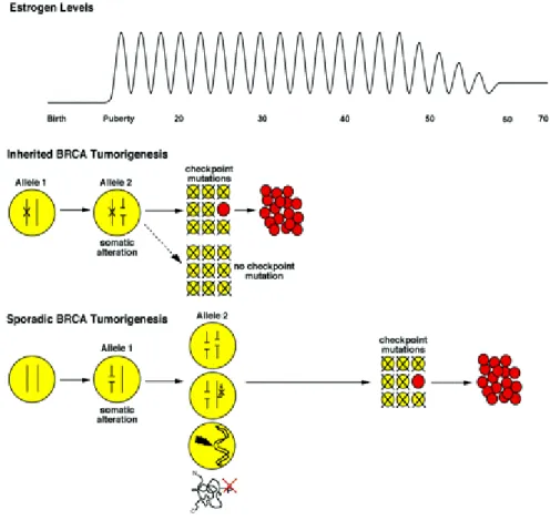
1.3 Mechanisms of BRCA-mediated breast and ovarian cancer formation
Relationship between mutation events and formation of breast or ovarian carcinomas are summarized in Figure 1. Estrogen mediated proliferation of breast and ovarian epithelial cells and the distinctive genomic context (high percentage of repetitive sequences) of the BRCA genes are critical components of this pathway. During puberty estrogen stimulates breast epithelial cells to proliferate. Among somatic mutations that appear in these rapidly dividing cells, alterations of BRCA genes are likely to be relatively frequent because of the density of repeats in these genes. Inactivation of the second allele in such a cell would generally result in cell death. Tumorigenesis would require inactivation of both alleles in a cell capable of escaping cell cycle checkpoints. Unlike inherited disease, inactivation of genes critical to cell cycle checkpoints could occur prior to inactivation of the second allele. Therefore, BRCA1-mediated sporadic tumorigenesis would be less likely. Also, because more mutational events are required, tumours occur later in a woman’s life.
Various genetic mechanisms may be responsible for somatic inactivation of both alleles by generation of large deletions of BRCA1 and BRCA2 in both inherited and somatic tumours. Deletion of a chromatin loop containing all or a large portion of BRCA genes may be mediated by homologous recombination between repeat sequences. Repeat mediated loss of chromatin formed at different points in the cell cycle will yield deletion of different sizes. If this process occurs in both copies of the BRCA1 region, deletions including the BRCA gene will overlap but not be identical. If genomic deletions overlap, they may incorrectly appear to define one region of LOH. This hypothesis may explain the large deletions observed in BRCA1 tumours (Welcsh and King, 2001).
Figure 1: Relationship between mutation events and formation of breast or ovarian cancers. (Welcsh and King, 2001).
Estrogen levels in women are cyclic from menarche until menopause when peak estrogen levels decrease. In response to estrogen at puberty and pregnancy, breast epithelial cells proliferate. Among women with a germline mutation in BRCA1 or BRCA2, somatic inactivation of the remaining allele will result in repair-deficient cells. These cells will be unable to repair damaged DNA in the following cell cycle and will ultimately die due to activation of critical cell cycle checkpoints. The rare repair-deficient cell (shown in red) that escapes death by checkpoint may acquire mutations at other sites including critical checkpoint genes, ultimately resulting in tumour formation. In inherited BRCA tumorigenesis, somatic inactivation of the second BRCA allele likely occurs early in a young woman’s life, leading to early onset of breast cancer. In women without a BRCA germline mutation (sporadic BRCA tumorigenesis), somatic alteration of one allele of BRCA1 or BRCA2 in breast epithelial cells is likely to occur during estrogen-mediated proliferation, due to the density of repetitive elements in these genes. Inactivation of a second BRCA allele in a cell would generally result in cell death.
Alternatively it has been proposed that inactivation of a BRCA allele could result in an overall decrease in BRCA function (haplo-insufficiency). Presence of only one BRCA allele is clearly sufficient for normal growth and development, because persons heterozygous for BRCA mutations have normal phenotypes apart from their cancer predisposition. However under conditions of cellular stress in breast and ovarian epithelium, caused by estrogen-stimulated proliferation, it is possible that even a modest decrease in BRCA function in cells with one somatically inactivated allele could increase the risk for additional cancer promoting mutations.
Third, transcriptional silencing may inactivate BRCA alleles, by loss of proteins that positively regulate their expression or by an increase in negative regulatory proteins. Table 1 summarizes the known BRCA1 regulatory proteins, and the nucleotide sites at which they function if the regulation is direct:
BRCA1 regulatory protein Binding domain on BRCA1 (L78833) Reference
Brn-3b -400 to –1 (2944-3344) (Budhram-Mahadeo et al., 1999) GA binding protein α/β -204 to –159 (3140-3185) (Atlas et al., 2000)
RB-E2F -23 to 15 (3321-3339) (Wang et al., 2000a) p53 indirect (Arizti et al., 2000) TGFβ (requires RB) ? (Satterwhite et al., 2000) Id4 indirect (Beger et al., 2001) Table 1: BRCA1 regulatory proteins
Among the BRCA1 regulatory proteins, Id4 (inhibitor of DNA binding 4) inversely regulates BRCA1 expression (Beger et al., 2001). Overexpression of Id4 (and concomitant reduction of BRCA1 expression) is associated with anchorage-independent growth, a critical characteristic of tumour cells. The relationship between Id4 and BRCA1 may be important for breast cancer because estrogen reduces Id4 expression, hence increasing expression of BRCA1. Conversely, breast epithelial cells that are no longer responsive to estrogen may overexpress Id4, with consequent reduction of BRCA1 expression. If this occurs in a cell that has lost one BRCA1 allele through somatic inactivation, these cells would then be BRCA1 deficient.
Transcriptional silencing of one BRCA1 allele could be accomplished through methylation of CpG islands, which are often found in the regulatory region of many genes. The 5’ regulatory region of the BRCA1 gene contains a TATA-less promoter with high cytosine-guanine content, making BRCA1 an excellent candidate for CpG methylation. In 5-10% of sporadic breast tumours, BRCA1 transcription is silenced by the methylation of CpG residues, which leads to absence of protein expression (Esteller et al., 2000). In a recent report, the BRCA1 promoter was methylated in 15% of sporadic ovarian tumours (Baldwin et al., 2000).
BRCA1 might be inactivated post-transcriptionally by failure of phosphorylation or other posttranslational modifications. Phosphorylation of BRCA1 is required for normal functioning of BRCA1. The roles of ATM, ATR and Chk2 in phosphorylating BRCA1 in response to DNA damage are likely to be most critical to carcinogenesis.
1.4 BRCA1
Hereditary breast cancer families account for only 5-10% of all breast cancer cases. Mutations in BRCA1 are responsible for nearly all of the hereditary breast and ovarian cancer families and up to 40-50% of families with hereditary breast cancer only (Easton et al., 1993a). BRCA2 is strongly linked to hereditary breast cancer in both males and females (Wooster et al., 1995; Wooster et al., 1994). Together BRCA1 and BRCA2 account for the majority, although not all, of hereditary breast cancer cases.
1.4.1 Mutational spectrum of BRCA1
Since the isolation of BRCA1, more than 700 sequence variations have been identified. Initially eight disease-associated mutations were described within the gene (Futreal et al., 1994; Miki et al., 1994), followed shortly by an increasing number of mutations (Castilla et al., 1994; Friedman et al., 1994; Simard et al., 1994). Most are frameshift mutations, but several missense mutations are known to alter protein
function. Mutational spectrum of BRCA genes is summarized in Table 2. Most known mutations are listed in the Breast cancer Information Core (BIC) and are available on the Internet (http://www.nhgri.nih.gov/Intramural_research/Lab_transfer/Bic). Several founder mutations have been identified in BRCA1.
Gene BRCA1 BRCA2
Mutation type Frameshifts 70% 68% Nonsense mutations 20% 12% Splice site 5% 7% Missense 5% 13%
Table 2: Mutation types in BRCA1 and BRCA2 (Bowcock, 1999)
1.4.2 BRCA1 gene and protein structure
BRCA1 gene is localized on chromosome 17q21 and spans 100 kb genomic sequence. BRCA1 is a large gene with a total mRNA of 5711 base pairs, divided into 24 exons, of which 22 are coding. Exon 1 is not coding and exon 4 is an Alu repetitive motif. The coding region begins in exon 2. Exon 11 constitutes more than 60% of the coding region, while most of the other exons are relatively small (Miki et al., 1994). The gene encodes a nuclear phosphoprotein of 220 kDa (Chen et al., 1996a) and smaller splice variants have been described (Wang et al., 1997). Full-length BRCA1 protein is localized in the nuclear foci of epithelial cells (Wilson et al., 1999) while a splice variant, BRCA1 delta 11b, might be cytoplasmic (Thakur et al., 1997).
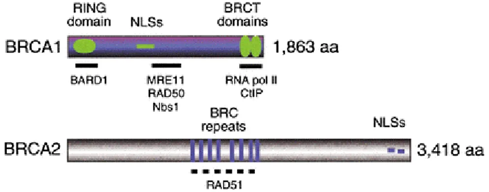
Functional domains have been identified in BRCA1 (Figure 2). The most phylogenetically conserved region is the C3HC4 zinc binding RING-finger domain at the amino terminus of the protein (Chen et al., 1996a; Saurin et al., 1996). Several other human proteins with zinc finger motifs have suggested this domain to be a region of protein-protein interactions and such motifs are found in a number of regulatory proteins. In addition, BRCA1 has two nuclear localization signals in exon
11 (Chen et al., 1995; Miki et al., 1994). The “BRCT” (BRCA1 Carboxyl-terminal) domain is an evolutionary region in BRCA1 (Callebaut and Mornon, 1997; Koonin et al., 1996). This region resembles the transactivation domain of a number of transcription factors (Abel et al., 1995) and contains two BRCT domains, which are situated in tandem at amino acids 1646-1736 and 1760-1855. BRCT motifs have been described in a number of proteins such as 53BP1, RAD9, RAD4, Crb2 and RAP1, that have function in cell cycle control and DNA damage repair pathways (Callebaut and Mornon, 1997; Koonin et al., 1996), consistent with recent data suggesting an important role for BRCA1 in cellular responses to DNA damage.
Figure 2: Features of the human BRCA Proteins (Venkitaraman, 2002) BRCA1
contains an N-terminal RING domain, nuclear localization signals (NLSs), and two C-terminal BRCT domains of ~110 residues (also found in several proteins with functions in DNA repair or cell cycle control). BRCA2 contains eight repeats of the ~40 residue BRC motifs. Six of the eight motifs in human BRCA2 can bind directly to RAD51 when expressed in vitro.
1.4.3 Cellular expression of BRCA1
In normal cells BRCA1 is a nuclear protein. BRCA1 protein expression is reduced or absent in most sporadic, advanced (grade III) ductal breast carcinomas (Wilson et al., 1999). Nuclear localization signals have been identified on BRCA1 (Thakur et al., 1997), which interact with importin-α, a subunit of the nuclear transport signal receptor.
BRCA1 mRNA and protein are prefentially expressed during the late G1-early S phase of the cell cycle (Gudas et al., 1996). In mitotic cells, BRCA1, BRCA2 and RAD51 interact and co-localize in a punctate pattern in the nucleus during the S phase of the cell cycle, whereas in meiotic cells all three proteins associate with unsynapsed axial elements along developing synaptonemal complexes (Chen et al., 1998b; Scully et al., 1997b). BRCA1 function is regulated by phosphorylation; it is hyperphosphorylated during the late G1 and S phases by an endogenous kinase activity from S1497 and T967 residues, minimally composed of the CDK2-cyclin complex that controls the G1-S transition, with dephosphorylation occurring at the M phase (Ruffner et al., 1999). A function of BRCA1 at the G1-S transition might be to arrest cell cycle progression by binding hyperphosphorylated retinoblastoma protein (Aprelikova et al., 1999).
BRCA1 might also regulate the G2-M checkpoint by controlling the assembly of mitotic spindles and the appropriate segregation of chromosomes to daughter cells. Mouse embryonic fibroblasts carrying a targeted deletion of Brca1 exon 11 had an intact G1-S checkpoint but were unable to arrest at the G2-M checkpoint (Xu et al., 1999). In a significant fraction of cells centrosomes were amplified, resulting in abnormal chromosomal segregation and aneuploidy. Proteins that regulated the G2-M checkpoint, including p53 and pRB, localize to centrosomes. BRCA1 also localizes to centrosomes during mitosis and interacts with γ tubulin, a component of the centrosome (Hsu and White, 1998). Mutant BRCA1 might induce genetic instability by disrupting centrosome duplication.
BRCA1 and BRCA2 are expressed ubiquitously with the highest levels in the thymus and testis (Hakem et al., 1996; Miki et al., 1994). In developing mouse embryos, both genes are highly expressed in rapidly dividing, differentiating tissues and most notably during mammary epithelial proliferation and differentiation (Rajan et al., 1997; Rajan et al., 1996). In the mammary gland the expression of both messages is developmentally regulated and is induced during puberty and pregnancy and reduced during lactation.
1.4.4 BRCA1-deficient cells and animal models
To understand how the loss of BRCA1 leads to breast cancer formation, mouse genetic models for Brca1 mutation have been established. This work has revealed that Brca1 homozygous deletions are lethal at early embryonic day E6.5 (Hakem et al., 1996; Liu et al., 1996; Ludwig et al., 1997). Gowen et al generated a distinct mutation in Brca1 and observed that embryos survived until E13.5 and exhibited defects in neural development, including anencephaly and spina bifida (Gowen et al., 1996). Another group generated a mouse model with targeted deletion of Brca1 exon 11 and the resulting embryo died at E12-E18.5 (Xu et al., 1999). Collectively these findings imply a role for the BRCA1 gene product in growth and/or differentiation during mouse embryogenesis.
Both BRCA1 and BRCA2-deficient cells are characterized by cumulative chromosomal abnormalities including chromosomal breaks, aberrant mitotic exchanges and aneuploidy (Lee et al., 1999; Xu et al., 1999). BRCA1 and BRCA2 deficient mice harbor inactivating mutations in p53 and mitotic checkpoint genes (Lee et al., 1999; Xu et al., 1999). Mutational inactivation of p53, which governs the G1/S cell cycle checkpoint, may thus circumvent the growth arrest that is normally induced upon DNA damage and also inhibit p53-mediated apoptosis, thereby permitting the survival of cells with severe chromosomal damage. Consistently, the embryonic lethality associated with brca1-null mutations can be partially rescued by targeted deletion of p53 or p21 (Hakem et al., 1997). On the other hand, inactivation of mitotic checkpoint genes could bypass mitotic arrest and permit aberrant chromosomes to segregate into progeny cells.
BRCA1-deficient HCC1937 cells, BRCA1-null embryonic stem (ES) cells and Brca1-exon 11 deletion MEF (fibroblast) cells are all characterized by radiation hypersensitivity. Increased sensitivity to the radiomimetic agent methyl methane-sulfonate (MMS) and ionising radiation (IR), but not to ultraviolet (UV) radiation, has also been observed in BRCA1-deficient cells (Gowen et al., 1998; Scully et al., 1999; Xu et al., 1999; Zhong et al., 1999). Reintroduction of a wild type BRCA1 allele, but not clinically validated BRCA1 missense mutant alleles, can complement the MMS and IR sensitivity of BRCA1-deficient cells, suggesting that cellular response to DNA damage is compromised in breast cancer patients carrying BRCA1 mutations (Scully et al., 1999; Zhong et al., 1999)
1.4.5 BRCA1 and p53
One of the key checkpoint genes for BRCA1-mediated tumorigenesis is p53, which is the most commonly mutated gene in many common human cancers. There are interesting parallels between p53 and BRCA1. BRCA1, like p53, is a cell cycle regulated nuclear phosphoprotein and has also been implicated in DNA damage response and repair pathways. In addition BRCA1 interacts with some of the major proteins involved in DSB repair and HR and participates in TCR. Both p53 and BRCA1 are post-translationally altered by phosphorylation in response to DNA damage. p53 regulates BRCA1 expression in response to stress conditions (Arizti et al., 2000). BRCA1 associates with the C-terminus of wild-type p53 (Ouchi et al., 1998) and stimulates transcription from p53-responsive promoters; whereas tumour associated mutants of BRCA1 are deficient in this coactivating activity (Somasundaram et al., 1999; Zhang et al., 1998). p53 coimmunoprecipitates with BRCA1. Interaction domains of p53 on BRCA1 were identified both at the N-terminus, overlapping the Rad50 binding domain, and in the second BRCT motif at the C-terminus. While BRCA1 is a potent enhancer of p53-mediated transcription, BRCA1 mediated stabilization of wild type p53 protein occurs through transcriptional activation of the p14 ARF gene and effects on the phosphorylation of p53 through the presence of BRCA1. Indeed, cells with deletions of exon 11 of BRCA1 are defective in the rapid stabilization of p53 following DNA damage (Xu et al., 2001). It is likely that BRCA1 participates in p53 stabilization in vivo. Like p14ARF, p53 has been
shown to repress the transcription of BRCA1, acting in a negative feedback loop of its own stabilization (Arizti et al., 2000; MacLachlan et al., 2000).
Evidence from conditional knockout mice suggests that loss of BRCA1 in mammary epithelial cells leads to incomplete proliferation, apoptosis and tumours at a low frequency (Xu et al., 1999). In these mice additional heterozygous mutations in p53 leads to many more mammary tumours, most of which have lost the remaining p53 allele. Tumours in patients with germline BRCA1 or BRCA2 mutations are frequently associated with somatic mutations of p53. In the proposed model, mutant p53 would inactivate a cell cycle checkpoint and lead to uncontrolled growth and invasive growth. Cells that have successfully escaped death by checkpoint will probably accumulate multiple mutations. Amplification of MYB oncogene and reduction of anti-apoptotic gene Bcl-2 are characteristics of most breast tumours from BRCA1 mutation carriers (Freneaux et al., 2000; Kauraniemi et al., 2000).
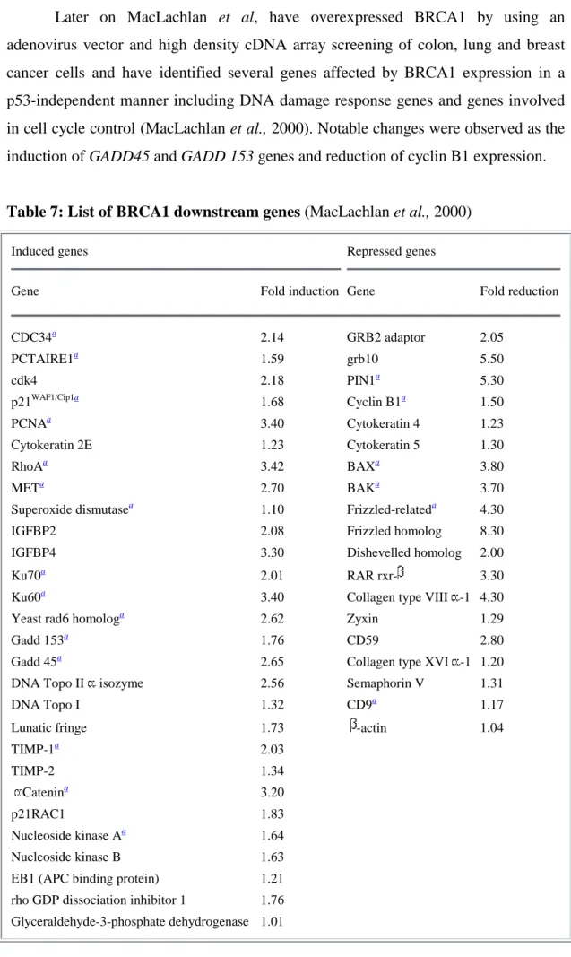
A recent report indicated that BRCA1 selectively coactivates the p53 transcription factor towards the genes that direct DNA repair and cell cycle arrest but not towards those that direct apoptosis (MacLachlan et al., 2002). Because BRCA1 overexpression stabilizes wild-type p53 but does not lead to apoptosis in most cell lines, the selectivity of BRCA1 for p53 dependent target gene activation has been analyzed using arrays containing known targets. It has been found that BRCA1-stabilized p53 regulates transcription of DNA repair and growth arrest genes, while p53 stabilized by DNA damaging agents induces genes involved in apoptosis.
1.5 Functions of BRCA1
Cancer susceptibility gene mutations fall into two general classes (Kinzler and Vogelstein, 1997). Genes whose mutation or altered expression relieves normal controls on cell division, death or lifespan, promoting the outgrowth of cancer cells, have been termed “gatekeepers”. Those whose disruption causes genome instability, increasing the frequency of alterations in gatekeeper genes, work instead as “caretakers”. BRCA genes are essential for preserving chromosome structure,
suggesting that, in their role as tumour suppressors, they behave as caretakers, suppressing genome instability (Figure 3).
BRCA1 may function as a scaffold in the assembly of a multi-protein complex, which is important in maintenance of genomic integrity by functioning in gene transcription, chromatin remodelling, checkpoint activation, DNA damage repair and transcription-coupled DNA-damage repair. Some of the functions in which the BRCA1 protein has been implicated are summarized in Figure 4. Figure 5 shows the putative roles of BRCA1 based on reported interactions. BRCA1 interacting proteins are summarized in Table 3. BRCA1 performs its tumour suppressor function in many ways:
Figure 3: Repair genes as caretakers of the genome. (Khanna and Jackson, 2001)
When cells lack one of the repair proteins, they fail to repair the DSBs correctly. This leads to genetic instability, gross chromosomal rearrangements and accumulation of mutations. These events then trigger cell-cycle checkpoints resulting in permanent growth arrest or death of affected cells. If the checkpoints are inactivated by mutations, this leads to tumorigenesis.
Figure 4: Multiple cellular functions of BRCA1
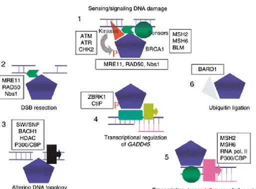
Figure 5: Putative roles of BRCA1 (Venkitaraman, 2002)
BRCA1 works as a signal processor (1) during DNA damage responses in complex with proteins that bind to aberrant DNA structures (sensors), and the kinases that signal their presence. Phosphorylation of BRCA1 may be essential for local functions [control of DSB resection (2), altering DNA topology (3)] near a DNA lesion, as well as for distant functions such as transcriptional control of checkpoint genes (4) (e.g., GADD45) or targets of estrogen receptor signalling, or transcription-coupled DNA repair (5) BRCA1 works with BARD1 (6) works as an ubiquitin ligase
BRCA1
BRCA1-P
DNA damage
Transcriptional regulation
DNA repair by homologous recombination Checkpoint activation and apoptosis Transcription-coupled DNA repair
Table 3: BRCA1 interacting proteins (Modified from Welcsh and King, 2002)
BRCA1 interacting protein or complex Function of interacting protein Interacting domain on BRCA1 (aa) Reference
RAD51 DSB repair exon 11 (758-1064) Scully et al., 1997
RAD50 DSB repair exon 11 (341-748) Zhong et al., 1999
BRCA2 DSB repair BRCT domain (1314-1863) Scully et al., 1997
BASC (ATM, BLM, MSH2, MSH6, MLH1, RCF) Mismatch repair BRCA1 part of complex Wang et al., 2000
ATP-MSH2 Mismatch repair ? Wang et al., 2000
H2AX Signals DNA damage ? Paull et al.,2000
p53 transcription factor, tumor suppressor Exon 11 and BRCT domain (244-500 and 1760-1863) Zhang et al., 1998;Ouchi et al., 1998; Chai et al., 1999
pRB cell cycle regulator, tumor suppressor exon 11 and BRCT domain (304-394 and 1536-1863) Aprelikova et al., 1999
c-Myc transcription factor, oncogene N-terminus and exon 11 (175-303 and 433-511 Wang et al., 1998
ZBRK1 transcription factor, represses GADD45 exon 11 (341-748) Zheng et al., 2000
ATF transcription factor RING (1-101) Houvras et al., 2000
STAT1 signal transducer, transcriptional activation exon 11 (502-802) Ouchi et al.,2000
E2F transcription factor, cell cycle regulator N-terminus (1-76) Wang et al., 1997
RNA Pol II Holoenzyme (hRPB10a;hRPB2) transcription BRCT domain (1650-1800) Scully et al., 1997; Schlegel et al.,2000
RNA helicase A component of RNA PolII holoenzyme BRCT domain (1650-1800) Anderson et al., 1998
Estrogen receptor ligand responsive transcription factor N-terminus (1-300) Fan et al., 1999;2001 Androgen receptor ligand responsive transcription factor exon 11 and BRCT domain (758-1064 and 1314-1863) Yeh et al., 2000
CtIP binds CtBP; transcriptional co-repressor BRCT domain (1651-1863) Li et al., 1999; Yu et al.,1998; Wong et al.,1998
p300/CBP transcriptional co-activator RING and BRCT domain (1-303 and 1314-1863) Pao et al.,1998
SWI/SNF chromatin remodelling complex exon 11 (260-553) Bochar et al., 2000
HDAC l and 2 Histone deacetylation, chromatin remodelling BRCT domain (1563-1863) Yarden and Brody, 1999
Centrosome (p53, pRB, Nm23) chromosome segregation BRCA1 part of complex Hsu and White, 1998
BRAP2 cytoplasmic retention NLS (303-701) Li et al.,1998
Vasolin containing protein (VCP) ATPase exon 11 (303-625) Zhang et al.,2000
BARD1 Ubiquitination? Polyadenylation via CstF-50? RING (1-101) Wu et al.,1996
BAP1 Deubiquitinating enzyme RING (1-101) Jensen et al., 1998
1.5.1 BRCA1 in damage signalling
An important step in the cellular response to DNA damage is to transduce damaged signals to downstream effectors involved in the arrest of cell cycle and repair of damaged DNA (figure 6). In mammalian cells, several kinases such as, ATM, ATR, DNA-PK, Chk1 and Chk2 are activated in response to DNA damage. BRCA1 becomes hyperphosphorylated in response to treatment of cells with a variety of DNA damaging agents including UV, hydroxyurea, mitomycin C, MMS, IR, H2O2 and adriamycin (Chen et al., 1996a; Li et al., 1999; Scully et al., 1997b). In vitro and in vivo phosphorylation sites in BRCA1 have been identified. The data from these studies have suggested that phosphorylation of BRCA1 by these kinases is necessary for BRCA1-mediated DNA damage response and multiple kinase activities are responsible for the DNA damage-induced hyperphosphorylation of BRCA1. Table 4 shows the in vivo phosphorylation sites on BRCA1. In addition to ATM, ATR and Chk2, DNA-PK have been shown to phosphorylate BRCA1 in vitro (Lim et al., 2000)
BRCA1 kinases In vivo phosphorylation sites Reference
ATM S1189, S1457, S1524, S1542 (Li et al., 2000)
ATR S1423 (Tibbetts et al., 2000)
Chk2 S988 (Lee et al., 2000)
Table 4: BRCA1 modifying proteins
Besides its roles in maintenance of genomic integrity, chromatin remodelling, and transcriptional regulation, BRCA1 has also been implicated in cell cycle/checkpoint control. BRCA1 regulates the G2/M checkpoint by activating Chk1 kinase upon DNA damage. BRCA1 regulates the expression of both Wee1 kinase, an inhibitor Cdc2/Cyclin B kinase and 14-3-3 family of proteins that sequesters phosphorylated Cdc25C and Cdc2/cyclin B kinase in the cytoplasm, leading to a conclusion that BRCA1 regulates key effectors that control the G2/M checkpoint and is therefore involved in regulating the onset of mitosis (Yarden et al., 2002).
Figure 6: Signaling of DSBs. (Khanna and Jackson, 2001)
a, The general organization of the DNA-damage response pathway. The presence of
DSBs is recognized by a sensor, which transmits the signal to a series of downstream effector molecules through a transduction cascade to activate signaling mechanisms for cell-cycle arrest and induction of repair, or cell death if the damage is irreparable.
b, A central role for ATM in the cellular response to DSBs. ATM is activated in
response to DSBs. Activated ATM signals the presence of DNA damage by phosphorylating targets involved in cell-cycle arrest, DNA repair and stress response. p21 inhibits the activity of cdk2/cyclinE and 14-3-3 inhibits the activity of cdc2/cyclin B and effecting cell-cycle arrest. c-Abl activates stress-activated protein kinase (SAPK) for transcriptional regulation of stress-response genes.
Phosphorylation of BRCA1 is apparently important for a proper DNA damage response. However, it remains unclear how phosphorylation modulates activities of BRCA1. Since BRCA1 forms a complex with RAD50/MRE11/NBS1 and colocalizes to DNA damage sites following ionizing radiation, it has been speculated that BRCA1 is involved in double strand break repair (Cortez et al., 1999; Lee et al., 2000). Alternatively, phosphorylation of BRCA1 may influence the transcriptional regulation activities of BRCA1 leading to cell cycle checkpoint. ATM phosphorylates CtIP in vivo and in vitro following IR treatment (Li et al., 2000). This ATM dependent phosphorylation is required for dissociation of the CtIP/CtBP corepressor complex from BRCA1 and subsequently leads to relieving BRCA1 mediated repression of GADD45 transcription and p21 expression (Li et al., 2000). These studies suggest that CtIP mediates one of the functional links between ATM and BRCA1 in DNA damage signaling pathways.
1.5.2 BRCA1 in chromatin remodelling
BRCA1 plays an important role in the cellular response to double strand breaks. Within minutes of DNA damage, the histone H2A family member H2AX becomes extensively phosphorylated and forms foci at break sites. BRCA1 is recruited to these foci several hours before other factors such as RAD50 and RAD51, suggesting that BRCA1 and H2AX initiate repair by modifying local chromatin structure, thereby allowing DNA repair proteins access to damaged sites (Paull et al., 2000). In support of this role for BRCA1 in response to DNA damage, it has been shown that BRCT domains of BRCA1 bind double strand breaks (Yamane et al., 2000).
BRCA1 interacts with the SWI/SNF chromatin remodelling complex (Bochar et al., 2000), with regulators of histone acetylation/deacetylation (Pao et al., 2000; Yarden and Brody, 1999), with a novel helicase BACH1 (Cantor et al., 2001) and RecQ homolog encoded by the Bloom’s syndrome gene, BLM helicase (Wang et al., 2000b). How these interactions may assist DSB repair is currently speculative. Chromatin changes mediated by histone modification may make DNA more accessible by the helicases and repair machinery.
1.5.3 BRCA1 in DNA repair
BRCA1 appears to participate in the cellular DNA damage response at multiple stages. In normal cells, responses to DNA damage include sensing damaged DNA, transducing DNA damage signals, relocating repair machinery to damage sites, completing a repair process and coordinating cell cycle progression with the DNA repair process. Accumulating evidence suggests that BRCA1 functions not only in association with the DNA repair machinery, but also in DNA damage induced cell cycle checkpoint control. BRCA1 may additionally regulate the expression of genes involved in DNA damage repair and also directly participates in the repair process itself. Finally phosphorylation of BRCA1 upon DNA damage implies a role for BRCA1 in DNA damage-induced signal delay.
BRCA1 interacts with the RAD50/MRE11/NBS1 protein complex containing the mammalian homologs of yeast molecules known to participate in double strand break repair, comigrants to sites marked by phospho-H2AX (Zhong et al., 1999). The equivalent of this complex in yeast, the Rad50/Mre11/Xrs2 complex functions in both nonhomologous end joining (Ivanov et al., 1992) and homologous recombinational repair of DNA double strand breaks (Bressan et al., 1999). Rad50/Mre11/Xrs2 complex has been shown to be involved in other cellular processes including chromatin configuration and telomere maintenance (Chamankhah et al., 2000; Gerecke and Zolan, 2000; Ohta et al., 1998). It has been proposed that Rad50/Mre11/Xrs2 is responsible for end processing of double strand breaks (Tsubouchi and Ogawa, 1998). In support with this idea, recombinant MRE11 proteins and purified human RAD50/MRE11/NBS1complexes exhibit exonuclease and endonuclease activities (Paull and Gellert, 1998). MRE11 encodes a nuclease activity, which resects flush DSB ends to generate ssDNA tracts. Under certain in vitro conditions, BRCA1 can inhibit this activity of MRE11 (Paull et al., 2001), regulating the length and presumably the persistence of ssDNA generation at sites of DNA breakage. It has been proposed that RAD50 may be a chromatin-associated protein and participate in chromatin structural reconfiguration (Alani et al., 1989). NBS1 is the product of the gene mutated in Nijmegen Break Syndrome (Carney et al., 1998).
BRCA1 appears to interact with the RAD50/MRE11/NBS1 complex directly through RAD50 (Zhong et al., 1999). Similar to the formation of the RAD50/MRE11/NBS1 complex, the association of BRCA1 with this complex does not change in response to DNA damage. Rather, the nuclear partitioning of this BRCA1-containing complex changes and BRCA1 forms ionising radiation-induced foci (IRIF), which is also a characteristic of RAD50, MRE11 and NBS1 (Maser et al., 1997).
Besides RAD50/MRE11/NBS1, other components involved in DNA damage repair, such as MSH2, MSH6, MLH1, ATM and BLM have been found to reside in a large BRCA1-containing DNA repair complex (Wang et al., 2000b). In addition, DNA replication factor C and PCNA (proliferating cell nuclear antigen) were also found in this complex. This complex has been proposed to present a BRCA1-associated genome surveillance complex (BASC) since many of its constituent proteins individually recognize distinctly abnormal DNA structures such as double strand breaks, base pair mismatches and stalled replication forks. Therefore the association of BRCA1 with these proteins suggests that BRCA1 may also participate in the resolution of aberrant DNA structures that occur during DNA replication or when DNA replication is stalled (Wang et al., 2000b). Consistent with this notion, a previous observation that BRCA1 foci at S phase disperse in response to DNA damage or replication blocks, and relocalize to PCNA-containing structures (Scully et al., 1997a), suggestive of a role for BRCA1 in replicational DNA repair.
BRCA1 has also been shown to interact and colocalize with RAD51 (Chen et al., 1998b; Scully et al., 1997b). Eukaryotic RAD51 proteins are homologs of bacterial RecA and are required for recombination during mitosis and meiosis and for recombinational repair of DNA double strand breaks. In response to DNA damage, BRCA1 undergoes distinctive phosphorylation that differs from the phosphorylation during G1-S transition (Scully et al., 1997a). The association of RAD51 and BRCA1 is supported by the observation that BRCA1 foci disperse during the S phase, and relocalize to PCNA-containing structures in response to UV-treatment or replication block by hydroxyurea (Scully et al., 1997b). BRCA1 has also been observed in IR-induced RAD51 foci; however such foci are distinct from those comprising BRCA1 and the RAD50/MRE11/NBS1 complex (Zhong et al., 1999). While the
RAD50/MRE11/NBS1 complex has been proposed to function in end processing, an early step in both homologous recombination and non-homologous end joining based repair of DNA double strand breaks, Rad51 is involved in strand exchange, a later step in homologous recombination. Homologous recombination is defective in BRCA1-deficient cells, but non-homologous end joining is unaffected (Wang et al., 2001). The biological implications underlying the dual participation of BRCA1 in two distinct steps of homology based recombinational DSB remain to be resolved.
On the other hand, besides DSB repair BRCA1 specifically enhances the global genomic repair pathway, independent of p53, and can induce p53 independent expression of the nucleotide excision repair genes XPC (a gene defective in xeroderma pigmentosum group C), DDB2 (a gene defective in xeroderma pigmentosum group E cells and the p48 protein) and GADD45 (gene involved in growth arrest and DNA damage). (Hartman and Ford, 2002). This work has suggested a mechanism for the effect of BRCA1 on global genome repair involving transcriptional regulation of nucleotide excision repair genes. BRCA1 can activate GADD45 promoter independent of p53, supporting the notion that BRCA1 activates GADD45 and maintains global genome repair in p53 deficient cells. This finding pointed to an important role for BRCA1 in DNA repair and the maintenance of genomic stability suggesting a specific mode of action for BRCA1 in DNA damage response pathways.
Transcription coupled repair: Brca1-deficient mouse embryonic stem cells have
been shown to be defective in the ability to carry out transcription-coupled repair (TCR), a process in which DNA damage is repaired more rapidly in transcriptionally active DNA than in the genome as a whole (Gowen et al., 1998). DNA damage induced by UV as well as oxidative damage caused by IR or H2O2 can be repaired by TCR. It has been demonstrated that Brca1-deficient cells are defective in TCR of oxidative DNA damage, but not in TCR of UV-induced DNA damage. Consistent with this observation, these Brca1-deficient cells are hypersensitive to oxidative DNA damage. Presently, it is not clear whether BRCA1 itself participates directly in TCR or, alternatively, whether, it functions as a transcription factor essential for the expression of genes whose products are required for TCR of oxidative damage (Gowen et al., 1998).
1.5.4 BRCA1 as a modulator of gene transcription
A role for BRCA1 in transcriptional regulation was initially indicated by the identification of an acidic domain near the carboxyl-terminus of BRCA1 with an inherent transactivation function that is sensitive to cancer predisposing mutations (Chapman and Verma, 1996; Chen et al., 1996a; Monteiro et al., 1996). This segment of BRCA1 polypeptide is rich in acidic amino acid residues, a character found in the activation domain of many transcription factors. When fused to a heterologous DNA-binding domain, the carboxyl terminal of BRCA1 was observed to exhibit strong transcriptional activity in mammalian cells and this activity was completely abolished by familial breast cancer derived BRCA1 mutations (Chapman and Verma, 1996; Chen et al., 1996a; Monteiro et al., 1996). The presence of an autonomous transactivation function within BRCA1, coupled with the absence of demonstrable sequence-specific DNA-binding activity, has led to the hypothesis that BRCA1 functions as a co-regulator of transcription.
BRCA1 interacts with several transcription factors such as p53, CtIP, c-myc, ZBRK1, ATF, E2F and signal transducer STAT1 (Zheng et al., 2000) and modulates their activity. In these studies, it appears that BRCA1 can activate transcription in some cases and repress transcription in others. For example, BRCA1 can bind to p53 and enhance its transcription activity at the promoter of p21WAF/CIP or BAX (Ouchi et al., 1998; Somasundaram et al., 1997; Zhang et al., 1998). Furthermore BRCA1-induced cell cycle arrest is dependent on functional p21. A recent study identified the transactivation of cyclin dependent kinase inhibitor p27Kip1 by BRCA1 (Williamson et al., 2002).
BRCA1-mediated transcriptional activation
The initiation of RNA polymerase II transcription represents a critical step for gene expression regulation within the cell. Gene-specific activators function to stimulate the rate of transcription initiation largely through the recruitment of either chromatin remodelling activities and/or the general transcription machinery in order to override nucleosome-mediated promoter repression and assemble