doi:10.30569.adiyamansaglik.753398
Bu eser, Creative Commons Atıf-GayriTicari 4.0 Uluslararası Lisansı ile lisanslanmıştır. Telif Hakkı © 2020 Adıyaman Üniversitesi Rektörlüğü
Research Article/Özgün Araştırma
Adverse effects of high-dose paracetamol on thyroid gland of female rats
Yüksek dozda parasetamolün dişi sıçanların tiroid bezi üzerine olumsuz etkileri
Banu EREN1 , Sare ULUBAY1 , Dilek SAĞIR2 , Burcu Demirel YILMAZ3 , SevcanMERCAN4
1Department of Biology, Faculty of Arts and Sciences, Ondokuz Mayis University,55139, Samsun-Turkey 2Department of Nursing, Faculty of Health Sciences, Sinop University, 57000, Sinop-Turkey
3Akkuş Vocational School, Ordu University, 52950, Ordu-Turkey
4Pathology Technician, Vocational School of Health Services, Ondokuz Mayıs University, 55139, Samsun-Turkey Atıf gösterme/Cite this article as: Eren B, Ulubay S, Sağır D, Yılmaz BD, Mercan S. Adverse effects of high-dose
paracetamol on thyroid gland of female rats. ADYÜ Sağlık Bilimleri Derg. 2020;6(3):311-319. doi:10.30569.adiyamansaglik.753398
Abstract
Aim: The aim of this study is to determine whether
paracetamol, an analgesic whose mechanism of action is not yet fully known but used unconsciously, causes toxicity on the thyroid gland.
Materials and Methods: A total of 25 female Wistar
albino rats divided into five groups as Control (C), Paracetamol 7 days (P7), Paracetamol 14 days (P14), Paracetamol 21 days (P21) and Paracetamol 28 days (P28). The Paracetamol groups were given 750 mg/kg/day paracetamol via oral gavage administration until the day they were sacrificed. Routine histological procedures were applied to the removed thyroid glands. Thyroid tissue sections were evaluated morphometrically and histopathologically.
Results: Cytoplasmic vacuolization and deterioration
in follicle and colloid structures were detected in follicular epithelial cells in thyroid tissue sections of groups given paracetamol. The mean of follicle diameter measurement of the P7 group was significantly decreased compared to the control group (p<0.05). In all paracetamol groups, the mean follicular epithelium height increased significantly compared to the control group (p<0.05).
Conclusion: These results show that high doses of
paracetamol cause toxic effects on the thyroid gland depending on the duration of use.
Keywords: Paracetamol (acetaminophen); Thyroid;
Histopathology.
Öz
Amaç: Bu çalışmanın amacı, etki mekanizması henüz
tam olarak bilinmeyen ancak bilinçsizce kullanılan bir analjezik olan parasetamolün tiroid bezi üzerinde toksisiteye neden olup olmadığını belirlemektir.
Gereç ve Yöntem: Toplam 25 adet dişi Wistar albino
rat kontrol (K), Parasetamol 7 gün (P7), parasetamol 14 gün (P14), Parasetamol 21 gün (P21) ve parasetamol 28 gün (P28) olarak 5 gruba ayrıldı. Parasetamol gruplarındaki sıçanlara sakrifiye edilecekleri güne kadar gavaj ile 750 mg/kg/gün parasetamol verildi. Çıkarılan tiroid bezlerine rutin histolojik prosedürler uygulandı. Tiroid dokusu kesitleri morfometrik ve histopatolojik olarak değerlendirildi.
Bulgular: Parasetamol verilen grupların tiroid dokusu
kesitlerinde folikül epitel hücrelerinde sitoplazmik vakuolizasyon, folikül ve kolloid yapılarında bozulma saptandı. P7 grubuna ait folikül çap ölçümü ortalaması kontrol grubuna kıyasla anlamlı azaldı (p<0,05). Tüm parasetamol gruplarında folikül epitel yüksekliği ortalaması kontrol grubuna kıyasla anlamlı olarak arttı (p<0.05).
Sonuç: Bu sonuçlar, yüksek dozda parasetamolün
kullanım süresine bağlı olarak tiroid bezi üzerinde toksik etkilere neden olduğunu göstermektedir.
Anahtar Kelimeler: Parasetamol (asetaminofen);
Tiroid; Histopatoloji.
Yazışma Adresi/Address for Correspondence: Dilek SAĞIR, Department of Nursing, Faculty of Health Sciences, Sinop
University, 57000, Sinop-Turkey E-mail: [email protected]
Geliş Tarihi/Received:16.06.2020 Kabul Tarihi/Accepted:03.11.2020 Yayım Tarihi/Published online:03.12.2020
https://dergipark.org.tr/tr/pub/adiyamansaglik
JOURNAL OF HEALTH SCIENCES OF ADIYAMAN UNIVERSITY
Bu makale araştırma ve yayın etiğine uygun hazırlanmıştır. intihal incelemesinden geçirilmiştir.
312
Introduction
Paracetamol is currently one of the most widely used drugs for analgesic and antipyretic purposes.1 It is considered to be one of the safest analgesic/antipyretic drugs in medical use, particularly in special groups such as children, elderly, and pregnant women and today, thousands of preparations throughout the world contain paracetamol. In Turkey, it is present in more than 300 pharmaceutical preparations as of 2015. It is used unconsciously as it is a cheap and easily accessible drug. Paracetamol intoxication is the most encountered one among intentional overdoses.2
Although paracetamol is widely used and over a hundred years have passed since its synthesis, its mechanism of action is still not fully understood. A small part of paracetamol is known to be excreted unchanged, but a large part is known to be excreted by undergoing biotransformation to a great extent in the liver and to a certain extent in the kidneys. Paracetamol is excreted as sulfate and glucuronide conjugation and some of it is converted to n acetyl-β-benzoquinone imine (NAPQI), a reactive metabolite, which is formed during the metabolism of paracetamol in the cytochrome P450 system in the liver.3,4
However, many studies have shown that high dose or frequent use of paracetamol lead to damage to various tissues, particularly in liver and kidney.5-8 Furthermore, its toxicity
potential has not yet been fully established. Therefore, there are many details that need to be investigated about paracetamol toxicity. There is a limited number of studies investigating the effects of paracetamol on thyroid gland in the literature. The aim of this study was to investigate the effects of high doses of paracetamol used for different duration on thyroid gland and to raise awareness regarding the adverse effects of unconscious use of analgesics on health.
Materials and Methods Study design
Before starting this study, approval was obtained from Local Ethics Committee for Animal Experiments. A total of 25 adult
female Wistar albino rats were used in the study. All animals were kept in a sterile experimental animal unit (55–60°C humidity and 19–22°C) and maintained under a 12:12 hours light/dark cycle. Animals in all groups were allowed to use ad-libitum unlimited feed and tap water. The animals were divided into five groups, each consisting of an equal number of animals: Control (C), Paracetamol 7 days (P7), Paracetamol 14 days (P14), Paracetamol 21 days (P21) and Paracetamol 28 days (P28). Control group was not subject to any procedure. Paracetamol groups were given 750 mg/kg per day via oral gavage administration until the day they were sacrificed.9 Animal experiments were
performed in accord with the National Guidelines for the Use and Care of Laboratory Animals.
Histochemical analyses
On the designated days, all rats were perfused and thyroid glands were removed. The removed tissues were fixed in 10% neutral buffered formalin solution and then examined under the light microscope. After routine histological tissue procedure, tissue samples were embedded in paraffin for sectioning. Then, sections were stained with hematoxylin-eosin (H-E) to measure the intrafollicular diameter and to assess epithelial height with periodic acid Schiff
(PAS) to evaluate the amount of
intrafollicular colloid in the sections obtained.
Morphometric analyses
In the sections taken for morphometric assessment of the thyroid gland, 10 regions
were randomly determined at 4x
magnification under the light microscope. Then, five follicles were randomly selected at 10x magnification from each of the identified regions, and their diameters were measured and recorded. Epithelial heights of follicles, the diameter of which was measured at 10x magnification, were measured at 40x magnification. Measurements were made at five different points in a follicle and averages were taken to calculate the height of the follicle epithelium. Photographs of the measured areas were taken using Leica DM 100 light microscope. For a single rat,
313 diameters of 50 follicles were measured using
Leica image transfer apparatus.
Scoring was performed considering the degree of staining in PAS-positive thyroid follicles to evaluate the PAS staining intensity. Staining intensities were divided into four categories: 0 (negative staining), 1 (weak staining), 2 (moderate staining), and 3 (strong staining)
Statistical analysis
Statistical analysis was performed using SPSS version 23.0 software. Data were expressed as mean±standard deviation. Differences between groups were determined with normality and homogeneity tests and
were evaluated using one-way ANOVA and Tukey tests. A p value of <0.05 was considered statistically significant.
Results
Histological findings
Hematoxylin-eosin staining results
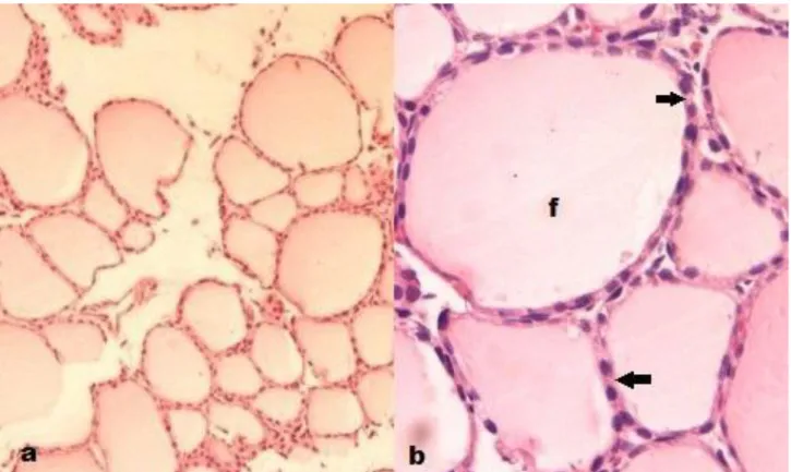
When the thyroid sections of the control group were examined, the general structure of the gland was seen to consist of units called follicles (Figure 1). The thyroid follicle lumen was full of colloid and the lumen was covered with single-layer epithelial cells.
Figure 1. a, b. Control group, thyroid follicles (x100, x400), f; follicle, arrow; follicle epithelium, H-E. There was a cytoplasmic vacuolization
showing significant cellular swelling in some regions of the thyroid follicle epithelium belonging to paracetamol groups. Besides the degenerative changes observed in epithelial cells, there were also deterioration in follicle epithelium continuity in some areas, epithelial cells or cell debris spilled into the follicle lumen, follicular-colloidal degeneration, and interfollicular haemorrhage (Figure 2).
Periodic acid Schiff staining results
PAS staining was performed to evaluate the colloid in the follicle. The PAS staining intensity of the intra-follicular colloid was weaker in the paracetamol groups compared to the control group (Figure 3, Figure 4). Statistical evaluation of scores determined according to staining intensity showed that staining difference was statistically significant (p<0.05).
314 Figure 2. Paracetamol groups, thyroid gland section. a,b. Group P7 (x100, x400), c,d. Group P14 (x100, x400), e,f.
Group P21 (x100, x400), g,h. Group P28 (x100, x400), red arrow; cytoplasmic vacuolization, black arrow; epithelium shedding and degeneration, white arrow; interfollicular haemorrhage, *; colloidal degeneration, H-E
315 Figure 3. a,b,c, Control group, thyroid gland. (x200, x400, x1000), f; follicle, *; follicle epithelium, PAS.
Morphometric results Bodyweight results
The bodyweights of the animals in all groups were evaluated both before and after the experiment (Table 1). There was a significant increase in the bodyweight of animals in the control group as a result of normal development (p<0.05). On the other hand, there was a decrease in the bodyweights of the animals in paracetamol groups. These decreases were found to be statistically significant in P7, P21 and P28 groups (p<0.05), but it was not statistically significant in the P14 group (p>0.05) (Table 2).
Morphometric results of the thyroid gland Evaluation of the data according to the results of one-way ANOVA
A significant difference was observed between the control group and P7, P14, P21, and P28 groups in terms of follicle epithelial height (p<0.05) (Table 3). When the staining intensity of colloid was evaluated, it was found to be weaker in the paracetamol groups than the control group and the results were statistically significant (p<0.05).
Evaluation of follicle diameter measurement according to post hoc Tukey test results
The histological appearance of follicular diameters were seen to be normal in the control group. The mean follicle diameter of the tissue samples belonging to the control group was measured to be 56.63±5.14. The mean follicle diameter of the tissue samples belonging to the P7 group was measured to be 44.51±4.39. The follicular diameter was found to be smaller in the P7 group compared to the control group, and P14, P21, and P28 groups. Statistical comparison of these groups showed that there was a significant difference between P7 and control groups in terms of mean follicle diameter. However, although there was no significant difference between P7 and other groups (P14, P21, P28) in terms of follicle diameter, there was a decrease in follicle diameter in the P7 group (p>0.05) (Table 1).
Evaluation of follicle epithelial height according to Post hoc Tukey test results
The mean follicle epithelial height of the thyroid tissue samples belonging to the control group was 2.19±0.20 (Table 1).
316 Figure 4. Paracetamol groups thyroid gland section a,b. Group P7 (x200, x400), c,d. Group P14 (x200, x400), e,f.
Group P21 (x200, x400), g,h. Group P28 (x200, x400), black arrow; vacuolization in the epithelium,*; colloidal vacuolization and degeneration, fd; follicular degeneration, PAS.
317 Comparison of the paracetamol groups and
control group showed that there was a significant increase in the mean follicle
epithelial height in the paracetamol groups (p<0.05).
Table 1. Morphometric results (The groups are different in follicle epithelial heights: P7 v C, p<0.01**; P14 v C,
p=0.01* ; P21 v C, p<0.01** ; P28 v C, p<0.05*; in follicle diameter: P7 v C, p<0.05*). Morphometric results N Follicle Diameter (±SD) Follicle Epithelial Height (±SD) Body Weight (g) Pre-experimental Post-Pre-experimental P7 5 44.51±4.39 3.20± 0.36 235.4 226.4 P14 5 48.80±9.94 2.82 ±0.25 236.6 233.6 P21 5 48.50±1.75 2.99±0.27 261.6 244.6 P28 5 49.88±5.36 2.80±0.14 238.2 227.2 CONTROL 5 56.63 ± 5.14 2.19 ± 0.20 226.4 232
Table 2. Comparison of body weights measured before and after the experiment according to Wilcoxon T test results
(statistical significance level was accepted as ** p<0.01, * p<0.05).
Test Statisticsa p7 post-experiment - p7 pre-experiment p14 post-experiment - - p14 pre-experiment p21 post-experiment - p21 pre-experiment p28 post-experiment - p28 pre-experiment control post-experiment - control pre-experiment Z -2.032 -1.095 -2.023 -2.032 -2.032
Asymp. Sig. (2-tailed)
p .042* .273 .043* .042* .042*
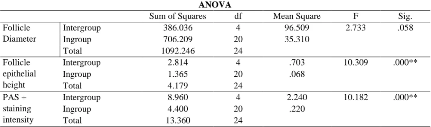
Table 3. Results of one-way analysis of variance in comparison of groups (statistical significance level was accepted as
** p<0.01, * p<0.05).
ANOVA
Sum of Squares df Mean Square F Sig.
Follicle Diameter Intergroup 386.036 4 96.509 2.733 .058 Ingroup 706.209 20 35.310 Total 1092.246 24 Follicle epithelial height Intergroup 2.814 4 .703 10.309 .000** Ingroup 1.365 20 .068 Total 4.179 24 PAS + staining intensity Intergroup 8.960 4 2.240 10.182 .000** Ingroup 4.400 20 .220 Total 13.360 24 Discussion
Paracetamol (acetaminophen) is one of the
most commonly used analgesic and
antipyretic drugs throughout the world. Its use has been reported to increase considerably in recent studies1. The widespread use of a drug
can cause intoxication and toxicity. Many studies have shown that high dose or frequent use of paracetamol lead to damage to various tissues, particularly in liver and kidney.10-12 This study investigated the effects of high doses of paracetamol on the thyroid gland depending on time.
In this study, the mean follicle diameters in thyroid glands of rats in all groups were measured. According to the data obtained, the follicle diameters were smaller in all paracetamol groups than in the control group, however, only the mean follicle diameter in the P7 group was significantly smaller than the other groups. Another important finding in the study was that the follicle epithelium height was higher in all paracetamol groups than in the control group. The highest increase in epithelial height was in the P7 group. In a study by İbrahim13 investigating
the effects of 500 mg/kg paracetamol on the thyroid gland, paracetamol at this dose has
318 been reported to cause an increase in follicle
cell height and a decrease in thyroid hormones and mean follicle diameter. The results of the present study are compatible with this study.
In other studies, paracetamol treatment has been reported to cause cyclic adenosine monophosphate (cAMP) inhibition, leading to a significant reduction in thyroid hormones. The decrease in the thyroid-stimulating hormone (TSH) levels is known to inhibit the stimulating effect of TSH on follicular cells in the thyroid gland. In this study, the increase in the length of follicular cells and the decrease in thyroid diameter in paracetamol-treated groups can be explained by this mechanism. In other words, it can be said that high doses of paracetamol cause a decrease in TSH level and this decrease inhibits the stimulating role of TSH on the follicular cell in the thyroid gland, eventually leading to an increase in the length of follicular cells and a decrease in thyroid diameter in the paracetamol-treated group.
Studies on the effects of paracetamol on the thyroid gland are limited, but many studies have shown that different agents such as medications14,15, stress16 heavy metals17 and pesticides18 have structural and functional effects on the thyroid gland. In the present study, colloidal degeneration of thyroid glands, epithelial cell deposition in the lumen, deformity of follicles, disturbances in the colloidal area, and follicle epithelium spills were observed in the thyroid glands of paracetamol groups.
We also evaluated the PAS staining intensity of colloid. Staining intensity was found to be weaker in the paracetamol groups than the control group and the results were statistically significant. It can be said that the amount of thyroglobulin in the structure of the glycoprotein in the colloid decreases in paracetamol groups. In a study by Gerard et al. involving old mice, active follicles have been reported to have a round core cubic or
cylindrical epithelium whereas
hypofunctional follicles are surrounded by squamous epithelial cells. They have shown that the lumen is filled with dark dense colloid in hypofunctional follicles whereas the
lumen is composed of colloid that are more clearly stained in the active follicles.19
One of the remarkable findings of the present study is that the bodyweight of animals in the control group showed a significant increase as a result of the normal development process, but there was a decrease in body weight in the paracetamol groups. This is one of the clinical signs of hyperthyroidism. Hyperthyroidism is a catabolic condition associated with increased energy expenditure20,21 increased lipolysis22,23 and increased protein turnover.24,25 These
metabolic effects lead to loss of body weight due to a decrease in both fat stores and lean body mass.26
As a result of the findings of the present study, paracetamol can be said to cause hyperactivity in the thyroid gland due to the decrease in body weight of female rats, an increase in the height of follicle epithelial cells and weaker staining of colloid in paracetamol groups.
Conclusion
These results of this study indicate that high doses of paracetamol can cause to toxic effects along with degeneration on thyroid gland depending on the duration of use but more detailed stereological and biochemical studies must be performed. The results presented in the literature are of great importance in terms of gaining awareness regarding the use of paracetamol that is widely used at the present time as it is an easy-to-access and cheap drug. Therefore, the society should be informed about the possible side effects of paracetamol to avoid over-the-counter use of paracetamol.
Furthermore, the results will contribute to the scientific literature on the effect of paracetamol on the thyroid gland and will be used as data in the studies to be performed in this regard. There is a need for further studies on the toxicity potential of paracetamol, which has not been fully understood yet.
Ethics Committee Aproval
The study was approved by the local ethics committee for animal experiments of Ondokuz Mayıs University (2014/23).
319
Author Contributions
The authors contributed equally to the study.
Conflict of Interest
The authors declared no conflict of interest.
Financial Disclosure
This work was supported by the project
numbered PYO.FEN.1904.15.001 of
Ondokuz Mayıs University Project
Management Office.
Statements
This study International Congress on 2nd International Congress of Forensic
Toxicology, 26-30 May 2016, at
Ankara/TURKEY has been presented as an poster presentation.
Peer-review
Externally peer-reviewed.
References
1. Ajith TA, Hema U, Aswathy MS. Zingiber officinale Roscoe prevents acetaminophen-induced acute hepatotoxicity by enhancing hepatic antioxidant status, Food Chem Toxicol. 2007; 45(11):2267-72.
2. Emet M, Yayla M. Asetaminofen (Parasetamol) Zehirlenmesi.
Turkiye Klinikleri J Emerg Med-Special Topics. 2016; 2.1:
51-7.
3. Küçükardalı ,Cinan, U, Acar HV, Özkan S, Top C, Nalbant S, Danacı M. Comparison of the Therapeutic Efficacy of 4Methylpyrazole and N-Acetylcysteine on Acetaminophen (Paracetamol) Hepatotoxicity in Rats. Current Medical
Research and Opinion. 2002; 18(2): 78-81.
4. Pacifici GM, Allegaert K. Clinical Pharmacology of Paracetamol in Neonates: A Review. Current Therapeutic
Research. 2015; 77: 24-30.
5. Tripathi D, Trilochana Y. Combined Hepatoprotective Effect of Leaves and Flowers of Bassia latifolia Roxb in Paracetamol Hepatotoxic Rats. Indian Journal of Research in Pharmacy and
Biotechnology (IJRPB). 2019; 7(4).
https://doi.org/10.31426/ijrpb. 2019.7.4.7411.
6. Udom GJ, Yemitan OK, Umoh EE, Mbagwu HO, Ukpe EE, Thomas PS. Hepatoprotective Properties of Ethanol Seed Extract of Citrus paradisi Macfad (Grape Fruit) Against Paracetamol-Induced Hepatotoxicity in Wistar Rats. Journal of
Herbal Drugs (An International Journal on Medicinal Herbs) 8(4), 219-225.
7. Fadda L, Ali HM, Aldrees GA, Alquraishi NM. Nano ubiquinone: Promising candidate for treatment of renal toxicity induced by over dose of paracetamol. Toxicology reports, 2019;
6, 712-717.
8. El-Maddawy ZK, El-Sayed YS. Comparative analysis of the protective effects of curcumin and N-acetyl cysteine against paracetamol-induced hepatic, renal, and testicular toxicity in Wistar rats. Environmental Science and Pollution Research, 2018; 25(4), 3468-3479.
9. Battal D. Postmortem rat serum ve dokularında parasetamol dağılımı ve stabilitesinin araştırılması, Doktora Tezi, Çukurova Üniversitesi, Sağlık Bilimleri Enstitüsü, Adli Tıp ABD, Adana, 2009.
10. Kolgazi M. Asetaminofen ile oluşturulan karaciğer hasarı patojenezinde nitrik oksit sentaz (nos) ve siklooksijenaz (cox)
sistemlerinin rolünün araştırılması, Doktora Tezi, Marmara Üniversitesi, Sağlık Bilimleri Enstitüsü, Fizyoloji Anabilim Dalı, İstanbul, 2013.
11. Majeed SK, Ramadhan MA, Monther W. Long-term toxicological effects of paracetamol in rats, Iraqi Journal of
Veterinary Sciences. 2013; 27 (1): 65-70.
12. Yılmaz I, Cetın A, Bılgıc Y. Hepatoprotective effects of apricot against acetaminophen induced acute hepatotoxicity in rats. Am
J Pharma Sci. 2015; 3.2: 44-48. DOI:10.12691/ajps-3-2-3
13. Ibrahim IR. Effect of Paracetamol and caffeine in structure and function of thyroidgland in male rats. Journal of College of
Education for Pure Science. 2011; 1(5), 89-102.
14. Ashour O.M., Sawan A.S. Celecoxib toxicity on rat’s thyroid gland, El-Mınıa Med. Bul. 2009; Vol. 20, No. 1.
15. Abeer S.M., Fayroz E.I.N. Effect of chlorpyrifos on thyroid gland of adult male albino rats, Egypt. J. Histol. 2010; Vol. 33,
No. 3: 441 – 450.
16. Gedikli S. Kronik stresin adrenal bez ve tiroid bezi üzerine etkilerinin morfometrik ve histokimyasal yöntemlerle sıçan modeli üzerinde araştırılması, Yüksek Lisans Tezi, Atatürk Üniversitesi, Sağlık Bilimleri Enstitüsü, Erzurum, 2010. 17. Karabulut-bulan Ö., Koyutürk M., Bolkent Ş., Yanardağ R.,
Tabakoğlu-oğuz A., Sıçan tiroid bezinde kadmiyum hasarına karşı c vitamini, e vitamini ve selenyumun kombine kullanımının etkileri, Cerrahpaşa Tıp Dergisi.2004; 35 (4). 18. Manal A.H., Marwa S. Lycopene reduces deltamethrin effects
induced thyroid toxicity and DNA damage in albino rats, The
Journal of Basic & Applied Zoology. 2013; 66; 155–163.
19. Gerard AC, Xhenseval V, Colin IM, Many MC, Denef JF. Evidence for coordinated changes between vascular endothelial growth factor and nitric oxide synthase III immunoreactivity, the functional status of the thyroid follicles, and the microvascular bed during chronic by low iodine and propylthiouracyl in old mice. Eur J Endocrinol. 2000; 142: 651660
20. Silva JE. Thyroid hormone control of thermogenesis and energy balance. Thyroid 1995;5:481–492.
21. Danforth E Jr, Burger A. The role of thyroid hormones in the control of energy expenditure. Clin Endocrinol Metab 1984;13:581–595.
22. Riis ALD, Gravholt CH, Djurhuus CB, Nørrelund H, Jørgensen JOL, Weeke J et al. Elevated regional lipolysis in hyperthyroidism. J Clin Endocrinol Metab. 2002;87:4747– 4753.
23. Muller MJ, Seitz HJ. Thyroid hormone action on intermediary metabolism. Part II: Lipid metabolism in hypo- and hyperthyroidism. Klin Wochenschr 1984;62:49–55.
24. Riis ALD, Jørgensen JOL, Ivarsen P, Frystyk J, Weeke J, & Møller, N. Increased protein turnover and proteolysis is an early and primary feature of shortterm experimental hyperthyroidism in healthy women. J Clin Endocrinol Metab 2008;93:3999–4005.
25. Muller MJ, Seitz HJ. Thyroid hormone action on intermediary metabolism. Part III. Protein metabolism in hyper- and hypothyroidism. Klin Wochenschr 1984;62:97–102.
26. Peterson, M. E., C. A. Castellano, and Mark Rishniw. "Evaluation of body weight, body condition, and muscle condition in cats with hyperthyroidism." Journal of veterinary