103 Research Article
An Application of Pulse Shape Discrimination Method to Obtain Pure
Photopeak
Elif Ebru ERMİŞ1*, Cüneyt ÇELİKTAŞ2
*Sorumlu yazar: [email protected]
1Izmir Tınaztepe University, Vocational School of Health Services, Medical Imaging Techniques Department,
IZMIR
Orcid No: 0000-0001-6309-5973 / [email protected]
2Ege University, Faculty of Science, Physics Department, IZMIR
Orcid No: 0000-0001-8608-066X / [email protected]
Abstract: A pulse shape discrimination setup was developed and the pulse shape discrimination method
was applied to γ-rays emitted from 137Cs radioisotope to test its discrimination performance. The setup was used
to discriminate the photopeak radiations from the non-photopeak ones arising from scattering, backscattering, background, low-energy X-rays and electronic noise etc. To perform this, pulse shape discrimination method which is a kind of timing process was used. The discrimination was performed in the region very close to the photopeak of the radioisotope. The developed setup showed its success in the discrimination of non-photopeak signals from the original photopeak ones.
Keywords: Gamma rays, pulse shape discrimination, NaI(Tl) scintillation detector, 137Cs
Net Fotopiki Elde Etmek İçin Puls Şekli Ayırma Yönteminin Bir
Uygulaması
Öz: Bu çalışmada, bir puls şekli ayırma devresi geliştirilmiştir ve bu devreyi test etmek için 137Cs
radyoizotopunun yayımladığı gamma ışınları üzerine puls şekli ayırma yöntemi uygulanmıştır. Bu devre, elektronik gürültü, düşük enerjili X-ışınları, tabi fon, geri saçılma ve saçılmalar nedeniyle ortaya çıkan diğer etkileri fotopik radyasyonlarından ayırmak için kullanılmıştır. Bunu gerçekleştirmek için bir çeşit zamanlama işlemi olan puls şekli ayırımı metodu kullanılmıştır. Ayırma işlemi radyoizotopun fotopikine yakın enerjideki bölgede gerçekleştirilmiştir. Geliştirilen devre, orijinal fotopik sinyallerinden fotopike ait olmayan sinyallerin ayırt edilmesinde başarılı olduğunu göstermiştir.
Anahtar Kelimeler: Gama ışınları, puls şekli ayırma yöntemi, NaI(Tl) sintilasyon dedektörü, 137Cs
1. Introduction
Sensing differences in the shape of output pulses from a radiation detector may provide valuable information on the radiation striking on the detector. The pulse-shape information can be used to identify the type of radiation, reduce the background events, discriminate between particles of
different range, discard defective pulses, and so on (Nakhostin, 2018).
An ideal spectrometer should have a big sensitivity to incident particles and gives a high detection efficiency and a good energy resolution in a wide energy range, low sensitivity to background and any noise components from the electronic devices in the system. As is known, additional counts
Ermiş and Çeliktaş, Ekim (2020) 46 (2): 103-109
104 due to environmental radiations such as background, scattering from detector surrounding material, backscattering from the radiation source etc. are always recorded in the energy spectra of any specific measurement. Since environmental radiations affect the original energy spectra of measurement particles, these radiations must be eliminated or isolated from the original energy spectra. To perform this firmly, the pulse shape discrimination (PSD) technique was applied to gamma ray spectrometer.
Background and noise, shortly unwanted signals are always observed in the energy spectra. These effects cause some difficulties in observing pure particle spectra and prevent seeking signals from particles, especially in low-energy regions. In addition, chance coincidences from cosmic rays are effective in the high-energy region, as well. To eliminate even these effects, for this reason, PSD technique was used.
PSD is one of the methods to increase the spectrum resolution, and the technique has wide range of application in radiation physics experiments (Pai et al., 1989; He et al., 1993; Jordanov et al., 1996; Knoll, 2010). It is the name given to a process that differentiates pulses produced by different types of particles in the same detector (Tsoulfanidis, 2015). To perform this process, the shapes of the signals of different particles in the detector are
analyzed. The shapes of the signals are characterized by a quantity named ‘rise time’. Rise time is the time between 10% and 90% of the amplitude on the rising edge of a signal. For this reason, PSD can be called as a kind of timing measurement process. PSD can be used in neutron-gamma discrimination (Senoville et al., 2020; Doucet et al., 2020; Yanagida et al., 2019), positron annihilation lifetime spectroscopy (Petschke and Staab, 2019), pulse pile-up effect reduction (Nakhoskin, 2020) and various electronics and detector technologies (Recker et al., 2020; Langeveld et al., 2020). Gridin et al. (2018) and Wahl et al. (2014) were studied on the energy resolution improvement of 137Cs gamma spectrum. To improve pulse shape discrimination performance of a pixelated plastic scintillator a digitization method was proposed by Cieslak et al. (2019). Xue et al. (2019) were examined the energy resolution performance of various digitizers on 662 keV-energy peak using different digital requirements. Energy improvement studies for 137Cs gamma spectrum were performed by Qin et al. (2018) using digital shaping filter.
The subject of the present work can be summarized in this way: pulse shape discrimination method was applied to gamma rays emitted from 137Cs radioisotope in a developed gamma spectrometer, which is composed of NaI(Tl) inorganic
105 scintillation detector and related electronic devices, in order to discriminate the photopeak radiations from the non-photopeak radiations. In this way, a different method to be increased the energy resolution performance of the spectrometer was proposed. Obtained experimental PSD spectrum was given.
2. Material and Methods
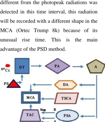
A PSD circuit is purposed to produce an output that reflects a variable property in the shape of input pulses. Fig. 1 shows the schematic block diagram of the experimental setup used in this work in this manner.
The experimental setup allows us to record the discriminated photopeak spectrum in multichannel analyzer (MCA). This is a kind of coincidence spectrum between energy signals and time signals of the photopeak of 137Cs. Gamma rays emitted from the radioisotope were detected by a Bicron 3"x3" NaI(Tl) scintillation detector which was placed inside a lead cover for passive background reduction. The detector signal was sent to a preamplifier (Ortec 113: PA) and then further amplified by another amplifier (Ortec 451: A). The bipolar output of the amplifier was split into two branches in order to gate the photopeak of 137Cs
radioisotope. The process was performed by a timing single channel analyzer (Ortec 420A: TSCA) and a delay amplifier (Ortec
427A: DA). The unipolar output of the amplifier was fed into a pulse shape analyzer (Ortec 552: PSA). This device generates two timing output signals. First signal is produced when the pulse has the amount of 10% of its maximum in the leading edge of the input signal. This signal is the ‘start’ signal for time to amplitude converter (Ortec 566: TAC). The second is generated when the pulse has the amount of 90% of its maximum in the leading edge of the input signal. This signal forms the ‘stop’ signal of TAC. The time between these signals is called rise time of the input signal, which is measured by TAC. If any radiation different from the photopeak radiations was detected in this time interval, this radiation will be recorded with a different shape in the MCA (Ortec Trump 8k) because of its unusual rise time. This is the main advantage of the PSD method.
Figure 1. Schematic diagram of the spectrometer:
DT: Detector, PS: Power supply, PA: Preamplifier, A: Amplifier, PSA: Pulse shape analyzer, TAC: Time to amplitude converter, TSCA: Timing single channel analyzer, DA: Delay amplifier, MCA: Multichannel analyzer.
Ermiş and Çeliktaş, Ekim (2020) 46 (2): 103-109
106 The radioactive source used in the measurement (5 µCi) is 137Cs radioisotope
which was embedded in a mylar disk of diameter of 25 mm and thickness of 5 mm.
3. Results and Discussion
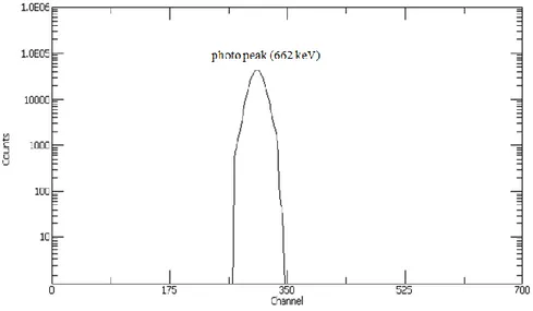
The spectra, which were acquired in a 300 s run time at room temperature of 291 K, were collected in the MCA card and then stored in a computer. The experimental pulse height spectrum of 137Cs is shown in Fig. 2.
Figure 2. Gamma ray pulse height spectrum of 137Cs (counts in log scale).
Fig. 3 illustrates the gated photopeak of 137Cs before the discrimination process.
Figure 3. Gated photopeak of 137Cs (counts in log scale).
Final PSD spectrum is also shown in Fig. 4. The discriminated radiations from the photopeak radiations can be seen on the left
edge of the photopeak. Since the pulses arrive with a constant time difference, one observes a single peak in the MCA. One
107 then switches the time delay in the start or stop channel by a known amount, and finds the peak shift on the MCA display (Lakowicz, 2006). This is the reason of the peak shift in Fig. 4.
The energy resolution of 5% was found for the isolated photopeak in the PSD spectrum in order to test the spectrometer performance.
A PSD setup was developed in order to obtain pure source spectrum, and the PSD method was applied to the photopeak of a γ radiation source (137Cs). For this reason, the
discrimination process was performed successfully in the present work.
Setting up the 10% and 90% of maximums on the leading edge of the input signal in the PSA was difficult in shape since the maximum values of leading edge of signals were changing at the rise time interval. For this reason, an approximate time interval (0.1 µs) was fixed. This is the reason why non-photopeak signals could not be separated (but discriminated) from the original photopeak signals in the final PSD spectrum.
Figure 4. PSD spectrum of the photopeak (counts in log scale).
The proposed detection system combines the timing signals with the energy signals simultaneously which it is different from the conventional ones. Thus, it is concluded from the experimental results that this method in this work will be helpful not only for obtaining pure source spectrum but
also for increasing the detector resolution with the further experiments. It is believed that the technique followed here can successfully be applied not only to gamma detection but also to alpha and beta particle measurements for obtaining pure spectra.
108 Consequently, theoretical spectra of the source particles interacting with a medium and the calculations of the reaction parameters will be more reliable with the experimental pure source spectra. In this connection, the data from the experimental setup presented here may contribute to improve the results obtained from the theoretical calculations. In addition, to analyze the different type of particles emitted from a radioactive source and to separate them from each other with high resolution,
and the determination of source activity can be possible through the presented setup here.
Acknowledgement
This work was supported by TUBITAK, the Scientific and Technological Research Council of TURKEY under project numbers 197T087, 104T379 and by EBILTEM, Center of Science and Technology, Ege University under Project No. 99 BIL 001.
References
Cieslak MJ, Gamage KAA, Glover R, Taylor CJ (2019). Pulse shape discrimination performance of a pixelated plastic scintillator (EJ-299-34) for a coded-aperture based dual particle imaging system. JINST 14: P07017.
Doucet E, Brown T, Chowdhury P, Lister CJ, Morse C, Bender PC, Rogers AM (2020). Machine learning n/ discrimination in CLYC scintillators. Nucl Instrum and Methods Phys Res A 954: 161201.
Gridin S, Onken DR, Williams RT, Swiderski L, Mianowska Z, Syntfeld-Kazuch A, Moszynski M, Gayshan V, vasiukov S, Gektin A (2018). Pulse shape analysis of individual gammaevents—Correlation to energy resolution and the possibility of its improvement. J Appl Phys 124: 154504.
He Z, Bird AJ, Ramsden D (1993). A ratio pulse-shape discriminator. Nucl Instrum and Methods A 336: 236–245.
Jordanov VT, Pantazis JA, Huber AC (1996). Compact circuit for pulse rise-time discrimination. Nucl Instrum and Methods A 380: 353–357.
Knoll GF (2010). Radiation Detection and Measurement, Wiley, New York.
Langeveld WGJ, Glenn AM, Sheets SA, Trellis DA, Zaitseva NP (2020). Comparison of pulse shape discrimination performance of stilbene and liquid scintillator under high count-rate active interrogation conditions. Nucl Instrum and Methods Phys Res A 954: 161204.
Lakowicz JR (2006). Principles of fluorescence spectroscopy, Springer.
Nakhoskin M (2020). A technique for the reduction of pulse pile-up effect in pulse-shape discrimination of organic scintillation detectors. Nucl Eng and Tech 52: 360–365. Nakhostin M (2018). Signal processing for radiation detectors, John Wiley and Sons Inc.,
109
discriminator. Nucl Instrum and Methods A 278: 749–754.
Qin ZJ, Chen C, Luo JS, Xie XH, Ge LQ, Wu QF (2018). A pulse-shape discrimination method for improving Gamma-ray spectrometry based on a new digital shaping filter. Radiat Phys Chem 145: 193–201.
Petschke D, Staab TEM (2019). A supervised machine learning approach using naive Gaussian Bayes classification for shape-sensitive detector pulse discrimination in positron annihilation lifetime spectroscopy. Nucl Instrum and Methods Phys Res A 947: 162742.
Recker MC, Cazalas EJ, McClory JW (2020). Pulse shape discrimination with a low-cost digitizer using commercial off-the-shelf components. Nucl Instrum and Methods Phys Res A 954: 161479.
Senoville M, Delaunay F, Parlog M, Achouri NL, Orr NA (2020). Neutron- discrimination with organic scintillators: Intrinsic pulse shape and light yield contributions. Nucl Instrum and Methods Phys Res A 971: 164080.
Tsoulfanidis N (2015). Measurement and Detection of Radiation, Taylor and Francis.
Wahl CG, Bernard EP, Lippincott WH, Nickel JA, Shin Y, McKinsey DN (2014). Pulse-shape discrimination and energy resolution of a liquid-argon scintillator with xenon doping. JINST 9: P06013.
Xue T, Zhu J, Wen J, Cang J, Zeng Z, Wei L, Jiang L, Liu Y, Li J (2020). Optimization of energy resolution and Pulse Shape Discrimination for a CLYC detector with integrated digitizers. JINST 15: P02018.
Yanagida T, Watanabe K, Okada G, Kawaguchi N (2019). Neutron and gamma-ray pulse shape discrimination of LiAlO2 and LiGaO2. Nucl Instrum and Methods Phys Res A