The expression of caspase-3, caspase-7, caspase-9
and cytokeratin AE1/AE3 in goats with enzootic nasal
adenocarcinoma: an immunohistochemical study
A. Aydogan, M. Haligur, O. Ozmen
Faculty of Veterinary Medicine, University of Mehmet Akif Ersoy, Burdur, Turkey
ABSTRACT: The aim of this study was to examine the expression of caspase-3, caspase-7, caspase-9 and cytokeratin
AE-1/AE-3 using the avidin-biotin complex (ABC) immunoperoxidase technique in 20 goats with enzootic nasal adenocarcinoma (ENA). Clinically, dyspnoea and nasal discharge were observed in all cases. Macroscopically, polypoid and sessile masses were seen in the ethmoidal area. At the histopathological examination, tubular, papil-lary and mixed patterns of ENA were diagnosed. Immunohistochemically, strong positive reactions were generally seen for caspase-3, while strong to moderate and slight reactions were observed for caspase-7 and caspase-9 in the cytoplasm of the tumour cells. Positive reactions for cytokeratin AE-1/AE-3 were only seen in epithelial cells. In addition, the causative agent of ENA, retrovirus, was detected immunohistochemically in tumour cells.
Keywords: enzootic nasal adenocarcinoma; caspase-3; caspase-7; caspase-9; cytokeratin AE-1/AE-3; goat
Enzootic nasal adenocarcinoma (ENA) is a chron-ic and contagious disease, characterised by neo-plastic proliferation of secretory epithelial cells in the ethmoidal region of the respiratory tract and it have been reported in sheep, goats, cattle, pigs, horses and buffaloes (Wilson and Dungworth 2002; De las Heras et al. 2003). Genetic, breed and sex predispositions were not reported in any published study (McKinnon et al. 1982; De las Heras et al. 1998). The causative agent of ENA is a simple ret-rovirus and it induces unilateral or bilateral neo-plastic growth in the mucosal nasal glands of the ethmoidal area (De las Heras et al. 1993, 2003).
Caspases are interleukin-1β-converting enzyme family proteases and play a vital role in the induc-tion of apoptosis. While some caspases are initia-tors (caspase-1, -2, -4, -5, -8, -9 and -10), others are effectors (caspase-3, -6 and -7) (Harvey and Kumar 1998; Stegh and Peter 2001). In mammals, apoptosis is mediated by the extrinsic pathway or intrinsic pathway. The extrinsic pathway is initiated by ligation of death receptors leading to activa-tion of caspase-8, while the intrinsic pathway is activated by cellular stress and leads to activation of caspase-9. Caspase-3 is a downstream effector
protease in the each of these pathways (Salvesen and Dixit 1997; Thornberry and Lazebnik 1998; Denault and Salvesen 2002). Overexpression and loss of expression of different caspases has been reported in some human tumours such as hepato-cellular carcinomas and prostate cancers (Winter et al. 2001; Persad et al. 2004; Yoo et al. 2010). In addition, caspase-3 activity indicative of apoptosis has been reported in tumour cells of enzootic na-sal adenocarcinoma (Ozmen et al. 2010). However, caspase expression is not fully characterised in animal tumors, especially in ENA. In this study, retrovirus antigen, caspase-3, caspase-7, caspase-9 expression, and cytokeratin AE-1/AE-3 reaction were immunohistochemically examined in twenty goats naturally affected with enzootic nasal adeno-carcinoma in Turkey.
MATERIAL AND METHODS
Tissue Samples and histopathology. Samples from ENA cases (12 male and eight female hair goats (Capra hircus)) were taken from the ar-chive of the Department of Pathology, Faculty of
Veterinary Medicine, University of Mehmet Akif Ersoy, Burdur, Turkey. The ages of the goats were between one and seven years. Macroscopically, uni-lateral (13 cases) and biuni-lateral (seven cases) tumour masses were found in the nasal cavity of the goats. For histopathological examination, tumour sam-ples were fixed in 10% neutral buffered formalin. After routine procedures, sections were stained with Haematoxylin-Eosin (H&E), and examined microscopically.
Immunohistochemistry. The routine streptavi-din biotin technique was used for the detection of Retrovirus antibody [Retrovirus, Idexx, Maine USA (ready for use)]; Caspase-3 [Neomarker, Caspase-3 (CPP32) Ab-4, Rabbit Polyclonal Antibody (1/100 dilution)]; Caspase-7 [Santa Cruz, Caspase-7, sc-8512, Goat Polyclonal Antibody (200 µg/ ml)]; Caspase-9 [Abcam, Anti-Caspase-9 Rabbit Polyclonal Antibody, ab52298, (1/100 dilution)]; Cytokeratin AE-1/AE-3 [Neomarker, Keratin, Pan Ab-1, Clone AE1/AE3, Mouse Monoclonal Antibody (1/100 dilution)] using immunohisto-chemistry. For the immunohistochemical staining, tumour sections were routinely processed accord-ing to the manufacturer’s recommendations. These sections were counterstained with Harris haema-toxylin. Non-tumour ethmoidal tissues provided from five healthy goats were used as control. To evaluate the degree of the immunohistochemical reaction of the tumour cells semiquantitative analy-sis was performed using an arbitrary visual scale with a grading score ranging from (–) to (+++) as follows: (–) = negative, (+) = weak staining, (++) = moderate staining, (+++) = strong staining. RESULTS
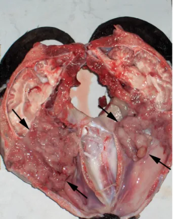
Clinically, respiratory distress, seromucous exu-dates around the nares and cachexia were seen in all of the effected goats. Macroscopically, tumour masses were found in the ethmoidal area of the nasal cavity (Figure 1). These masses were irregular in shape, soft to firm, polypoid (1–2.5 cm in length), and sessile (0.5–3 cm in diameter) and covered with seromucous exudate. The surface and cut surface of the masses were homogeneous and pinkish-white in colour. The gross and microscopic examination did not reveal metastasis in any organ.
Microscopically, enzootic nasal adenocarcino-ma was diagnosed according to the characteristic microscopic features. Case findings were similar
in all examined tumours. The tumour cells were arranged in tubular, papillary and mixed patterns (Figure 2). The tubular pattern was seen in nine cases; the papillary pattern was noted in 8 cases; mixed pattern was observed in three cases. The tumour masses consisted of uniform tumour cells and were well vascularised. Marked atypia in the tumour cells was seen but mitotic figures were not common. The tumour cells had a cuboidal shape Figure 1. Gross appearance of ENA. Tumour mass fills the nasal cavity (arrows)
Figure 2. Histopathological appearance of the mixed type of ENA. Haematoxylin and eosin; bar = 100 μ
with large round nuclei. Metastasis and invasion were not detected. In the tumour stroma, lympho-cytes, neutrophils, plasma cells and macrophage infiltrations were commonly seen. In some areas of the tumours, necrosis and fibrinopurulent exudate were also observed.
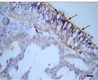
Immunohistochemically, strong positive reactions for retrovirus-related antigens were found especially on the surface of secretory epithelial cells of ethmoid region (Figure 3). Immunohistochemical staining for caspase-3 revealed strong (+++) staining in 16 cases (Figure 4), and moderate (++) staining in four cases in the cytoplasm of tumour cells. Immunostaining of caspase-7 gave a moderate signal (++) in sev-en cases (Figure 5) and was weak (+) in 13 cases.
Immunoreactivity of caspase-9 was strong (+++) in three cases, moderate (++) in 12 cases (Figure 6), and weak (+) in five cases in the cytoplasm of tumour cells. Immunostaining of cytokeratin AE-1/AE-3 was strong (+++) in eight cases and moderate in 12 cases in epithelial cells. Control sections showed negative or weak immunostaining confirming the specificity of the primary antibodies.
DISCUSSION
ENA arises from secretory epithelial cells in the ethmoid turbinate of the respiratory tract (De las Heras et al. 2003). Clinically, profuse seromucous Figure 3. Immunolabeling of retrovirus-related antigen
on the surface of secretory epithelial cells of the ethmoid region (arrows). Streptavidin biotin method. Harris haematoxylin counterstain; bar = 100 μ
Figure 4. Caspase-3 expression in the cytoplasm of tumour cells (arrows). Streptavidin biotin method. Harris haematoxylin counterstain; bar = 100 μ
Figure 5. Caspase-7 immunoreaction in the cytoplasm of tumour cells (arrows). Streptavidin biotin method. Harris haematoxylin counterstain; bar = 200 μ
Figure 6. Caspase-9 immunostaining in the cytoplasm of tumour cells (arrows). Streptavidin biotin method. Harris haematoxylin counterstain; bar = 200 μ
nasal exudates, dyspnoea and stertorous breath-ing have been noted in affected animals which lose weight and eventually die (De las Heras et al. 1991a). In this study, the clinical findings included respira-tory distress, seromucous exudates around the nares and cachexia. These findings are similar to those reported in the veterinary literature (De las Heras et al. 1991a; Svara et al. 2006; Ozmen et al. 2010). Histopathologically, ENA is classified as having pap-illary, mucinous, tubular and acinar patterns, but in goats carcinomas with papillary, tubular or acinar patterns are also interpreted to be well differenti-ated (Wilson and Dungworth 2002). In this study, histopathologically, ENA was classified as having tubular, papillary and mixed patterns. According to the histopathological findings, the tumour was classified as a low grade adenocarcinoma. No metas-tases were detected in regional lymph nodes, brain or other organs and tissues.
In recent years, the aetiology of ENA has been re-ported widely. According to these reports, viral-like particles, compatible with a type-D retrovirus, are the cause of ENA. For detection of the causative agent of ENA, different techniques such as electron micros-copy, immunohistochemistry, polymerase chain reac-tion (PCR), reverse transcriptase-PCR and western blotting have been used (De las Heras et al. 1991a,b, 1998, 2003; Cousens et al. 1996, 1999; Ozmen et al. 2010). In the present study, Retrovirus-related anti-gens were detected using the streptavidin-biotin com-plex peroxidase technique on the surface of secretory epithelial cells of the ethmoid region.
Cytokeratin is expressed in epithelial cells and constituted the cytoskeleton of these cells (Chu and Weiss 2002). In the present study, in all cases a posi-tive immunoreaction was observed for cytokeratin AE1/AE3 immunostaining. This indicates an epithe-lial origin of the tumour cells in ENA.
Apoptosis is mediated by a family of intracellular cysteine proteases named caspases (Ghavami et al. 2009). Caspases are normally present in healthy cells as inactive precursor enzymes (Taylor et al. 2008), and are divided into initiator caspases (caspase-1, -2, -4, -5, -8, -9 and -10) that are responsible for triggering caspase activation; and effector caspases (-3, -6, -7) that are responsible for the breakdown of the cell in the later stages of apoptosis (Harvey and Kumar 1998; Stegh and Peter 2001; Logue and Martin 2008). In mammals, there are two main pathways for the induction of apoptosis, extrinsic and intrinsic (Salvesen and Dixit 1997; Denault and Salvesen 2002). The extrinsic pathway is induced
by death ligands and death receptors such as Fas and results in activation of caspase-8. By contrast, the intrinsic pathway is regulated by the Bcl-2 fam-ily which causes an alteration in the mitochondrial membrane potential leading to mitochondrial mem-brane permeabilisation and downstream activation of caspase-9 (Reed 2000). A downstream effector of apoptosis in both pathways is caspase-3 (Salvesen and Dixit 1997; Denault and Salvesen 2002). Previous immunohistochemical studies on human cancers, like breast cancers and gastric adenocarcinomas, have shown that expression of caspass-3 and cas-pase-9 in tumour cells is higher than in normal cells (Yoo et al. 2002). According to a previous study, ex-pression of caspase-3 was found in more than 50% of tumour cells and caspase-9 in more than 10% of tumour cells in human anaplastic astrocytomas and glioblastomas (Bodey et al. 2004). Similarly, in stomach cancer both initiator and effector caspases are highly expressed (Yoo et al. 2002). In this study, strong immunostaining for caspase-3 was found in 16 cases and moderate immunostaining was seen in four cases. Caspase-9 expression was strong in three cases, moderate in 12 cases and weak in five cases. These findings indicate that apoptotic molecules of the mitochondria-dependent death pathway may play important roles in ENA.
Caspase-3 and caspase-7 are effector caspases and are highly homologous to each other (Degterev et al. 2003). However, in this study, the expression levels of caspase-3 and caspase-7 were not observed to be similar to each other. While caspase-3 gave strong immunoreactivity in 16 cases and moderate in four cases, immunoreactions of caspase-7 were moder-ate in seven cases and weak in 13 cases. Although caspase-3 and caspase-7 are highly homologous to each other, caspase-3 is more significant for apop-totic signaling in ENA compared to caspase-7.
In conclusion, to the best of our knowledge, this is the first immunohistochemical study to report the expression of caspase-7 and caspase-9 in goats with ENA.
REFERENCES
Bodey B, Bodey V, Siegel SE, Nasir A, Coppola D, Hakam A, Kaiser HE (2004): Immunocytochemical detection of members of the caspase cascade of apoptosis in high-grade astrocytomas. In Vivo 18, 593–602. Chu PG, Weiss LM (2002): Keratin expression in human
Cousens C, Minguijon E, Garcia M, Ferrer LM, Dalziel RG, Palmarini M, De las Heras M, Sharp JM (1996): PCR-based detection and partial characterization of a retrovirus associated with contagious intranasal tumors of sheep and goats. Journal of Virology 70, 7580–7583. Cousens C, Minguijon E, Dalziel RG, Ortın A, Garcia M,
Park J, Gonzalez L, Sharp JM, De las Heras M (1999): Complete sequence of enzootic nasal tumor virus, a retrovirus associated with transmissible intranasal tu-mors of sheep. Journal of Virology 73, 3986–3993. De las Heras M, Garcia de Jalon JA, Sharp JM (1991a):
Pathology of enzootic intranasal tumor in thirty-eight goats. Veterinary Pathology 28, 474–481.
De las Heras M, Sharp JM, Garcia de Jalon JA, Dewar P (1991b): Enzootic nasal tumour of goats; demonstra-tion of a type D-related retrovirus in nasal fluids and tumours. Journal of General Virology 72, 2533–2535. De las Heras M, Sharp JM, Ferrer LM, Garcia de Jalon
JA, Cebrian LM (1993): Evidence for a type D-like re-tovirus in enzootic nasal tumor of sheep. Veterinary Record 132, 441.
De las Heras M, Minguijon E, Ferrer LM, Ortin A, Dewar P, Cebrian LM, Pascual Z, Garcia L, Garcia de Jalon JA, Sharp JM (1998): Naturally occuring enzootic nasal tu-mor of sheep in Spain: pathology and associated retro-virus. European Journal of Veterinary Pathology 4, 11–16. De las Heras M, Ortin A, Cousens C, Minguijon E, Sharp
JM (2003): Enzootic nasal adenocarcinoma of sheep and goats. Current Topics in Microbiology and Im-munology 275, 201–223.
Degterev A, Boyce M, Yuan J (2003): A decade of cas-pases. Oncogene 22, 8543–8567.
Denault JB, Salvesen GS (2002): Caspases: keys in the ig-nition of cell death. Chemical Reviews 102, 4489–4500. Ghavami S, Hashemi M, Ande SR, Yeganeh B, Xiao W,
Eshraghi M, Bus CJ, Kadkhoda K, Wiechec E, Halayko AJ, Los M (2009): Apoptosis and cancer: mutations within caspase genes. Journal of Medical Genetics 46, 497–510.
Harvey NL, Kumar S (1998): The role of caspases in ap-optosis. Advances in biochemical engineering/bio-technology 62, 107–128.
Logue SE, Martin SJ (2008): Caspase activation cascades in apoptosis. Biochemical Society Transactions 36, 1–9.
McKinnon AO, Thorsen J, Hayes MA, Misener CR (1982): Enzootic nasal adenocarcinoma of sheep in Canada. Canadian Veterinary Journal 23, 88–94. Ozmen O, Sahinduran S, Haligur M, Demir N (2010):
Clinical, pathological, immunohistochemical and ul-trastructural observations on enzootic nasal adeno-carcinoma in five goats. Kafkas University, Faculty of Veterinary Medicine Journal 16, 633–639.
Persad R, Liu C, Wu TT, Houlihan PS, Hamilton SR, Diehl AM, Rashid A (2004): Overexpression of cas-pase-3 in hepatocellular carcinomas. Modern Pathol-ogy 17, 861–867.
Reed JC (2000): Mechanisms of apoptosis. American Journal of Pathology 39, 1415–1430.
Salvesen GS, Dixit VM (1997): Caspases: intracellular signaling by proteolysis. Cell 91, 443–446.
Stegh AH, Peter ME (2001): Apoptosis and caspases. Cardiology Clinics 19, 13–29.
Svara T, Gombac M, Vrecl M, Juntes P, Kostanjsek R, Pogacnik A, Pogacnik M (2006): Enzootic nasal adeno-carcinoma of sheep in Slovenia. Journal of Veterinary Medicine A 53, 26–29.
Taylor RC, Cullen SP, Martin SJ (2008): Apoptosis: con-trolled demolition at the cellular level. Nature reviews: Molecular Cell Biology 9, 231–241.
Thornberry NA, Lazebnik Y (1998): Caspases: enemies within. Science 281, 1312–1316.
Wilson DW, Dungworth DL (2002): Tumors of the res-piratory tract. In: Meuten DJ (ed.): Tumors in the Do-mestic Animals. 4th ed. Iowa State Press, Iowa. 374–375. Winter RN, Kramer A, Borkowski A, Kyprianou N
(2001): Loss of caspase-1 and caspase-3 protein ex-pression in human prostate cancer. Cancer Research 61, 1227–1232.
Yoo NJ, Kim HS, Kim SY, Park WS, Kim SH, Lee JY, Lee SH (2002): Stomach cancer highly expresses both ini-tiator and effector caspases; an immunohistochemical study. Acta Pathologica, Microbiologica et Immuno-logica Scandinavica 110, 825–832.
Yoo, NJ, Kim MS, Park SW, Seo SI, Song SY (2010): Ex-pression analysis of caspase-6, caspase-9 and BNIP3 in prostate cancer. Tumori 96, 138–142.
Received: 2012–07–01 Accepted after corrections: 2013–08–29
Corresponding Author:
Ahmet Aydogan, DVM, PhD, University of Mehmet Akif Ersoy, Faculty of Veterinary Medicine, Department of Pathology, Istiklal Yerleskesi, 15030, Burdur, Turkey