RESEARCH ARTICLE
28
THE INVESTIGATION OF THE ACTION MECHANISM OF AMMONIUM PYRROLIDINE DITHIOCARBAMATE ON RAT BLADDER SMOOTH MUSCLE
CONTRACTION-RELAXATION RESPONSES Hayri DAYIOĞLU1
, Ayhan YILMAZ2, Aysun ERDOĞAN3, Fatih ALAN4 and Sinan DARCAN5 1Kütahya Dumlupınar University, Faculty of Science and Literature, Department of Biology, 43270, Kütahya,
[email protected], ORCID: 0000-0002-9270-8561 2
Kütahya Dumlupınar University, Faculty of Science and Literature, Department of Biology, 43270, Kütahya,
[email protected], ORCID: 0000-0003-0410-8687
3Kartaltepe Mah. Tıp Fakültesi Cad. No: 211, 06620 Mamak, Ankara,aysn_erdoğ[email protected],
ORCID: 0000-0002-4738-8132
4General Directorate of Presidential Protection Services, Presidential Complex, 06530, Beştepe, Ankara,
[email protected],ORCID: 0000-0002-0561-6192
5Kütahya Health Science University, Evliya Çelebi Campus, Tavşanlı Yolu 10. Km, 43100, Kütahya,
[email protected], ORCID: 0000-0002-2135-4807
Received Date: 20.04.2020 Accepted Date: 04.11.2020
ABSTRACT
Ammonium pyrrolidine dithiocarbamate (PDTC), the specific inhibitor of NF-κB, is a thiol compound with anti-viral, anti-apoptotic, anti-inflammatory and antioxidant effects proven in many studies. There is not sufficient literature in what fashion that APDTC has produced these effects. We aimed to investigate the action mechanism of APDTC in rat bladder smooth muscle in our vitro study. In our study, 70 Wistar albino male rats were used. The rats were euthanized by cervical dislocation under the anesthesia and then abdomens were opened and bladders were isolated. After the removal of the connective tissues around the bladders, they were placed in the organ bath containing Krebs solution. KCl, ACh, APDTC, Atropine, Phentolamine, Propranolol, Nifedipine and TEA were applied to the bath with an appropriate protocol. The obtained data were evaluated with Kruskal Wallis and Mann-Whitney U tests. ACh induced bladder produced APDTC relaxation response in its own smooth muscle. Cholinergic receptor blocker Atropine, β-adrenergic receptor blocker Propranolol, L-type calcium channel blocker Nifedipine and atropine+phentolamine+propranolol mix have not changed the relaxation response that APDTC produced. In the presence of potassium channel blocker TEA, APDTC produced contraction response, but this was not a significantly important grade response. In the presence of α-adrenergic receptor blocker Phentolamine, APDTC produced significant contractile response. As a result, we think that APDTC shows its effect on rat bladder smooth muscle via α-adrenergic receptors and NANK system.
Keywords: adrenergic, APDTC, bladder, calcium channels, cholinergic, NANK system, potassium channels, TRP.
Dayıoğlu et all., Journal of Scientific Reports-A, Number 45, 28-49, December 2020.
29 1. INTRODUCTION
The bladder is a hollow, muscular organ that acts as a temporary reservoir for urine storage [1]. It is located inside the pelvic cavity, behind the symphysis pubis and under the peritoneum. In the male, it extends posteriorly to the rectum and in the woman, it contacts with the anterior walls of the uterus and vagina [2]. The main responsible system for bladder emptying is the parasympathetic system and acetylcholine is the main neurotransmitter in this duty [3]. Adenosine 5'-triphosphate (ATP) released in the bladder with acetyl choline (ACh) activates its specific P2x receptors. Pyrrolidine
dithiocarbamate, a thiol compound derived from dithiocarbamates, has generally been described as an antioxidant [4]. KCl-treated bladder smooth muscle contractile responses were decreased, and the mechanism of action of CP (yarrow) extract and compounds was shown to be through voltage-sensitive L-type Ca2+ channels [5]. Silodosin has an inhibitory effect on ACh-induced contractions in the bladder, and also demonstrated that palonosetron inhibits ACh-induced in vitro bladder smooth muscle contractions [6]. Raflumilast in pig bladder neck increases endogenous H2S production and
EFS relaxation, and facilitates increased neuronal cAMP, NO and H2S mediated bladder neck
inhibitory nerve conduction following FDE4 (phosphodiesterase 4) inhibition [7]. While hexamethonium (non-specific nAChR antagonist), mesamylamine (α3β4 nAChR antagonist), dihydro-β-erythroidin (α4β2 nAChR antagonist) inhibit nicotine-mediated increase of EFS-induced contraction responses, α-bungarotoxin (α7 nAChR antagonist) partially inhibited this effect of nicotine [8]. These findings have demonstrated that EFS-induced neurogenic contractions in the rabbit urinary bladder smooth muscle lanes are mediated by purinergic and cholinergic transmissions and contribute to the therapeutic effect of nicotine on EFS-induced contractile responses of the a4β2, a3β4, and a7 subtypes of nAChRs. Stimulation of muscarinic receptors in the rat bladder induces the release of urothelial NO that inhibits detrusor contraction in the case of cystitis [9]. As a result of the present findings, it is pointed out that a muscarinic receptor other than M1 or M3 is the main mediator of this effect. Darblade et al. (2006) showed that the phasic contractions in the human detrusor are due to calcium input via L-type calcium channels, and that the BKCa and SKCa channels play an important role in regulating the phasic contractility of human detrusor smooth muscle [10].
Muscarinic receptor 5 subtypes (M1-M5) have been found in the bladder of different species, including human [11]. Although the M3 receptor is considered the most important subtype functionally fit for detrusor contraction, it is thought to have a dual effect between the activation of M3 and M2 receptors [12]. Atropine is the prototype of the antagonist that competes with acetylcholine to bind to the muscarinic-type receptor [13]. Atropine competes with ACh and other muscarinic agonists for a common binding site on the muscarinic receptor [14]. Atropine is a mixture of D and L hyosamine [15]. Atropine is highly selective for muscarinic receptors. Atropine does not distinguish between M1, M2 and M3 subgroups of muscarinic receptors [16]. Atropine relaxes the bladder detrusor smooth muscles, increases the tone of the trigone and sphincter smooth muscles, thus creating voiding difficulties [17]. Adrenergic receptors are divided into alpha adrenergic receptors and beta adrenergic receptors. Alpha adrenergic receptors are of two types: alpha1 and alpha2 receptors. Beta adrenergic receptors are divided and called as beta1, beta2 and beta3 receptors [18].
Phentolamine is the imidazoline-derived α-adrenergic receptor blocker [17]. Propranolol has equal affinity for β1 and β2 adrenergic receptors; thus, it is a non-selective β adrenergic receptor antagonist [14]. Nifedipine is a prototype of the dihydropyridine family calcium channel blockers [16] and is a uncharged hydrophobic substance [13]. Several K+ channels with different molecular bases contribute to the regulatory base K+ conductance in smooth muscle cells. These; voltage-gated K+ channels (Kv),
Dayıoğlu et all., Journal of Scientific Reports-A, Number 45, 28-49, December 2020.
30
large conductivity Ca2+ activated K+ channels (BKCa), small conductivity Ca2+ activated K+ channels (SKCa), inward rectifier K+ channels (Kir), two porous area K+ channels (K2P), tension-dependent K+ channels and ATP sensitive K+ channels (KATP) [19]. Almost all potassium channels are blocked by externally supplied TEA (Tetraethyl Ammonium). However, sensitivity to TEA is different in different types of potassium channels [20]. Bladder contraction and relaxation are caused by adrenergic, cholinergic and non-adrenergic non-cholinergic (NANK) systems. NANK system and purinergic system are thought to be effective on bladder with neurotransmitters such as nitric oxide, transient receptor potential (TRP) channel, histamine, seratonin, VIP (vasoactive intestinal polypeptide), neuropeptide Y, substance P, calcitonin gene-associated peptide (CGRP), gamma amino butyric acid (GABA) and bradykinin [21]. Purinergic receptors in the cell membrane are divided into adenosine (P1) receptors and ATP receptors (P2X and P2Y receptors). The P1 and P2 receptors have various subtypes. Adenosine receptors and P2Y receptors mediate their responses through G proteins, while P2X receptors are a subfamily of ligand gated ion channels [14]. Nitric oxide (NO) is an unstable free radical gas that acts as a neurotransmitter and modulator in the central and peripheral nervous system [22]. Transient receptor potential (TRP) channels are class of ion channels characterized by variable activation mechanisms [23]. The TRP superfamily is one of the largest ion channel families and consists of several protein groups. In mammals, about 28 genes encode TRP ion channel subunits [24]. This class of ion channels known as TRPV (vaniloid), TRPC (canonical), TRPM (melastatin), TRPP (polysistin), TRPML (mucolipin) and TRPA (ankyrin) are six subtypes [25]. Pyrrolidine dithiocarbamate (PDTC) is a stable pyrrolidine derivative of dithiocarbamates [26]. Therefore, we aimed to investigate whether Ammonium Pyrrolidine Dithiocarbamate (APDTC), whose mechanism of action is not fully elucidated, blocks α-adrenergic and β-adrenergic receptors, cholinergic receptors, L-type calcium channels and potassium channels in bladder smooth muscle contraction and relaxation responses or these receptors and channels are effective in the mechanism. 2. MATERIALS AND METHODS
This study was carried out at Dumlupınar University Experimental Animal Breeding Application and Research Center and Dumlupınar University Faculty of Science and Arts Biology Department Physiology Laboratory. The study was approved by Kütahya Dumlupınar University Animal Experiments Local Ethics Committee (HADYEK) with the decision number of 2015.12.04. Animals used in the experiments obtained from Saki Yenilli Test Animals Production Center were 70 male Wistar albino rats weighing 300-350 gr and 6-7 months old. All the experimental groups and procedures are shown in Table 1, 2 and 3.
Table 1. Organ bath preparation for experimental procedures. Steps Procedure
1
2
3
Rats under standard conditions was kept in plastic cages with the ventilation (15 times/hour and 100% fresh air), at a humidity of 50±5%, at room temperature of 20±2°C for 12 hours night/12 hours daytime.
↓
Isolated organ bath was used to maintain the viability of smooth muscle strips prepared from rat bladder and to test their biological activity against active substances.
↓
Isolated organ bath consists of Data Acquisition analysis system (MP36, USA), Isometric transducers (Biopac, USA), Organ bath (Commat, Turkey), Water bath (WBC3044V3, May, Turkey), A double-chamber wells and O2-CO2 mixture tube.
Dayıoğlu et all., Journal of Scientific Reports-A, Number 45, 28-49, December 2020. 31 4 5 6 7 8 9 10 11 12
Four wells each with a capacity of 10 ml were used in the experiments. ↓
The wells are double chambers with distilled water circulation heated in the outer chambers of the water bath.
↓
The inner chamber contains Krebs solution. ↓
The strips prepared from the rat bladder are placed in the inner chamber and all experimental procedures are performed here.
↓
Krebs solution in the chamber were gassed with a mixture of 95% O2 and 5% CO2 gas at the bottom of the chamber during the experiment.
↓
The isometric transducer (Biopac, USA) transmits the responses of the bladder strips to the applied substance to the Data Acquisition analysis system (MP36, USA) by converting them into electrical signals.
↓
These recordings were analyzed at the end of the experiment and the resulting contraction relaxation responses before and after administration of the active substance in the bladder smooth muscles were determined.
↓
70 Wistar albino rats weighing 250-300 g and ages between 6-7 months were placed into cages in groups of 10.
↓
There was no restriction in the maintenance and feeding conditions of the rats during the experiments.
Table 2. Test groups with used chemicals. Group Addition
1 Ammonium Pyrrolidine Dithiocarbamate group (control)
2 Cholinergic receptor antagonist (atropine)+Ammonium Pyrrolidine Dithiocarbamate
3 α adrenoreceptor antagonist (phentolamine)+Ammonium Pyrrolidine Dithiocarbamate
4 β adreneseptor antagonist (proprimanol)+Ammonium Pyrrolidine Dithiocarbamate
5 L-type Ca2+ channel blocker (nifedipine)+Ammonium Pyrrolidine Dithiocarbamate
6 K+ channel blocker (tetraethylammonium)+Ammonium Pyrrolidine Dithiocarbamate
7 Both adrenergic and cholinegic receptor antagonist
(phentolamine+propranol+atropine)+Ammonium Pyrrolidine Dithiocarbamate
Dayıoğlu et all., Journal of Scientific Reports-A, Number 45, 28-49, December 2020.
32
Table 3. Experimental protocol for rat bladder smooth muscle contraction and relaxation response measurements.
Steps Experiment
1
2
3
4
5
6
7
8
9
10
11
12
13
Rats were euthanized by cervical dislocation method.
↓
The abdomen was opened from the median line and the bladder was removed and taken to the petri dish containing Krebs Henseleit solution.
↓
Connective tissues around the bladder were removed.
↓
The bladder was then opened with a longitudinal incision from the neck to the top.
↓
The bladder was sectioned 8 mm in length and 2 mm in width and bladder strips were prepared.
↓
The prepared strips were tied with ropes.
↓
The bladder smooth muscle strips were inserted into the organ bath having a temperature of 37°C and a supplied gas mixture of 95% O2 and 5% CO2 with Krebs solution by attaching to the transducer with steel hooks at the upper and lower ends.
↓
Bladder strips suspended in the isolated organ bath were stabilized for 45 minutes by washing with a Kreps solution every 15 minutes by applying 1 g of tension.
↓
After the equilibration period, viability of the organs was tested with 6×10⁻² M KCl.
↓
After waiting 5 minutes, KCl was removed from the organ by washing 3 times in a row with Krebs solution.
↓
After 10 minutes, 10-6 M Ach was given to the bath for pre-contraction.
↓
After 10 min, the groups have received as follows: 2.5×10⁻⁵ M APDTC to the control group, 10-6 M atropine to the 2nd group, 10-5 M phentolamine to the 3rd group, 10-6 M propranolol to the 4th group, 10-6 M nifedipine to the 5th group, 10-3 M tetraethylammonium to the 6th group and 10-5 M phentolamine+10-6 M propranolol+10 -6 M atropine to 7th group. Then, 2.5×10⁻⁵M APDTC was applied to all groups except control group after 15 minutes.
↓
The data obtained from the experiments were evaluated by Kruskal Wallis and Mann-Whitney U tests. p <0.05 values was considered statistically significant.
Bath recording lasted 1 hour 43 minutes. To compare, the phenylephrine and antagonist agent range was used, and then the antagonist PDTC range. The calculation was performed as follows. When phenylephrine is 100, % value was calculated by establishing the ratio of what would be the antagonist and what would be APDTC. Simultaneous experiments were carried out in four baths. SPSS program was used for statistical analysis. When statistical analysis was made in SPSS, both standard error and standard deviation were calculated and used in statistical processing. We drew the graphics according to our SPSS results in excel files. We also drew according to bladder
Dayıoğlu et all., Journal of Scientific Reports-A, Number 45, 28-49, December 2020.
33
phenylephrine values in the 100 file. We created p-p values according to this file. First we found the p-p values. Then we calculated how the drugs affected when the contraction was 100%. Then, we set the proportion to compare them with phenylephrine, such as what is the value of atropine when phenylephrine is 100. It also gave us statistical results if there were any significant results. The exact sig. or asymp. sig. from the mann-whitney test results made us decide whether the difference is meaningful or meaningless. The data obtained from the experiments were evaluated by applying mann-whitney u and kruskal wallis tests. p<0.05 values were considered statistically significant.
Figure 1. Rat bladder. 3. RESULTS
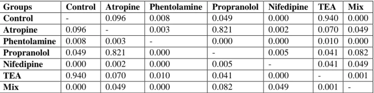
In our study, there are seven different groups as control, atropine, phentolamine, propranolol, nifedipine, tetraethyl ammonium and phentolamine+propranolol+atropine (Mix). Contraction-relaxation responses of 10-4 M ACh treated bladder smooth muscles against ammonium pyrrolidine dithiocarbamate in the presence and absence of antagonists or blockers were investigated in the groups (Table 4.).
Table 4. Comparison of APDTC between groups.
Groups Control Atropine Phentolamine Propranolol Nifedipine TEA Mix
Control - 0.096 0.008 0.049 0.000 0.940 0.000 Atropine 0.096 - 0.003 0.821 0.002 0.070 0.049 Phentolamine 0.008 0.003 - 0.000 0.000 0.010 0.000 Propranolol 0.049 0.821 0.000 - 0.005 0.041 0.082 Nifedipine 0.000 0.002 0.000 0.005 - 0.041 0.049 TEA 0.940 0.070 0.010 0.041 0.000 - 0.001 Mix 0.000 0.049 0.000 0.082 0.049 0.001 -
There was a statistically significant difference (p <0.05) on the ACh-induced contractions between the effect of atropine in the 10-6 M dose treated group and the groups of phentolamine, nifedipine and mix. It was found that there was a statistically significant difference (p <0.05) between the effect of phentolamine treated group with the 10-5 M dose and all other groups on the ACh-induced contractions. It was found that there was a statistically significant difference (p<0.05) between the effect of propranolol treated group with the 10-6 M dose and the control, phentolamine, nifedipine and
Dayıoğlu et all., Journal of Scientific Reports-A, Number 45, 28-49, December 2020.
34
TEA groups on the ACh-induced contractions. It was found that there was a statistically significant difference (p <0.05) between the effect of nifedipine treated group with the 10-6 M dose and all other groups on the ACh-induced contractions. There was a statistically significant difference (p <0.05) on the ACh-induced contractions between the effect of TEA in the 10-3 M dose treated group and the groups of phentolamine, propranolol, nifedipine and mix. It was found that there was a statistically significant difference (p <0.05) between the effect of Mix of known doses of the group administered simultaneously and the other groups except propranolol on the ACh-induced contractions (Figure 2.).
Figure 2. Effects of APDTC on bladder treated with 10-4 M Ach in the presence and absence of antagonist or blocker.
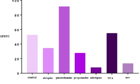
There was a statistically significant difference between ACh contracted bladder smooth muscle responses and treated APDTC in the presence of atropine. There was also a significant difference between antagonist atropine and APDTC (p <0.05). The bladder smooth muscle was relaxed with atropine and continued to relax when APDTC was administered. However, there was no statistically significant difference (p> 0.05) between the ACh-contracted APDTC-treated control group and the APDTC-treated group in the presence of atropine. Bladder smooth muscle contraction responses were not changed after the atropine treatment (Table 5. and Figure 3).
Table 5. Comparison of APDTC in the presence of atropine and atropine.
Atropine ACh Antagonist APDTC
ACh - 0.015 0.000
Antagonist 0.015 - 0.010
Dayıoğlu et all., Journal of Scientific Reports-A, Number 45, 28-49, December 2020.
35
Figure 3. The effect of APDTC in the presence of atropine and atropine on bladder smooth muscle contraction treated with ACh.
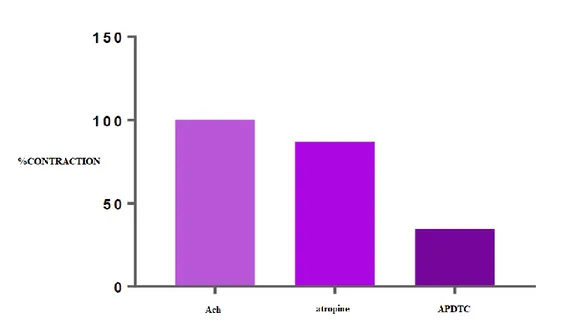
There was a statistically significant difference (p <0.05) between ACh-contracted bladder smooth muscle responses and APDTC in the presence of phentolamine. The bladder smooth muscle was relaxed with phentolamine and contracted when APDTC was administered in the presence of phentolamine. There was a statistically significant difference (p<0.05) between the ACh-contracted APDTC-treated control group and the APDTC-treated group in the presence of phentolamine. The antagonist phentolamine changed bladder smooth muscle contraction responses (Table 6 and Figure 4).
Table 6. Comparison of APDTC in the presence of phentolamine and phentolamine.
Phentolamine ACh Antagonist APDTC
ACh - 0.001 0.001
Antagonist 0.001 - 1.000
Dayıoğlu et all., Journal of Scientific Reports-A, Number 45, 28-49, December 2020.
36
Figure 4. The effect of APDTC in the presence of phentolamine and phentolamine on bladder smooth muscle contraction treated with ACh.
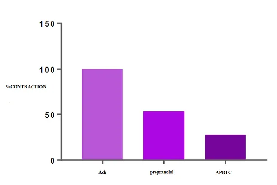
There was a statistically significant difference (p<0.05) between ACh-contracted bladder smooth muscle responses and APDTC treatment in the presence of propranolol. The bladder smooth muscle is relaxed with propranolol and continued to relax when APDTC was administered in the presence of propranolol. There was a statistically significant difference (p <0.05) between ACh-contracted and APDTC treated control group and APDTC treated group in the presence of propranolol. β- adrenoceptor antagonist propranolol changed bladder smooth muscle responses (Table 7 and Figure 5).
Table 7. Comparison of APDTC in the presence of Propranolol and Propranolol. Propranolol ACh Antagonist APDTC
ACh - 0.001 0.000
Antagonist 0.001 - 0.070
Dayıoğlu et all., Journal of Scientific Reports-A, Number 45, 28-49, December 2020.
37
Figure 5. The effect of APDTC in the presence of propranolol and propranolol on bladder smooth muscle contraction treated with ACh.
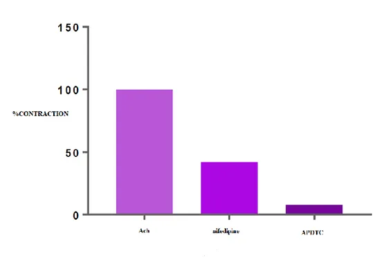
There was a statistically significant difference between ACh-contracted bladder smooth muscle responses and administered APDTC in the presence of nifedipine. There was a statistically significant difference (p<0.05) between APDTC applied in the presence of nifedipine and calcium channel blocker nifedipine. The bladder smooth muscle is relaxed with nifedipine and continued to relax when APDTC was administered. There was a statistically significant difference (p<0.05) between ACh-contracted APDTC-treated control group and APDTC-treated in the presence of nifedipine group. Bladder smooth muscle responses were altered by nifedipine administration (Table 8 and Figure 6). Table 8. Comparison of APDTC in the presence of nifedipine and nifedipine.
Nifedipine ACh Antagonist APDTC
ACh - 0.000 0.000
Antagonist 0.000 - 0.000
Dayıoğlu et all., Journal of Scientific Reports-A, Number 45, 28-49, December 2020.
38
Figure 6. Effect of APDTC in the presence of nifedipine and nifedipine on ACh treated bladder smooth muscle contraction.
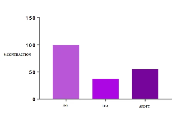
There was a statistically significant difference (p<0.05) between ACh-contracted bladder smooth muscle responses and APDTC applied in the presence of tetraethylammonium. The bladder smooth muscle was relaxed with tetraethyl ammonium and contracted when APDTC was administered in the presence of tetraethyl ammonium. There was no statistically significant difference (p>0.05) between ACh-contracted APDTC-treated control group and APDTC-treated group in the presence of tetraethylammonium. Potassium channel blocker tetraethylammonium did not alter bladder smooth muscle responses (Table 9 and Figure 7).
Table 9. Comparison of APDTC in the presence of TEA and TEA.
TEA ACh Antagonist APDTC
ACh - 0.000 0.001
Antagonist 0.000 - 0.290
Dayıoğlu et all., Journal of Scientific Reports-A, Number 45, 28-49, December 2020.
39
Figure 7. The effect of APDTC in the presence of tetraethyl ammonium and tetraethyl ammonium on bladder smooth muscle contraction treated with ACh.
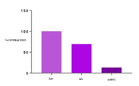
There is a statistically significant difference between ACh-contracted bladder smooth muscle responses and APDTC applied in the presence of a mix. There was also a statistically significant difference (p<0.05) between the mix of phentolamine+propranolol+atropine and APDTC applied in the presence of mix. Bladder smooth muscle was relaxed with mix and continued to relax when APDTC was applied in the presence of the mix. There was a statistically significant difference (p<0.05) between ACh-contracted APDTC treated control group and APDTC treated group in the presence of the mix. Administration of phentolamine+propranolol+atropine changed bladder smooth muscle responses (Table 10 and Figure 8).
Table 10. Comparison of APDTC in the presence of Mix and Mix.
Mix ACh Antagonist APDTC
ACh - 0.001 0.000
Antagonist 0.001 - 0.000
Dayıoğlu et all., Journal of Scientific Reports-A, Number 45, 28-49, December 2020.
40
Figure 8. The effect of APDTC in the presence of phentolamine+propranolol+atropine (mix) and phentolamine+propranolol+atropine (mix) on bladder smooth muscle contraction treated with ACh. 4. DISCUSSION AND CONCLUSION
Nuclear factor kappa B (NF-κB), a common transcription factor, regulates various genes encoding inflammatory mediators. NF-κB signaling is a classic signaling pathway that plays a role in the inflammatory response as well as regulation of cell proliferation and apoptosis [27]. Pyrrolidine dithiocarbamate (PDTC) is an antioxidant and has been shown to be effective in the treatment of various human diseases [28], although it inhibits activation of NF-κB [29]. APDTC affected the contractile-relaxation responses in bladder smooth muscle and showed that APDTC produces dose-dependent increased relaxation responses in ACh-contracted bladder smooth muscle [30]. Therefore, it is necessary to explain the anti-viral, anti-apoptotic, anti-inflammatory and antioxidant effects of APDTC compound seperately in following examples of studies. According to a study, the anti-viral effects of PDTC were reported as follows: Previously, it was shown that PDTC inhibits proteolytic polyprotein processing and replication of human rhinovirus by transporting metal ions into cells. It is shown that PDTC also inhibits replication of two other picornaviruses: coxsackievirus B3 (CVB3), a closely related virus that belongs to the genus Enterovirus, and mengovirus, an encephalomyocarditis virus strain that belongs to the genus Cardiovirus, and that this inhibition is due to the dithiocarbamate moiety of the compound. Making use of subgenomic replicons, evidence is provided that PDTC inhibits replication of these two viruses by disturbing viral RNA synthesis. Furthermore, it is shown that PDTC transports zinc ions into cells and that these zinc ions play an important role in the antiviral activity mediated by PDTC. Finally, it is shown that PDTC interferes with proteolytic processing of the polyproteins of both CVB3 and mengovirus, but that the underlying mechanism between these two viruses differs. In CVB3-infected cells, PDTC interferes strongly with the proteolytic activity of 3CD(pro), as shown by the impaired production of the mature capsid proteins as well as the autocleavage of 3CD(pro) into 3C(pro) and 3D(pol). In mengovirus-infected cells, however, PDTC
Dayıoğlu et all., Journal of Scientific Reports-A, Number 45, 28-49, December 2020.
41
had no effect on the proteolytic production of capsid proteins or the autocleavage of 3CD(pro). Instead, PDTC caused the accumulation of a high-molecular-mass precursor protein, due to an impairment in the primary 'break' that normally occurs at the 2A-2B junction. Thus, PDTC disturbs polyprotein processing and replication of two groups of picornaviruses, enteroviruses and cardioviruses, but the underlying mechanism is different [31]. According to an another study, the anti-apoptotic and anti-oxidant effects were reported as follows: Hypoxia induces vascular endothelial injuries; however, the mechanisms involved and effects of interventions remain unclear. Investigation was the inflammatory response and oxidative stress in co-cultured neutrophils and vascular endothelial cells, apoptotic changes in endothelial cells, and effects of the antioxidant, Tempol or the PDTC, upon endothelial cells under conditions of intermittent and/or continuous hypoxic exposure. Polymorphonuclear neutrophils co-cultured with human umbilical vein endothelial cells were subjected to the following conditions: intermittent normoxia (IN), intermittent hypoxia (IH), continuous hypoxia (CH), intermittent with continuous hypoxia (OS), OS+Tempol (OS+T), or OS+PDTC (OS+P) for 2, 5, or 8 h. Inflammatory factors, TNF-α and IL-6, the adhesion molecule, ICAM-1, CAT activity, and MDA concentrations in supernatants from the co-culture as well as pro-(Bak) and anti-(Bcl-xl) apoptotic gene expression levels in the endothelial cells were determined. Inflammatory factors, adhesion molecules, oxidative stress, and apoptosis genes in all groups showed significant, time-dependent increases as compared with the IN group. TNF-α, IL-6, ICAM-1, and MDA levels in the OS group were increased, while CAT was decreased as compared with that observed in the IH, CH, OS+T, and OS+P groups. Bcl-x1 expression and Bcl-x1/BAK ratios were decreased and BAX increased in the OS versus IH, CH, OS+T, or OS+P groups. Both pro- and anti-apoptotic proteins showed time-dependent increases, while the Bcl-x1/BAK ratio decreased over these times. Tempol and PDTC partially prevented these effects. Inflammation, oxidative stress, and apoptosis are all involved in vascular endothelial injury induced by OS. Anti-inflammatory and anti-oxidative interventions can partially improve effects of OS [32]. According also to an another study, the inflammatory effects were reported as follows: PDTC is a thiol compound that elicits anti-inflammatory effects by inhibiting NF-κB signaling. Study report that regulator of calcineurin activity 1 (RCAN1) expression is induced by PDTC treatment and that increased RCAN1 expression is dependent on the generation of reactive oxygen species (ROS) and activation of p38 MAPK and JNK signaling. It is also reported that the ability of PDTC to induce RCAN1 is mediated by activator protein-1 (AP-1)-dependent gene transcription, and identified a functional AP-1 binding site in the RCAN1 promoter by producing mutations and conducting chromatin immunoprecipitation (ChIP) analyses. Moreover, we show that the PDTC-mediated inhibitory effect on NF-κB signaling is significantly perturbed by knocking out RCAN1. The data provide the first evidence that PDTC prevents in vivo expression of pro-inflammatory cytokines by inducing RCAN1 expression [33].After all,since the mechanism of action of APDTC, which has been proven to relax bladder smooth muscle, has not been elucidated, we investigated the mechanism of action of APDTC in bladder smooth muscle in vitro.
In this study, seven different groups (control, atropine, phentolamine, propranolol, nifedipine, TEA and phentolamine+propranolol+atropine) were set to determine whether APDTC used some channels and receptors in rat bladder smooth muscle treated with ACh. 2.5×10⁻⁵ M APDTC produced a relaxation response in the bladder smooth muscle. According to the experimental results obtained from the atropine group, there was no significant difference in contraction responses between Ach treated rats with 2.5×10⁻⁵ M APDTC administration in the presence of 10-6 M atropine and 2.5×10⁻⁵ M
APDTC administration to the control group. In the presence of 10-6 M atropine, APDTC relaxed the bladder smooth muscle. Similarly, 10-6 M atropine has been shown to inhibit the Ach caused
Dayıoğlu et all., Journal of Scientific Reports-A, Number 45, 28-49, December 2020.
42
contraction of bladder rat bladder smooth muscle [34]. Admiministration of APDTC in the presence of atropine ensures that relaxation continues to increase significantly, indicating that 2.5×10⁻⁵ M doses
of APDTC do not use muscarinic-cholinergic pathways in the bladder smooth muscle. Whether M3 cholinergic receptor signal transduction pathway is involved in regulation of the activation of NF-κB and the expression of chemokine MOB-1, MCP-l genes in pancreatic acinar cells were investigated. Rat pancreatic acinar cells were isolated, cultured and treated with carbachol, atropine and PDTC in vitro. The MOB-1 and MCP-1 mRNA expression was detected by using RT-PCR. The activation of NF-κB was monitored by using electrophoretic mobility shift assay. The results showed that as compared with control group, M3 cholinergic receptor agonist (10-3 mol/L, 10-4 mol/L carbachol) could induce a concentration-dependent and time-dependent increase in the expression of MOB-1, MCP-1 mRNA in pancreatic acinar cells. After treatment with 10-3 mol/L carbachol for 2 h, the expression of MOB-1, MCP-1 mRNA was strongest. The activity of NF-κB in pancreatic acinar cells was significantly increased (p<0.01) after treated with M3 cholinergic receptor agonist (10-3 mol/L carbachol) in vitro for 30 min. Either M3 cholinergic receptor antagonist (10-5 mol/L atropine) or NF-κB inhibitor (10-2
mol/L PDTC) could obviously inhibit the activation of NF-κB and the chemokine MOB-1, MCP-1 mRNA expression induced by carbachol (p<0.05). This inhibitory effect was significantly increased by atropine plus PDTC (p<0.01). The results of these studies indicated that M3 cholinergic receptor signal transduction pathway was likely involved in regulation of the expression of chemokine MOB-1 and MCP-l genes in pancreatic acinar cells in vitro through the activation of NF-κB [35].
According to the results of the phentolamine group, the administration of 2.5×10⁻⁵ M dose of APDTC in the presence of 10-5 M phentolamine in the bath caused a statistically significant difference compared to 2.5×10⁻⁵
M APDTC administrated to the control group. In the presence of 10-5 M doses of phentolamine, APDTC increased bladder smooth muscle contractions. Reversed relaxation effect of phentolamine by APDTC was not significant. APDTC acted as antagonist against phentolamine. Phentolamine, a potent adrenergic receptor antagonist, blocks the adrenergic receptors to which phenylephrine binds to cause urethral smooth muscle contraction. When these receptors are blocked, phentolamine cannot trigger the expected contraction response and the contraction cannot be expected in the urethral smooth muscles [36]. The increase in contractions is not important, and so APDTC continues its relaxation effect. However, phentolamine has caused a decrease in the relaxation caused by APDTC, and therefore APDTC may be partially using α-adrenergic receptors. Epinephrine increased gene- and protein-expression of interleukin-6 (IL-6) and interleukin-11 (IL-11), which are capable of stimulating the development of osteoclasts from their hematopoietic precursors, in human osteoblast (SaM-1) and human osteosarcoma (SaOS-2, HOS, and MG-63) cell lines. An increase in IL-6 and IL-11 synthesis in response to epinephrine appeared to be a common feature in osteoblastic cells, but the magnitude of expression was different in these cell lines. In HOS cells treated with epinephrine, increases of IL-6 and IL-11 synthesis were inhibited by timolol (a beta-blocker), H-89 (N-[2-((p-bromocinnamyl)amino)ethyl]-5-isoquinolinesulfonamide; an inhibitor of protein kinase A (PKA)) and SB203580 [4-(4-fluorophenyl)-2-(4-methylsulfinylphenyl)-5-(4-pyridyl)1H-imidazole; an inhibitor of p38 mitogen-activated protein kinase (MAPK)], but not by phentolamine (an alpha-blocker), calphostin C [an inhibitor of protein kinase C (PKC)], or PD98059 (2'-amino-3'-methoxyflavone; an inhibitor of classic MAPK), suggesting a common pathway mediated by beta-adrenergic receptors in the PKA and p38 systems involved in the signal transduction of 6 and IL-11. Furthermore, expression of both genes was inhibited by curcumin [an inhibitor of activating protein-1 (AP-1) activation], but not by PDTC. The pharmacological study suggested that co-induction of the two genes in response to epinephrine occurred via activation of AP-1. The findings
Dayıoğlu et all., Journal of Scientific Reports-A, Number 45, 28-49, December 2020.
43
suggest that coinduction of IL-6 and IL-11 in response to epinephrine probably occurs via the PKA and p38 MAPK systems, leading to the transcriptional activation of AP-1 in human osteoblastic cells [37].
There was a statistically significant difference between 2.5×10⁻⁵ M APDTC in bladder smooth muscle in the presence of 10-6 M propranolol dose and 2.5×10⁻⁵ M APDTC applied to the control group. The dose of 10-6 M propranolol in the bladder smooth muscle treated with ACh produced relaxation. APDTC maintained its relaxation responses. APDTC and propranolol acted as agonists together, as relaxation continued to increase compared to control group results. Propranolol increases the amplitude of spinal reflex bladder contractions. It has been shown that α-1 and α-2 adrenergic receptors, possibly mediated by sympathetic efferent pathways in the hypogastric nerves or sympathetic chain, can suppress tonic contraction activation by affecting regions of the bladder smooth muscle [38]. Blocking of β-adrenergic receptors with propranolol increased the relaxation effect of APDTC. As a result, it cannot be said that APDTC exerts its effect through these receptors. Interaction of peroxisome proliferator-activated receptor γ coactivator-1α (PGC-1α) with other cellular signalling pathways plays an important role in training-induced mitochondrial adaptations. The study examined whether PDTC and propranolol would affect the PGC-1α-induced mitochondrial transcription factors, enzymes and proteins involved in energy metabolism and antioxidant defense in response to endurance training. Female Sprague-Dawley rats (aged 8 weeks) were randomly divided into two groups (n = 24), one subjected to 8 weeks of treadmill training and one remaining sedentary. Each group of rats was subdivided into three groups that were injected (i.p.) daily with PDTC (50 mg (kg body weight)-1), propranolol (30 mg kg-1) or saline as a control 1 h before the daily exercise session. Sedentary PDTC-treated rats showed 75% higher PGC-1α content (p<0.01) but lower mitochondrial transcription factor A and phosphorylated cAMP-responsive element binding protein (p-CREB) than control rats. Training increased PGC-1α by 57% (p<0.01), cytochrome c oxidase 4 by 30% (p<0.05) and p-CREB by 13% (p<0.05), whereas the mitochondrial mitofusin-2 level was decreased by 24% (p<0.01). Treatment with PDTC decreased PGC-1α and p-CREB content by 34 and 53% (p<0.05), respectively, in trained rats and abolished training effects on cytochrome c oxidase 4 and mitochondrial mitofusin-2. None of the training effects was abolished by propranolol treatment. Mitochondrial superoxide dismutase activity was decreased with PDTC, whereas training-induced glutathione peroxidase activity was unaltered by either drug. This indicates that nuclear factor-κB-inhibitory and antioxidant properties of PDTC can attenuate PGC-1α-mediated mitochondrial adaptation to endurance training, whereas the β-adrenergic pathway has little adverse effect [39]. There was a significant difference between 2.5×10⁻⁵ M APDTC in nifedipine 10-6 M dose group and the APDTC in ACh treated group. Nifedipine inhibited ACh contraction responses in the bladder, resulting in relaxation. L-type calcium channel blocker nifedipine was found to block 75-80% of contractile responses in isolated rat detrusor smooth muscle [40]. Nifedipine has been shown to reduce the number and amplitude of contractions on detrusor contractions and increase bladder capacity [41]. It is indicated that bladder Ca2+ in rats are collected from both intracellular and extracellular pools during both spontaneous and stimulated contractions. Nifedipine inhibits the tone and spontaneous activity of the isolated rat bladder [42]. The relaxation effect of nifedipine created continued with APDTC in statistically significant manner. Nifedipine increased the effect of APDTC even more, and blocking of the channels did not change this effect. We can say that blocking L-type calcium channels will increase the effect of APDTC. Ca2+/calmodulin-dependent protein kinase II (CaMKII) and NF-κB play crucial roles in pathogenesis of doxorubicin (DOX)-induced cardiomyopathy. Their activities are regulated by intracellular Ca2+. It is hypothesized that blockade of L-type Ca2+ channel (LTCC) could
Dayıoğlu et all., Journal of Scientific Reports-A, Number 45, 28-49, December 2020.
44
attenuate DOX-induced cardiomyopathy by regulating CaMKII and NF-κB. DOX activated CaMKII and NF-κB through their phosphorylation and increased cleaved caspase 3 in cardiomyocytes. Pharmacological blockade or gene knockdown of LTCC by nifedipine or small interfering RNA, respectively, suppressed DOX-induced phosphorylation of CaMKII and NF-κB and apoptosis in cardiomyocytes, accompanied by decreasing intracellular Ca2+ concentration. Autocamtide 2-related inhibitory peptide (AIP), a selective CaMKII inhibitor, inhibited DOX-induced phosphorylation of NF-κB and cardiomyocyte apoptosis. Inhibition of NF-κB activity by APDTC suppressed DOX-induced cardiomyocyte apoptosis. DOX-treatment (18 mg/kg via intravenous 3 injections over 1 week) increased phosphorylation of CaMKII and NF-κB in mouse hearts. Nifedipine (10 mg/kg/day) significantly suppressed DOX-induced phosphorylation of CaMKII and NF-κB and cardiomyocyte injury and apoptosis in mouse hearts. Moreover, it attenuated DOX-induced left ventricular dysfunction and dilatation. Our findings suggest that blockade of LTCC attenuates DOX-induced cardiomyocyte apoptosis via suppressing intracellular Ca2+ elevation and activation of CaMKII-NF-κB pathway. LTCC blockers might be potential therapeutic agents against DOX-induced cardiomyopathy [43].
There was no statistically significant difference between the study results of 2.5×10⁻⁵ M APDTC administered organ bath containing 10-3 M TEA and study results of the control group. The bladder was relaxed by TEA. APDTC reversed the effect of TEA and caused contraction. It has been shown that TEA is able to block the potassium channels of the inducible membranes that prolapse repolarization and the penetration of calcium ions [44]. TEA blocks the calcium-activated potassium channel, causing membrane depolarization and increased muscle contractions [45]. Potassium channel blocker TEA showed an inhibitory effect on bladder contraction. Contractions created with APDTC are not important enough to alter the relaxation response. It was suggested that the 2.5×10⁻⁵ M dose of APDTC was using different pathways other than potassium channels. These result is a new finding and there is no study so far in the literature.
To determine the effect of the NANK system on the mechanism of action of APDTC, 10-5 M phentolamine+10-6 M propranolol+10-6 M atropine (mix group) was applied to the bladder smooth muscle. 2.5×10⁻⁵ M APDTC was applied to the organ bath containing the mix and there was a significant difference between the control group. Mix mixture in bladder smooth muscle produced a relaxation response. APDTC continued to increase relaxation by creating agonistic effect with the mix group. The mix group had more relaxant effects than phentolamine, propranolol and atropine administered in separate groups on ACh-induced contractions. Increasing effect of APDTC as a result of the significant difference between the APDTC relaxant effect in the control group and the mix group showed that it did not use adrenergic and cholinergic routes.
In this study, we investigated the mechanism of action of APDTC with proven antioxidant and anti-inflammatory effects in bladder smooth muscle, and showed that APDTC created relaxation response in rat bladder smooth muscle induced by ACh. This response was altered by the α-adrenergic receptor antagonist phentolamine, suggesting that APDTC may be effective through the α-adrenergic receptor. On the other hand, the results suggested that the NANK system, which plays an important role in bladder smooth muscle contraction-relaxation, will help to clarify the APDTC mechanism hopefully in further studies.
Dayıoğlu et all., Journal of Scientific Reports-A, Number 45, 28-49, December 2020.
45 REFERENCES
[1] Martini, F. H., (2003), Fundamentals of Anatomy & Physiology, 6. Baskı, Yayımlayan Addison Wesley Longman, s. 1004.
[2] Hole, J.W., (1993), Human Anatomy and Physiology, 6. Baskı, William C. Brown Publisher, s. 770.
[3] Demirci, A., Canda, E.C., (2010), AĢırı Aktif Mesanenin Patofizyolojisi, Türk Üroloji Seminerleri, 1: 23-6.
[4] Tahata, S., Yuan, B., Kikuchi, H., Tagaki, N., Hirano, T., Toyoda, H., (2014), Cytotoxic effects of pyrrolidine dithiocarbamate in small-cell lung cancer cells, alone and in combination with cisplatin, International Journal of Oncology, 45(4): 1749-1759.
[5] Şengül, E., (2014), Achillea Millefolium (Civan perçemi) Ekstraktlarının ve Bazı Biyolojik Aktif Bileşiklerinin In Vitro Ortamda Rat Mesanesi Düz Kasları Üzerine Etkilerinin Araştırılması, Doktora Tezi, Atatürk Üniversitesi, Erzurum, s. 2, 101.
[6] Ege, H., (2016), Silodosin ve Palonosetronun İzole Sıçan Mesane Kasılmaları Üzerindeki Etkileri, Yüksek Lisans Tezi, Necmettin Erbakan Üniversitesi, Konya, s. 2, 42, 43, 44.
[7] Agis-Torres, A., Recio, P., López-Oliva, M.E., Martínez, M.P., Barahona, M.V., Benedito, S., Bustamante, S., Jiménez-Cidre, M.A., García-Sacristán, A., Prieto, D., Fernandes, V.S., Hernández, M., (2018), Phosphodiesterase type 4 inhibition enhances nitric oxide- and hydrogen sulfide-mediated bladder neck inhibitory neurotransmission, Scientific Reports, 8: 4711.
[8] Ozturk Fincan G.S., Vural I.M., Yildirim S.S., Isli F., Dilekoz E., Ercan S., Sarioglu Y., (2016), Role of nicotinic acetylcholine receptor subtypes on nicotine's enhancing effect on electrical field stimulation elicited contractile responses in rabbit urine bladder, European Review for Medical and Pharmacological Sciences, 20(8): 1636-1641.
[9] Andersson, M., Aronsson, P., Doufish, D., Lampert, A., Tobin, G., (2012), Muscarinic receptor subtypes involved in urothelium-derived relaxatory effects in the inflamed rat urinary bladder, Autonomic Neuroscience: Basic and Clinical, 170(1-2): 5-11.
[10] Darblade, B., Behr-Roussel, D., Oger, S., Hieble, J.P., Lebret, T., Gorny, D., Benoit, G., Alexandre, L., Giuliano, F., (2006), Effects of potassium channel modulators on human detrusor smooth muscle myogenic phasic contractile activity: potential therapeutic targets for overactive bladder, Urology, 68(2): 442-8.
[11] Giglio, D., Tobin, G., (2009), Muscarinic receptor subtypes in the lower urinary tract, Pharmacology, 83(5): 259-69.
[12] Hegde, S.S., Eglen, R.M., (1999), Muscarinic receptor subtypes modulating smooth muscle contractility in the urinary bladder, Life Sciences, 64(6-7): 419-28.
Dayıoğlu et all., Journal of Scientific Reports-A, Number 45, 28-49, December 2020.
46
[13] Lüllmann, H., Hein, L., Mohr, K., Bieger, D., (2005), Color Atlas of Pharmacology, 3. Baskı, Thieme Publishers New York, s. 6, 7, 54.
[14] Brunton, L., Chabner, B., Knollman, B., (2010), Goodman & Gilman’s The Pharmacological Basis of Therapeutics, 12. Baskı, McGraw-Hill Higher Education, s. 214-320.
[15] Altinkurt, O., (1981), Farmakoloji I, Ankara Üniversitesi Basımevi, Ankara, s. 35, 105.
[16] Katzung, B.G., Masters, S.B., Trevor, A.J., (2012), Basic & Clinical Pharmacology, 12.Baskı, McGraw-Hill Higher Education, s. 87, 152.
[17] Kayaalp, S.O., (2002), Rasyonel Tedavi Yönünden Tıbbi Farmakoloji, 10.Baskı, Hacettepe-Taş, Ankara, s. 1093-1138.
[18] Guyton A.C. and Hall, J.E., (2013), Tıbbi Fizyoloji (çev. Yeğen, B.), 12. Baskı, Nobel Tıp Kitabevleri, s. 91, 307, 308, 731, 773.
[19] Thorneloe, K.S. and Nelson, M.T., (2005), Ion channels in smooth muscle: regulators of intracellular calcium and contractility, Canadian Journal of Physiology and Pharmacology, 83: 215–242.
[20] Bisset, D. and Chung, S.H., (2008), Efficacy of external tetraethylammonium block of the KcsA potassium channel: molecular and Brownian dynamics studies, Biochimica et Biophysica Acta (BBA) - Biomembranes, 1778(10): 2273-82.
[21] Gür, S., (1998), Mesane’nin Non-adrenerjik Non-kolinerjik Kontrolü, Ankara Eczacılık Fakültesi Dergisi, 27(1): 51-60.
[22] Vesela, R., Aronsson, P., Andersson, M., Wsol, V., Tobin, G., (2012), The potential of non-adrenergic, non-cholinergic targets in the treatment of interstitial cystitis/painful bladder syndrome, Journal of Physiology and Pharmacology, 63(3): 209-16.
[23] Daly, D.M., 1, Collins, V.M., Chapple, C.R., Grundy, D., (2011), The afferent system and its role in lower urinary tract dysfunction, Current Opinion in Urology, 21: 268–274.
[24] Gohar, O., (2005), The Transient Receptor Potential (TRP) Ion Channels, Modulator, 20: 20-23. [25] Araki, I., Du, S., Kobayashi, H., Sawada, N., Mochizuki, T., Zakoji, H., Takeda, M., (2008),
Roles of mechanosensitive ion channels in bladder sensory transduction and overactive bladder, International Journal of Urology, 15(8): 681-687.
[26] Si, X., McManus, BM., Zhang, J., Yuan, J., Cheung, C., Esfandiarei, M., Suarez, A., Morgan, A., Luo, H., (2005), Pyrrolidine dithiocarbamate reduces coxsackievirus B3 replication through inhibition of the ubiquitin-proteasome pathway, Journal of Virology, 79(13): 8014–8023.
Dayıoğlu et all., Journal of Scientific Reports-A, Number 45, 28-49, December 2020.
47
[27] Xiang, N., Liu, J., Liao, Y., Huang, Y., Wu, Z., Bai, Z., Lin, X., Zhang, J., (2016), Abrogating ClC-3 Inhibits LPS-induced Inflammation via Blocking the TLR4/NF-κB Pathway, Scientific Reports, 6: 27583. 57
[28] Zhang, J., Jiang, W., Zuo, Z., (2014), Pyrrolidine dithiocarbamate attenuates surgery-induced neuroinflammation and cognitive dysfunction possibly via inhibition of nuclear factor kappaB, Neuroscience, 261: 1-10.
[29] Ivan, A.L., Campanini, M.Z., Martinez, R.M., Ferreira, V.S., Steffen, V.S., Vicentini, F.T., Vilela, F.M., Martins, F.S., Zarpelon, A.C., Cunha, T.M., Fonseca, M.J., Baracat, A.A., Georgetti, S.R., Verri, W.A.Jr., Casagrande, R., (2014), Pyrrolidine dithiocarbamate inhibits UVB-induced skin inflammation and oxidative stress in hairless mice and exhibits antioxidant activity in vitro, Journal of Photochemistry and Photobiology B: Biology, 138: 124-33.
[30] İnan, O. A., (2015), Sıçan Mesane’si Düz Kası Kasılma-GevĢeme Yanıtları Üzerine Ammonium Pyrrolidine Dithiocarbamate, SG-Benz, Caffeic Acid Phenil Ester, Atorvastatin Kalsiyum’ un Etkileri, Yüksek Lisans Tezi, Dumlupınar Üniveristesi Fen Bilimleri Enstitüsü, Kütahya, s. 39.
[31] K Lanke, BM Krenn, W J G Melchers, J Seipelt and FJM van Kuppeveld, (2007), PDTC inhibits picornavirus polyprotein processing and RNA replication by transporting zinc ions into cells, J Gen Virol. 88 (Pt 4): 1206-1217. doi: 10.1099 / vir.0.82634-0. PMID: 17374764 DOI: 10.1099 / vir.0.82634-0.
[32] Yanjie D., Qing Z., Laifang W., (2019), Pro-apoptotic and anti-inflammatory effects of araloside A on human rheumatoid arthritis fibroblast-like synoviocytes, Chem Biol Interact., 306:131-137. doi: 10.1016 / j.cbi.2019.04.025. PMID: 31004595 DOI: 10.1016 / j.cbi.2019.04.025.
[33] Eun H. L., Seon K. S., Su R. S., (2017), Pyrrolidine dithiocarbamate (PDTC) inhibits inflammatory signaling via expression of regulator of calcineurin activity 1 (RCAN1): Anti-inflammatory mechanism of PDTC through RCAN1 induction, Biochem Pharmacol.; 143:107-117. doi: 10.1016 / j.bcp.2017.07.011. PMID: 28712932 DOI: 10.1016 / j.bcp.2017.07.011 . [34] Han, J.S., Kim, S.J., Nam, Y., Lee, H.Y., Kim, G.M., Kim, D.M., Sohn, U.D., (2019), The
Inhibitory Mechanism on Acetylcholine-Induced Contraction of Bladder Smooth Muscle in the Streptozotocin-Induced Diabetic Rat, Biomolecules & Therapeutics, 27 (1): 101-106.
[35] Hai Z., Daoda C., Jinghui Z., Yuan T., (2004), Involvement of M3 cholinergic receptor signal transduction pathway in regulation of the expression of chemokine MOB-1, MCP-1 genes in pancreatic acinar cells, J Huazhong Univ Sci Technolog Med Sci.; 24 (2): 140-3, 157. doi: 10.1007 / BF02885413. PMID: 15315164 DOI: 10.1007 / BF02885413.
[36] Rembetski, B.E., Cobine, C.A., Drumm, B.T., (2018), Laboratory practical to study the differential innervation pathways of urinary tract smooth muscle, American Physiological Society, 1;42(2): 295-304.
Dayıoğlu et all., Journal of Scientific Reports-A, Number 45, 28-49, December 2020.
48
[37] A Kondo, M Mogi, Y Koshihara, A Togari., (2001), Signal transduction system for interleukin-6 and interleukin-11 synthesis stimulated by epinephrine in human osteoblasts and human osteogenic sarcoma cells, Biochem Pharmacol.; 61(3):319-26. doi: 10.1016/s0006-2952(00)00544-x. PMID: 11172736 DOI: 10.1016/s0006-10.1016/s0006-2952(00)00544-x.
[38] Rogers, M.J., Xiao, Z., Shen, B., Wang, J., Schwen, Z., Roppolo, J.R., de Groat, W.C., Tai, C., (2015), Propranolol, but not naloxone, enhances spinal reflex bladder activity and reduces pudendal inhibition in cats, American Physiological Society, 1;308(1): R42-9.
[39] Hong F., Chounghun K., Jonathan R. D., Ryan K., Iwalola A., Yong Z., Li L. J., (2013), Training-induced mitochondrial adaptation: role of peroxisome proliferator-activated receptor γ coactivator-1α, nuclear factor-κB and β-blockade, Exp Physiol.; 98 (3): 784-95. doi: 10.1113 / expphysiol.2012.069286. PMID: 23104933 DOI: 10.1113 / expphysiol.2012.069286.
[40] Zar, M.A., Iravani, M.M., Luheshi, G.N., (1990), Effect of nifedipine on the contractile responses of the isolated rat bladder, The Journal of Urology, 143(4): 835-9.
[41] Rud, T,. Andersson, K.E., Ulmsten, U., (1979), Effects of Nifedipine in Women with Unstable Bladders, Urologia Internationalis, 34: 421–429.
[42] Maggi, C.A., Manzini, S., Parlani, M., Conte, B., Giuliani, S., Meli, A., (1988), The effect of nifedipine on spontaneous, drug-induced and reflexly-activated contractions of the rat urinary bladder: evidence for the participation of an intracellular calcium store to micturition contraction, General Pharmacology: The Vascular System, 19(1): 73-81.
[43] Soichiro I., Shouji M., Kosuke O., Masataka I., Akihito I., Tomonori T., Nobuyuki E., Taishi Y., Masashi S., Hiroko D., Sachio M., Tomomi I., Hiroyuki T., (2019), Blockade of L-type Ca 2+ channel attenuates doxorubicin-induced cardiomyopathy via suppression of CaMKII-NF-κB pathway, Sci Rep.; 9(1):9850. doi: 10.1038 / s41598-019-46367-6. PMID: 31285514 PMCID: PMC6614470 DOI: 10.1038 / s41598-019-46367-6.
[44] Carpenter, F.G., (1978), Potentiation of nevre-induced bladder responses by tetraethylammonium in reletion to junctionaland extrajunctional muscarinic receptors, British journal of pharmacology, 64(3): 331-9.
[45] Oh, S.J., Ahn, S.C., (2003), Inhibitory effects of potassium channel blockers on carbachol induced contraction in rat detrusor muscle, Journal of Korean Medical Science, 18(5): 701-6.
Dayıoğlu et all., Journal of Scientific Reports-A, Number 45, 28-49, December 2020.
49
ATTACHMENTS