http://journals.tubitak.gov.tr/biology/ © TÜBİTAK
doi:10.3906/biy-1401-30
Bacterial toxin colicin N-T domain structure changes to ordered
state upon binding C-terminal domain of TolA
Yakup ULUSU1, Sema BİLGİN ŞENTÜRK2, Fatma GEDİKLİ2, Jeremy H. LAKEY3, İsa GÖKÇE4,*
1Department of Bioengineering, Faculty of Engineering, Karamanoğlu Mehmetbey University, Karaman, Turkey 2Department of Chemistry, Faculty of Art and Science, Gaziosmanpaşa University, Tokat, Turkey
3Institute for Cell and Molecular Biosciences, The Medical School, University of Newcastle, Newcastle upon Tyne, UK 4Department of Bioengineering, Faculty of Engineering, Gaziosmanpaşa University, Tokat, Turkey
1. Introduction
Colicins are plasmid-encoded bactericidal proteins produced by immune Escherichia coli that are active against sensitive E. coli and its closely related cells. Their toxic activities are of various types: some colicins form ion channels in the cytoplasmic membrane of sensitive cells, while others act as nucleases that degrade DNA or 16S RNA in the cytoplasm. One colicin, colicin M, inhibits the biosynthesis of murein (Lakey et al., 1994). Their toxic activities against target cells are known to occur in 3 distinct stages (Figure 1). First is receptor recognition and binding, where colicins bind to a specific receptor at the cell surface. Second is the translocation step, where colicins cross both the outer membrane and the periplasm to reach their cellular target (Lakey et al., 1994; Lazdunski, 1995). The final stage is the killing action, where colicins exert their lethal effects by forming a pore in the cytoplasmic membrane (Lazdunski, 1995), by DNAse or RNAse activity, or by inhibiting murein biosynthesis in the cytoplasm (Lakey et al., 1994).
Colicins have 3 linearly organized functional domains, each domain implicated in a specific stage of the colicin
activity. As shown in Figure 1, the central domain (R-domain) is responsible for the receptor-binding activity. The N-terminal domain (T-domain) is involved in translocation. The C-terminal domain (P-domain) carries out the lethal activity; this domain either forms a voltage-gated pore in the cytoplasmic membrane or digests nucleic acids in the cytoplasm (Raggett et al., 1998; Vetter et al., 1998; Papadokus et al., 2011).
Colicin N is a group A (Tol-dependent) pore-forming colicin whose target receptor is the E. coli outer membrane protein OmpF (Pugsley, 1987). It is composed of a largely unstructured T-domain linked by a glycine-rich sequence to a central R-domain containing a 6-stranded β-sheet structure. The R-domain is connected to the P-domain (a 10 α-helical structure) by means of a 65 Å α-helix (El-Kouhen et al., 1993; Gokce et al., 2000). While receptor binding and pore formation have been extensively studied, much remains unknown about the translocation step. Colicin N translocation requires 3 members of the Tol locus: TolA, TolQ, and TolR (Webster, 1991). TolQ and TolR are integral membrane proteins of the E. coli inner membrane and there is no evidence that they reach across
Abstract: Colicin N is a bacterial toxin that kills Escherichia coli and related cells. Its mode of action is of interest in protein import
and toxicology. Colicin N translocates across the E. coli outer membrane and periplasm by interacting with several receptors. The translocation process involves the interaction of the colicin N with the outer membrane porin OmpF and subsequently with the integral membrane protein TolA. The N-terminal domain of colicin N is involved in the import process. TolA consists of 3 domains. The N-terminal domain of colicin N interacts with the C-terminal domain of TolA at later stages of the translocation process. Our aim was to produce a large quantity of colicin N T-domains for spectroscopic and crystallization studies. These both require a correctly folded and stable protein. Here we present an expression of the complex between the N-terminal domain of colicin N and the C-terminal domain of TolA obtained by fusing these 2 domains with a flexible linker. Circular dichroism spectroscopy studies indicated that unstructured bacterial toxin colicin N T-domains changed to an ordered state upon binding to the C-terminal domain of TolA; this fusion protein has a secondary and tertiary structure.
Key words: Colicins, TolA, interaction, E. coli
Received: 08.01.2014 Accepted: 03.06.2014 Published Online: 05.09.2014 Printed: 30.09.2014 Research Article
the periplasm to the outer membrane. However, TolA is thought to span the periplasm by means of its extended central domain (TolA-II), which links the N-terminal TolA-1 domain in the cytoplasmic membrane to the C-terminal TolA-III domain (Levengood et al., 1991). The interaction between TolA-III and the N-terminal domain of colicin N is thought to be the principle event of translocation (Benedetti et al., 1991).
It has been shown that the 3-domain arrangement of bactericidal colicins is identical to that of the protein g3p of the filamentous phage. During infection of E. coli by this phage, TolA-III binds to the N-terminal domain of g3p protein (Holliger and Riechmann, 1997; Holliger et al., 1999). The structure of the g3p N-terminal domain is known (Holliger and Riechmann, 1997). Further NMR studies and the crystal structure of the g3p–TolA-III complex have shown that extensive conformational change in the N-terminal domain of g3p occurs on interaction with TolA-III (Lubkowski et al., 1999).
In contrast, there is very little information on the N-terminal domain of any colicin. The T-domain of colicin Ia was found to be largely unstructured. The crystal structure of colicin N lacked the N-terminal 90 residues. Further evidence of an unstructured colicin N T-domain came from structural studies of the isolated T-domain. Fluorescence studies of the isolated domain with TolA-III indicated that the T-domain acquired structure on interaction with TolA-III (Raggett, 1998; Raggett et al., 1998; Gokce et al., 2000; Gökçe and Lakey, 2003; Hecht et al., 2009). To investigate this further, a colicin N T-domain–TolA-III fusion was constructed. Its expression in E. coli and purification are presented in this paper. The structure of this complex will not only provide structural information on T-domain of colicins, but will also shed light on the colicin translocation process. By studying 1H–13C–15N NMR isotopically labeled T-domains interacting with unlabeled TolA-III (the C-terminal domain of TolA), Hecht et al. identified the TolA-binding epitope and showed that the extent of its disorder is reduced upon binding to TolA, although it does not fold into a globular structure with defined secondary structure elements. Residues upstream and downstream of the 27-residue TolA-binding epitope remain disordered in the TolA-bound T-domain, as they are in the free T-domain.
2. Materials and methods
2.1. Bacterial strains and plasmids
Shown in Figure 2, construction of pISA-TolA-III was completed in 2 stages. First, DNA encoding the TolA-III domain was amplified using 2 oligonucleotides (TOLAXHOSENSE: TTTTTCTCGAGCAACAATGGCGCATCAGGG and TOLABMLUREV: TTTTTGGATCCCCCACCCGG TTTGAAGTCCAATGGCGC) with an 18 bp matching sequence to TolA-III and plasmid pSK17 from Citrobacter freundii as a template. The pET8c vector was designed to introduce a 6-histidine and 2-serine linker at the N-terminus of this construct, which facilitates purification via nickel affinity chromatography (Politou et al., 1994; Sandallı et al., 2014). The DNA fragment encoding TolA-III and an extra BamHI restriction site was included to introduce the T-domain of colicin N into the pET8C vector using XhoI and MluI restriction sites. Second, DNA encoding for the colicin N T-domain was amplified using 2 oligonucleotides
(COLNBAMSENSE: TTTTTGGATCCGGTAGTAA TGG CGCAGATAAT and COLNMLUREV: TTTTTAC GCGTTATTA TCGATTTCCACTATTCCC)
with an 18 bp matching sequence to the colicin N T-domain. The pCol-N-T plasmid was used as a template (Raggett, 1998; Raggett et al., 1998). The PCR products were gel-purified and ligated into a purified, digested vector. The colicin N T-domain was introduced into a pISA TolA-III plasmid using BamHI and MluI restriction sites. This final construct (pISA-TolA-III-Col-N-T) was used initially to transform E. coli DH5α cells with ampicillin
N C
1 66 183 387
T R P
Colicin N domains
Figure 1. The domain structure of colicin N. These domains
deviate from the domain sizes determined by Pugsley (1987).
pIsaTolAColNT T O L A 3 C O L N T l a c O r i A m p T 7 XhoI BamHI MluI
Figure 2. Circular plasmid map of the pISA TolA-III–ColN-T
construct to produce TolA-III and colicin N-T domain fusion. XhoI, BamHI, and MluI restriction enzymes were used and their sites are shown. TOLA3: TolA-III domain gene, COLNT: colicin N-T domain gene, Amp: ampicillin resistance gene, and T7: promoter from the T7 phage.
selection. Successful transformants were selected on the basis of miniprep restriction digest analysis or analytical PCR, and a subsequent plasmid preparation was used for DNA sequencing and to transform E. coli BL21 (DE3) cells. DNA sequencing of this plasmid showed that both colicin N T-domains and TolA-III were correctly inserted. A circular plasmid map of this construct is shown in Figure 2.
2.2. Protein purification
E. coli BL21 (DE3) cells were transformed with pISA-TolA-III-Col-N-T and grown in 3 mL O/N cultures of LB medium containing ampicillin (200 µg/mL). A 3 mL O/N culture was introduced into 500 mL of LB containing ampicillin (200 µg/mL) and grown at 37 °C. The cells were induced at OD600 0.8–0.9 with isopropyl β-D-thiogalactopyranoside (1 mM) and grown for a further 3 h. The cells were harvested and resuspended in 20 mM phosphate and 300 mM NaCl (pH 7.4) containing RNAse (20 µg/mL) and DNAse (20 µg/mL). The cells were lysed by French press and the supernatant was obtained by ultracentrifugation at 40,000 rpm (45 Ti rotor used at 4 °C) for 1 h. The fusion protein was found to be soluble, with none remaining in the cell membrane pellet. The N-terminal 6X histidine tag facilitated purification of the fusion by means of a QIAGEN Ni-NTA affinity column (Hochuli et al., 1987; Chen et al., 2014; Sandallı et al., 2014). The fusion protein was washed onto the column with a 50 mM phosphate and 300 mM NaCl (pH 8.0) buffer, additionally washed with the same buffer containing 30 mM imidazole, and eluted in 300 mM imidazole (pH 7.0). The fusion protein was analyzed for purity by SDS-PAGE (Figure 3). Protein concentration was determined by UV absorption at 280 nm.
2.3. Circular dichroism spectroscopy
Far-UV circular dichroism (CD) spectroscopy was carried out on a Jobin Yvon CD6 spectrophotometer. Measurements were made at 25 °C using a 0.01 cm path length circular cuvette (Hellma) and protein concentration of 1 mg/mL. The protein was in a 50 mM phosphate and 300 mM NaCl (pH 7.4) buffer; this was also used for baseline subtraction. The CD signal measured was mean residue ∆ɛ (M–1 cm–1).
3. Results and discussion
Details on the construction of pISA-TolA-III-Col-N-T plasmid are given in Figure 2 and Section 2. The pET8c vector was used to clone the genes encoding the colicin N T-domain and TolA-III peptide fragments. This vector is under the tight control of the T7 promoter that facilitates the expression of toxic proteins (Studier and Moffat, 1986; Politou et al., 1994; Yike et al., 1996; Chen et al., 2014). Our construct also included an N-terminal polyhistidine tag (6 histidines) for simple and efficient protein purification. DNA sequencing of this constructed plasmid (Figure 2)
confirmed that the desired inserts were successfully cloned into the pET8C expression vector and also that the fusion constructs were in the correct reading frame.
The fusion protein was purified from the E. coli cell lysate as described in Section 2. Ni-NTA agarose resin was used to purify the fusion protein from the supernatant of E. coli cell lysate after ultracentrifugation. Proteins containing the histidine affinity tag were able to bind to the Ni-NTA agarose resin with a greater affinity than other E. coli proteins. Proteins that were not specifically bound were washed through the column without affecting the binding of His-tagged fusion protein. Elution of the fusion protein from the column was achieved by imidazole, which competes with the His tag for interaction with the Ni-NTA resin.
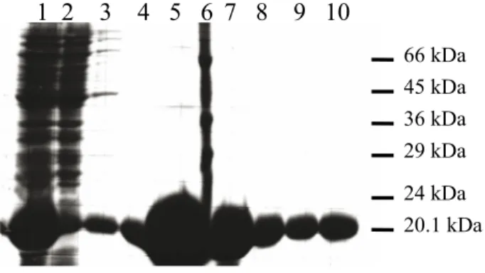
Eluted samples were analyzed on SDS-PAGE and showed a strong protein with a molecular weight of around 20.1 kDa. The calculated molecular weight of the fusion protein using the ProtParam tool was 19,505.1 kDa, which is very close to the experimental molecular weight. The extinction coefficient fusion at 280 nm is 17,780 and its theoretical pI is 9.2 (http://expasy.proteome.org.au/cgi-bin/protparam). The protein yields were calculated using UV absorbance at 280 nm and the extinction coefficient was 17,780 as taken from the ProtParam tool. One liter of BL21 (DE3) E. coli cell culture gave more than 100 mg (110–120 mg) of highly pure fusion protein.
The event central to the translocation process is the interaction of the colicin T-domain with TolA-III. Studies of colicin have shown that translocation is accelerated in urea, indicating that colicins are likely to unfold to expose the T-domain, allowing interaction with TolA
1 2 3 4 5 6 7 8 9 10
66 kDa 45 kDa 36 kDa 29 kDa 24 kDa 20.1 kDa Figure 3. TolA-III–colicin N-T domain fusion expression in E. coli BL21 (DE3). 1: Supernatant after ultracentrifugation (beforeNi-NTA resin). 2: Flow through Ni-NTA resin. 3: Wash with 20 mM imidazole, 50 mM phosphate, and 300 mM NaCl. 4: Elution fraction 1 in 300 mM imidazole, 50 mM phosphate, and 300 mM NaCl. 5: Elution fraction 2. 6: Molecular weight markers (66 kDa, 45 kDa, 36 kDa, 29 kDa, 24 kDa, 20.1 kDa). 7: Elution fraction 3. 8: Elution fraction 4. 9: Elution fraction 5. 10: Elution fraction 6.
(Benedetti et al., 1992). The structure of the T-domain of any colicin before and after interaction with TolA is still unknown. Despite obtaining the first crystals of residues 36–397 of colicin N (El-Kouhen et al., 1993), the final solved structure lacked N-terminal 90 residues and gave no structural information on the N-terminal domain structure of colicin N (residues 1–66). The only member of the colicin family whose full-length structure is known is colicin Ia (Wiener et al., 1997). While its pore-forming domain adopts a similar 10 α-helical fold to colicin N, its N-terminal domain appears largely unstructured. Recent structural studies of the colicin N T-domain substantiate the idea of an unfolded T-domain:
1) Its sequence scores poorly on most secondary structure prediction programs.
2) The CD spectrum of the isolated T-domain shows an unstructured domain.
3) Fluorescence studies of the purified domain shows aqueous exposure of T-domain tryptophans (Raggett, 1998; Papadokus et al., 2011).
Further CD studies with whole colicin N indicated that in the presence of the R- and P-domains, the T-domain may adopt a weak α-helical structure (Deprez et al., 2002) (Figure 4). Fluorescence studies showed that the addition of TolAII–III to the T-domain resulted in a burial of tryptophan, indicating that a conformational change in T-domain had occurred. It is possible that, similarly to colicin A, colicin N unfolds during translocation and releases the weakly structured T-domain, which acquires structure upon interaction with TolA. A substantial conformational change in phage g3p upon interaction with TolA was also observed (Holliger and Riechmann, 1997). Previous NMR studies on colicin N and the N1 domain of the filamentous phage g3p protein demonstrated that TolA-III changes its conformation upon binding to colicin N but retains its global fold (Riechmann and Holliger, 1997; Deprez et al., 2002, 2005; Hecht et al., 2009). The nature of these structural changes would be extremely useful in elucidating the events of translocation, and
therefore a construct of the T-domain TolA complex was constructed.
The fusion protein consists of 2 different proteins originating from different parent molecules, attached by a flexible glycine-rich linker. The construct we describe here (pISA-TolA-III–Col-N-T) permits the production of colicin N T-domain and TolA-III domain fusion under the tight control of the T7 promoter system. The construct was prepared in this manner to force both proteins to be present in the crystal at equimolar concentration and to achieve the formation of the complex. The addition of an extra histidine tag (6 His) was used for purification of this fusion protein by a single affinity chromatography step. The pET8c construct gives high yields of stable fusion protein and it is a critical step for the crystal structure studies. The secondary structure of colicin N-T–TolA-III fusion protein was determined by CD spectroscopy, which indicated an α-helix + β-sheet structure; therefore, the fusion is structured and further crystallographic work can proceed. Structural knowledge of this fusion will be useful for better understanding colicin N’s translocation through the cell envelope of gram-negative bacteria.
200 210 220 230 240 Wavelength/nm 2 1 0 –1 –2 Δε 250 Figure 4. Far-UV CD spectroscopy of the colicin N-T–TolA-III
fusion. Measurements were carried out in a 0.01 cm path length circular cuvette with a protein concentration of 1 mg/mL. The signal measured was mean residue ∆ɛ.
References
Benedetti H, Lazdunski C, Lloubes R (1991). Protein import into
Escherichia coli: colicins A and E1 interact with a component
of their translocation system. EMBO J 10: 1989–1995. Benedetti H, Lloubes R, Lazdunski, C, Letellier L (1992). Colicin A
unfolds during its translocation in Escherichia coli cells and spans the whole cell envelope when its pore has formed. EMBO J 11: 441–447.
Chen X, Huang Z, Zhou B, Wang H, Jia G, Qiao J (2014). Expression and purification of porcine Akirin2 in Escherichia coli. Turk J Biol 38: 339–345.
Deprez C, Blanchard L, Guerlesquin F, Gavioli M, Simorre JP, Lazdunski C (2002). Macromolecular import into Escherichia coli: the TolA C-terminal domain changes conformation when interacting with the colicin A toxin. Biochemistry 41: 2589– 2598.
Deprez C, Lloubes R, Gavioli M, Marion D, Guerlesquin F, Blanchard L (2005). Solution structure of the E. coli TolA C-terminal domain reveals conformational changes upon binding to the phage g3p N-terminal domain. J Mol Biol 346: 1047–1057. El-Kouhen R, Fierobe HP, Scianimanico S, Steiert M, Pattus F, Pages
JM (1993). Characterization of the receptor and translocator domains of colicin N. Eur J Biochem 214: 635–639.
Gokce I, Raggett EM, Hong Q, Virden R, Cooper A, Lakey JH (2000). The TolA-recognition site of colicin N. ITC, SPR and stopped-flow fluorescence define a crucial 27-residue segment. J Mol Biol 304: 621–632.
Gökçe İ, Lakey JH (2003). Production of an E. coli toxin protein; colicin A in E. coli using an inducible system. Turk J Chem 27: 323 –331.
Hecht O, Ridley H, Lakey JH, Moore GR (2009). A common interaction for the entry of colicin N and filamentous phage into Escherichia coli. J Mol Biol 388: 880–893.
Hochuli E, Dobeli H, Schacher AJ (1987). New metal chelate adsorbent selective for proteins and peptides containing neighbouring histidine residues. Chromatography 411: 177– 184.
Holliger P, Riechmann L (1997). A conserved infection pathway for filamentous bacteriophages is suggested by the structure of the membrane penetration domain of the minor coat protein g3p from phage fd. Structure 5: 265–275.
Holliger P, Riechmann L, Williams RL (1999). Crystal structure of the two N-terminal domains of g3p from filamentous phage fd at 1.9A: evidence for conformational lability. J Mol Biol 288: 649–657.
Lakey JH, Van der Goot FG, Pattus F (1994). All in the family: the toxic activity of colicins. Toxicology 87: 85–108.
Lazdunski C (1995). Colicin import and pore formation: a system for studying protein transport across membranes. Mol Microbiol 16: 1059–1066.
Levengood SK, Beyer WJ, Webster RE (1991). TolA: A membrane protein involved in colicin uptake contains an extended helical region. Proc Natl Acad Sci U S A 8: 5939–5943.
Lubkowski J, Hennecke F, Plückthun A, Wlodawer A (1999). Filamentous phage infection: crystal structure of g3p in complex with its coreceptor, the C-terminal domain of TolA. Structure 7: 711–722.
Papadokus G, Wojdyla JA, Kleanthous C (2011). Nuclease colicins and their immunity proteins. Quart Rev Biophy 16: 1–47.
Politou AS, Gautel M, Pfuhl M, Labeit S, Pastore A (1994). Immunoglobulin-type domains of titin: same fold, different stability. Biochemistry 33: 4730–4737.
Pugsley AP (1987). Nucleotide sequencing of the structural gene for colicin N reveals homology between the catalytic, C-terminal domains of colicins A and N. Mol Microbiol 1: 317–325.
Raggett EM (1998). Structure and function of translocation domain of colicin N. PhD, University of Newcastle upon Tyne, Newcastle upon Tyne, UK.
Raggett EM, Bainbridge G, Evans LJ, Cooper A, Lakey JH (1998). Discovery of critical Tol A-binding residues in the bactericidal toxin colicin N: a biophysical approach. Mol Microbiol 28: 1335–1343.
Riechmann L, Holliger P (1997). The C-terminal domain of TolA is the coreceptor for filamentous phage infection of E. coli. Cell 90: 351–360.
Sandallı C, Saral A, Ülker S, Karaoğlu H, Beldüz AO, Çiçek AÇ (2014). Cloning, expression, and characterization of a novel CTP synthase gene from Anoxybacillus gonensis G2. Turk J Biol 38: 111–117.
Studier FW, Moffatt BA (1986). Use of bacteriophage T7 RNA polymerase to direct selective high-level expression of cloned genes J Mol Biol 189: 113–130.
Vetter IR, Parker MW, Tucker AD, Lakey JH, Pattus F, Tsernoglou D (1998). Crystal structure of a colicin N fragment suggests a model for toxicity. Structure 6: 863–874.
Webster RE (1991). The tol gene products and the import of macromolecules into Escherichia coli. Mol Microbiol 5: 1005– 1011.
Wiener M, Freymann D, Ghosh P, Stroud RM (1997). Crystal structure of colicin Ia. Nature 385: 461–464.
Yike I, Zhang Y, Ye J, Dearborn DG (1996). Expression in Escherichia coli of cytoplasmic portions of the cystic fibrosis transmembrane conductance regulator: apparent bacterial toxicity of peptides containing R-Domain sequences. Prot Exp Purif 7: 45–50.