ILCs can be replenished and complemented, albeit only in part, through contributions of hema-togenously derived precursors or mature cells in situations of extended inflammation and tissue repair. Consistent with our findings, it has been reported that ILC subsets are elevated in the pe-ripheral blood of patients suffering from psoria-sis (30, 31). Furthermore, peripheral blood ILC2s have been shown to dynamically modulate the expression of molecules that regulate tissue hom-ing in mice and humans (20, 32). In addition, we have detected donor-derived lymphoid and ILC progenitors in parabiotic BM (fig. S8), raising the possibility that ILC progenitors can physi-ologically seed tissues not only during embryonic development (3) but also in adult mice. It remains to be determined whether this observation re-flects the physiologic migration of ILC progenitors or the engraftment of donor-derived hematopoietic stem cells (33, 34) giving rise to ILCs. Indepen-dent of these considerations, our data support a model in which ILCs are locally maintained and expanded as tissue-resident cells during homeo-stasis and acute infection. This“sedentary” lifestyle of ILCs in broadly differing microenvironments is consistent with the proposed roles of ILCs as sentinels and local keepers of tissue function.
R E FE R E N C ES A ND N OT ES
1. D. Artis, H. Spits, Nature 517, 293–301 (2015).
2. G. Eberl, M. Colonna, J. P. Di Santo, A. N. McKenzie, Science 348, aaa6566 (2015).
3. J. K. Bando, H. E. Liang, R. M. Locksley, Nat. Immunol. 16, 153–160 (2015).
4. E. Montaldo et al., Immunity 41, 988–1000 (2014). 5. M. G. Constantinides, B. D. McDonald, P. A. Verhoef,
A. Bendelac, Nature 508, 397–401 (2014). 6. C. S. Klose et al., Cell 157, 340–356 (2014). 7. R. A. Franklin et al., Science 344, 921–925 (2014). 8. P. Kamran et al., J. Vis. Exp. 2013, e50556 (2013). 9. H. Peng et al., J. Clin. Invest. 123, 1444–1456 (2013). 10. K. G. Anderson et al., Nat. Protoc. 9, 209–222 (2014). 11. M. H. Kim, E. J. Taparowsky, C. H. Kim, Immunity 43, 107–119
(2015).
12. C. C. Bain et al., Nat. Immunol. 15, 929–937 (2014). 13. E. C. Mackley et al., Nat. Commun. 6, 5862 (2015). 14. D. K. Sojka et al., eLife 3, e01659 (2014).
15. J. M. Kim, J. P. Rasmussen, A. Y. Rudensky, Nat. Immunol. 8, 191–197 (2007).
16. K. Liu et al., Science 324, 392–397 (2009).
17. M. Camberis, G. Le Gros, J. Urban Jr., Curr. Protoc. Immunol. (2003).
18. Y. Huang et al., Nat. Immunol. 16, 161–169 (2015). 19. B. J. Marsland, M. Kurrer, R. Reissmann, N. L. Harris, M. Kopf,
Eur. J. Immunol. 38, 479–488 (2008). 20. E. D. Tait Wojno et al., Mucosal Immunol. (2015). 21. J. E. Turner et al., J. Exp. Med. 210, 2951–2965 (2013). 22. G. Gasteiger, A. Y. Rudensky, Nat. Rev. Immunol. 14,
631–639 (2014).
23. G. Gasteiger, S. Hemmers, P. D. Bos, J. C. Sun, A. Y. Rudensky, J. Exp. Med. 210, 1179–1187 (2013).
24. B. Roediger et al., J. Allergy Clin. Immunol. (2015). 25. C. Wilhelm et al., Nat. Immunol. 12, 1071–1077 (2011). 26. A. B. Molofsky et al., Immunity 43, 161–174 (2015). 27. C. Vonarbourg et al., Immunity 33, 736–751 (2010). 28. J. H. Bernink et al., Immunity 43, 146–160 (2015). 29. S. A. van de Pavert et al., Nature 508, 123–127 (2014). 30. M. B. Teunissen et al., J. Invest. Dermatol. 134, 2351–2360
(2014).
31. F. Villanova et al., J. Invest. Dermatol. 134, 984–991 (2014).
32. J. M. Mjösberg et al., Nat. Immunol. 12, 1055–1062 (2011).
33. D. E. Wright, A. J. Wagers, A. P. Gulati, F. L. Johnson, I. L. Weissman, Science 294, 1933–1936 (2001). 34. J. L. Abkowitz, A. E. Robinson, S. Kale, M. W. Long, J. Chen,
Blood 102, 1249–1253 (2003).
AC K N OW L E D GM E N TS
We thank R. Franklin, S. Dadi, M. Li, and A. Chaudhry for help with parabiosis or cell isolations; D. Artis, L. Monticelli, and B. Hoyos for providing and maintaining worms; K. Wu and A. Bravo for general laboratory support; and T. O’Sullivan, J. Sun, A. Diefenbach, W. Kastenmüller, and members of the Rudensky and Gasteiger laboratories for critical discussions. Data from this study are tabulated in the main paper or in the supplementary materials. This work was supported by an Irvington Fellowship from the Cancer Research Institute (G.G.), NIH Medical Scientist Training Program grant T32GM07739 to the Weill Cornell/Rockefeller/Sloan-Kettering Tri-Institutional MD-PhD Program (X.F.), Cancer Center Support Grant P30CA008748 from the NIH National Cancer Institute, NIH grant R37AI034206 (A.Y.R.), the Ludwig Center at Memorial Sloan Kettering
Cancer Center, and the Hilton-Ludwig Cancer Prevention Initiative (Conrad N. Hilton Foundation and Ludwig Cancer Research) (A.Y.R.). G.G. is an investigator with the Deutsche Forschungsgemeinschaft Emmy Noether Programme, and A.Y.R is an investigator with the Howard Hughes Medical Institute.
SUPPLEMENTARY MATERIALS
www.sciencemag.org/content/350/6263/981/suppl/DC1 Materials and Methods
Figs. S1 to S8
5 July 2015; accepted 2 October 2015 Published online 15 October 2015 10.1126/science.aac9593
CANCER IMMUNOLOGY
Patrolling monocytes control tumor
metastasis to the lung
Richard N. Hanna,1* Caglar Cekic,2
Duygu Sag,3Robert Tacke,1Graham D. Thomas,1 Heba Nowyhed,1Erica Herrley,1Nicole Rasquinha,1Sara McArdle,4Runpei Wu,1
Esther Peluso,1Daniel Metzger,5Hiroshi Ichinose,6Iftach Shaked,1
Grzegorz Chodaczek,4Subhra K. Biswas,7Catherine C. Hedrick1*
The immune system plays an important role in regulating tumor growth and metastasis. Classical monocytes promote tumorigenesis and cancer metastasis, but how nonclassical “patrolling” monocytes (PMo) interact with tumors is unknown. Here we show that PMo are enriched in the microvasculature of the lung and reduce tumor metastasis to lung in multiple mouse metastatic tumor models. Nr4a1-deficient mice, which specifically lack PMo, showed increased lung metastasis in vivo. Transfer of Nr4a1-proficient PMo into Nr4a1-deficient mice prevented tumor invasion in the lung. PMo established early interactions with metastasizing tumor cells, scavenged tumor material from the lung vasculature, and promoted natural killer cell recruitment and activation. Thus, PMo contribute to cancer immunosurveillance and may be targets for cancer immunotherapy.
M
onocytes and monocyte-derivedmac-rophages play key roles in tumor pro-gression (1–4). Classical “inflammatory” monocytes (CCR2highLy6C+ in mice;
CCR2highCD14+CD16–in humans) are
recruited to tumor sites where they contribute to macrophage content and promote growth and metastasis (5, 6). In contrast, very little is known about the role of nonclassical “patrol-ling” monocytes (PMo) (CX3CR1highLy6C–in mice; CX3CR1highCD14dimCD16+in humans) in the early growth and metastasis of tumors. PMo are involved in the resolution of inflammation; they actively survey the endothelium of the
vas-culature, where they scavenge microparticles and remove cellular debris (7–9). The orphan nuclear receptor Nr4a1 (also known as Nur77/TR3/NGIFB) is highly expressed in PMo compared with other immune cells and functions as a master regulator for the development of PMo in mice (10).
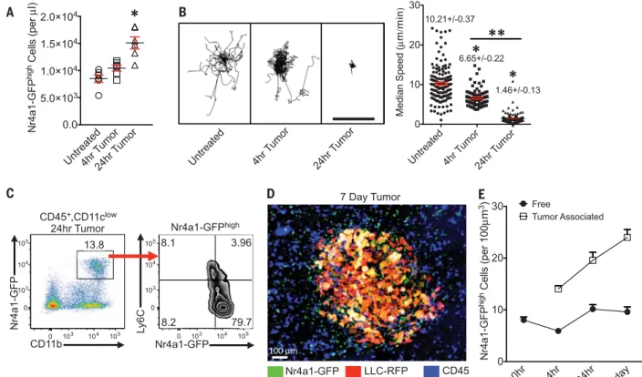
To investigate the actions of PMo during early tumor metastasis, we used mice expressing green fluorescent protein (GFP) under the control of the Nr4a1 promoter (Nr4a1-GFP mice). In these mice, PMo (but not Ly6C+classical monocytes) express high levels of GFP (GFPhigh) (10, 11). We focused our studies on the lung, which is a com-mon site of tumor metastasis and an important locus of PMo activity (12–14). We used flow cy-tometry to confirm that Nr4a1-GFPhighcells in the lung were PMo (fig. S1, A to C). Tracking of Nr4a1-GFPhighcells by confocal imaging in the
lungs allowed us to identify a large number of Nr4a1-GFPhighPMo patrolling the
microvascula-ture (movie S1 and Fig. 1A). Consistent with an important role for PMo in the lung vasculature, we found a three- to fourfold enrichment of Nr4a1-GFPhighPMo in the lung compared with
other tissues (fig. S1D).
To examine the interactions of PMo with tu-mors in vivo, we imaged Nr4a1-GFPhighPMo in the lung after intravenous (IV) injection of Lewis
1Division of Inflammation Biology, La Jolla Institute for
Allergy and Immunology, La Jolla, CA 92037, USA.
2Department of Molecular Biology and Genetics, Bilkent
University, Ankara, Turkey.3Izmir Biomedicine and Genome
Center, Dokuz Eylul University, Izmir, Turkey.4Microscopy
Core, La Jolla Institute for Allergy and Immunology, La Jolla,
CA 92037, USA.5Department of Functional Genomics and
Cancer, Institut de Génétique et de Biologie Moléculaire et Cellulaire (IGBMC), INSERM U964, CNRS UMR 7104,
Université de Strasbourg, Illkirch, France.6Graduate School
of Bioscience and Biotechnology, Tokyo Institute of
Technology, Yokohama, Japan.7Singapore Immunology
Network (SIgN), Agency for Science, Technology and Research (A*STAR), Singapore.
*Corresponding author. E-mail: [email protected] (R.N.H.); [email protected] (C.C.H.)
on September 28, 2017
http://science.sciencemag.org/
lung carcinoma cells expressing red fluorescent protein (LLC-RFP). The number of Nr4a1-GFPhigh
monocytes in the lung increased significantly 24 hours after injection, which implies that PMo are actively recruited to the lung tumor environ-ment (Fig. 1A). Within 4 hours after tumor in-jection, most Nr4a1-GFPhighmonocytes exhibited
decreased patrolling speed in the vasculature, and by 24 hours they had arrested near lung tumor sites (Fig. 1B). The majority of Nr4a1-GFPhighmonocytes isolated from the lung after LLC tumor transfer maintained their PMo phe-notype, which we further confirmed in vitro (Fig. 1C and fig. S1).
Nr4a1-GFPhighPMo were recruited to tumor cell clusters within 30 min after IV tumor in-jection, and recruitment continued for at least 7 days (Fig. 1, D and E, and movies S2 to S5). Nr4a1-GFPhighcells that were recruited to lung tumor sites were not positive for Ly6C/G (GR-1) in vivo, further confirming that these Nr4a1-GFPhighcells are not Ly6G+granulocytes or Ly6C+
classical monocytes (movie S4). The kinetics of PMo recruitment to the lung differed from that of Ly6C+monocytes (fig. S2A). At 7 days, there were
significantly higher numbers of Nr4a1-GFPhigh
PMo (~24/100mm3) associated with tumor areas compared with tumor-free areas, confirming ac-tive recruitment of PMo to the tumor (Fig. 1, D and E; movie S5; and fig. S1B).
Nr4a1-GFPhighmonocytes patrolling the
vas-culature 4 hours after tumor injection appeared to move toward and inhibit the attachment of tumor cells to the lung microvasculature (movie S3). We examined whether Nr4a1-GFPhighPMo could extravasate outside the vasculature. We found that by 4 hours after tumor injection, 10 to 20% of Nr4a1-GFPhighPMo had extravasated at tumor sites (fig. S2, B and C), and this increased to 40 to 50% PMo extravasation by 7 days. To-gether, these findings confirm that PMo establish early immune interactions with tumor cells and can extravasate and accumulate at tumor sites.
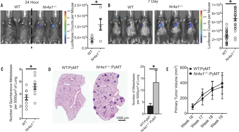
To determine whether PMo have a major role in regulating tumor invasion, metastasis, and growth in the lungs in vivo, we used Nr4a1 knock-out (Nr4a1−/−) mice, which exhibit selective loss of PMo (10) (fig. S3). Nr4a1−/−mice were IV in-jected with either syngeneic B16F10 melanoma cells expressing a luciferase reporter or LLC-RFP cells (Fig. 2, A and B, and figs. S4 and S5). As early as 24 hours and up to 21 days after IV injection of
B16F10 melanoma, we observed increased tumor invasion in the lungs ofNr4a1−/−mice compared with control mice (Fig. 2, A and B, and fig. S4, A and B). We observed no differences in either Ly6C+monocyte or Ly6G+granulocyte
popula-tions in the lungs of these mice 7 days after tu-mor injection (fig. S4C). B16F10 tutu-mor invasion appeared to be specific for the lung, as increased tumor metastasis was not observed in the liver (fig S4B). Additionally, increased spontaneous metastases to the lung were observed inNr4a1−/− mice after subcutaneous injection of B16F10 mela-noma, which suggests that Nr4a1 expression is important for suppressing primary tumor metastasis to the lung (Fig. 2C). A similar early and sustained increase in lung metastasis inNr4a1−/−mice was also observed after intravenous LLC tumor trans-fer (fig. S5). We did not detect diftrans-ferences in lung vascular permeability betweenNr4a1−/−and con-trol mice (fig. S6).
We next investigated the mouse mammary tu-mor virus–polyoma middle T (MMTV-PyMT) mod-el, in which female mice spontaneously develop mammary tumors that metastasize to the lung (15). To focus on Nr4a1 function exclusively in hematopoietic cells, we performed bone marrow
Fig. 1. Nr4a1-GFPhighmonocytes patrol the vasculature and interact with
tumor cells in the lung. (A) Quantification of Nr4a1-GFPhigh PMo per
microliter of blood volume in lung for control tissue (Untreated) or 4 or 24 hours after IV LLC-RFP transfer (n = 5 mice per group). (B) Quantification
of Nr4a1-GFPhighmonocyte movement in the lung before (Untreated), 4 hours
after, or 24 hours after LLC-RFP tumor injection. (Left) Monocyte tracks transposed to a common origin from a representative 20-min movie (scale
bar, 100mm). (Right) Quantification of median speed of monocytes (combined
speed data from analysis of three separate mice; *P < 0.001 lower than
untreated; **P < 0.001 lower than 4-hour tumor). (C) Representative gating
of Nr4a1-GFPhighCD11b+cells from all live CD45+CD11clowcells 24 hours
after IV LLC-RFP transfer. (D) Representative confocal image of
Nr4a1-GFPhighmonocytes (green) interacting with LLC-RFP cells (red) in the lung
7 days after IV LLC-RFP transfer. Immune cells in the vasculature were labeled with IV-injected antibody to CD45 (blue). (E) Quantification of free
(>100mm from tumor site) and tumor-associated (<50 mm from tumor
site) Nr4a1-GFPhighmonocytes in the lung at various time points after tumor
injection (combined analysis of five mice per group;P < 0.01 for each
tumor-associated area compared with tumor-free areas for each time point). Error bars indicate SEM.
on September 28, 2017
http://science.sciencemag.org/
transplants using either wild-type (WT) orNr4a1−/− bone marrow transferred into female recipient MMTV-PyMT mice. MMTV-PyMT mice receiving Nr4a1−/−bone marrow developed significantly
higher numbers of spontaneous metastases to the lung but no differences in primary mammary tumor growth compared to mice receiving WT bone marrow (Fig. 2, D and E).
We further tested hematopoietic Nr4a1 func-tion using B16F10 melanoma. Only mice receiving Nr4a1−/−bone marrow had increased B16F10
tu-mor metastases, confirming that Nr4a1 expres-sion in hematopoietic cells regulated tumor cell metastasis to the lung (fig. S7, A and B). Analysis of immune cells isolated from lung tumors verified a selective loss of PMo inNr4a1−/−bone marrow– transplanted mice (fig. S7C). In the 1:1 chimera mice, we observed equal reconstitution of im-mune cells from each donor (fig. S7D). However, PMo were derived almost exclusively from WT bone marrow, suggesting that the restoration of the Ly6C−monocyte population prevented tu-mor metastasis.
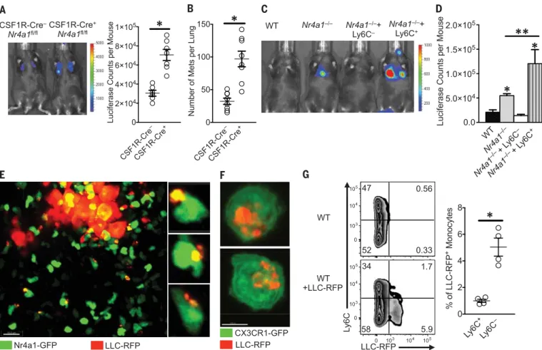
To confirm that Nr4a1 expressed in myeloid cells was regulating tumor metastasis to the lung, we examined two different myeloid-specific Nr4a1 conditional knockout models (CSF1R-Cre+Nr4a1fl/fland LysM-Cre+Nr4a1fl/fl). Deletion
of Nr4a1 using CSF1R-Cre+Nr4a1fl/fland LysM-Cre+Nr4a1fl/flmice significantly reduced the
num-ber of PMo in circulation (fig. S8) and increased tumor lung metastasis (Fig. 3, A and B, and fig. S9). Nr4a1 deletion using CSF1R-Cre or LysM-Cre also targets Nr4a1 in macrophages and Ly6C+
mono-cytes, so we cannot completely rule out effects of Nr4a1 in these cells. However, Nr4a1 expression in macrophages and Ly6C+monocytes is rela-tively low, which suggests limited Nr4a1 function (10, 16, 17). No differences in tumor metastasis were observed with T lymphocyte–specific Nr4a1 dele-tion (fig. S10). Collectively, our studies illustrate increased lung metastasis burden in the absence of Nr4a1 in myeloid cells in multiple cancer models. To confirm a direct role for PMo in regulating tumor metastasis, WT Ly6C+or Ly6C−monocytes were adoptively transferred into recipientNr4a1−/− mice before tumor injection. A substantial num-ber of the transferred monocytes could be found in the lungs (fig. S11, A and B). Reconstitution of PMo intoNr4a1−/−mice prevented lung tumor metastasis (Fig. 3, C and D). In contrast, transfer of Ly6C+monocytes intoNr4a1−/−mice actually
promoted tumor metastasis, consistent with known protumoral properties of this subset of mono-cytes (5, 18). The majority (80 to 90%) of trans-ferred Ly6C+ monocytes in circulation did not
lose Ly6C expression (fig. S11C). Transfer of PMo 24 hours after tumor injection intoNr4a1−/−mice did not suppress tumor metastasis (fig. S11D), sug-gesting that PMo must already be present and active in the vasculature to prevent early tumor metastasis. These data directly show that non-classical PMo inhibit tumor metastasis to the lung. Patrolling monocytes can act as“intravascular housekeepers” that scavenge microparticles and remove cellular debris from the microvascula-ture (7). Extracellular vesicles from tumors are important mediators of tumor metastasis, pro-gression, and immune suppression, and target-ing their removal is an emergtarget-ing focus for cancer therapy (19, 20). We used high-resolution con-focal imaging to determine whether PMo could engulf and remove tumor material from the lung vasculature. A sizable number of Nr4a1-GFPhigh PMo containing large amounts of LLC-RFP tumor material were observed at tumor sites in the lung 24 hours after IV tumor transfer (Fig. 3E). Co-culture assays of mouse PMo with fluorescently labeled tumor cells confirmed engulfment of large amounts of tumor material (Fig. 3F). Analy-sis of monocyte populations isolated from the lung at 24 hours after IV LLC-RFP injection indi-cated that PMo preferentially took up ~fivefold more tumor material than did Ly6C+classical
Fig. 2. Increased lung metastasis of tumors inNr4a1−/−mice. (A) (Left)
In vivo luciferase detection in WT control andNr4a1−/−mice 24 hours after IV
injection of 5 × 10⁵B16F10 melanoma cells expressing luciferase. (Right)
Luciferase quantification (*P < 0.03, representative experiment with five mice
per group). (B) In vivo luciferase detection (left) and quantification (right) in WT
andNr4a1−/−mice 7 days after IV injection with 3 × 10⁵B16F10-luciferase cells
(*P < 0.001, n = 18 mice per group combined from three separate experiments).
(C) Number of spontaneous tumor metastases per 5000mm2of lung surface
28 days after subcutaneous injection of 1 × 10⁵B16F10-YFP cells (*P < 0.01, n = 7
mice per group). (D and E) Lung tumor metastasis in MMTV-PyMT mice
reconstituted with WT (WT:PyMT) orNr4a1−/−(Nr4a1−/−:PyMT) bone marrow.
(D) (Left) Representative MMTV-PyMT mouse lung histology, stained with hematoxylin and eosin. (Right) Quantification of the number of spontaneous
lung metastases per 5000mm2of lung surface (*P < 0.05, n = 12 for WTand 15
forNr4a1−/−). (E) Quantification of primary breast tumor growth in MMTV-PyMT
mice (n = 12 for WTand 15 for Nr4a1−/−). Error bars indicate SEM.
on September 28, 2017
http://science.sciencemag.org/
monocytes (Fig. 3G). PMo also preferentially took up substantially more B16F10–yellow fluorescent protein (YFP) tumor material, with an average tumor material size of 1.39mm2and an average
total amount of tumor material per monocyte of 1.92mm2(fig. S12, A and B). The homologous
human CD14dimCD16+population of PMo, which
similarly has high Nr4a1 expression (8, 21), also engulfed a large quantity of tumor material in vitro, suggesting analogous tumor engulfment function (fig. S12C). Moreover, PMo actively en-gulfed tumor material within classic endocytic compartments (22) (Fig. 4A). Collectively, these results demonstrate that Nr4a1-dependent PMo rapidly and preferentially endocytose tumor material.
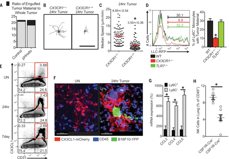
We then asked how PMo recognized tumor cells to prevent metastasis in the lung. The chemo-kine receptor CX3CR1 is highly expressed on PMo
and is important for their arrest at inflammatory sites (fig. S13A) (23–25). CX3CR1-deficient(Cx3cr1−/−) mice, which also have a significant reduction in PMo, exhibit a similar phenotype toNr4a1−/−mice (i.e., increased tumor burden and metastasis to the lung) (26, 27). Although PMo numbers were reduced in the lung vasculature ofCx3cr1−/−mice [~30 to 50% reduction (fig. S13B), confirming pre-vious reports] (27), a major proportion of the remaining CX3CR1-deficient PMo was observed patrolling the vasculature, as previously observed (7). Unlike Cx3cr1−/+or WT PMo,Cx3cr1−/−PMo did not arrest near LLC tumor cells and instead remained patrolling within the lung vasculature (Fig. 4, B and C, and movie S6).Cx3cr1−/−PMo were not recruited to the lung 24 hours after LLC tumor challenge, whereas Ly6C+recruitment was unaffected by the loss of CX3CR1 expression (fig. S13B).Cx3cr1−/−PMo present in the lung showed
defective engulfment of tumor material, indicat-ing that CX3CR1 expression on PMo is critical for mediating the sensing and uptake of tumor ma-terial (Fig. 4D).
CX3CL1, a ligand for CX3CR1, has been re-ported to be present in high levels in human and mouse lungs (28). Using a CX3CL1-mCherry re-porter mouse (29), we found that CX3CL1 was specifically expressed on CD31+endothelial cells (ECs) at low levels in the lung microvasculature (Fig. 4, E and F, and fig. S13C). CX3CL1 expres-sion was most prevalent in lung ECs compared with ECs in other tissues (fig. S13D), which may partially explain the enrichment and preferential function of PMo in the lung. CX3CL1 expression on lung ECs increased in response to tumor chal-lenge (Fig. 4E) and at sites of tumor metastasis (Fig. 4F), consistent with reports of increased CX3CL1 during lung inflammation (30).
Fig. 3. Nr4a1-expressing PMo reduce tumor metastasis and engulf tu-mor material in the lung. (A) In vivo imaging (left) and quantification (right)
of lung tumors in CSF1R-Cre−Nr4a1fl/fl(CSF1R-Cre−) or CSF1R-Cre+Nr4a1fl/fl
(CSF1R-Cre+) mice 7 days after IV injection of 3 × 10⁵B16F10-luciferase
tu-mor cells (n = 6 mice per group, *P < 0.01; experiment replicated twice). (B) Quantification of the number of tumor metastases per lung of
CSF1R-Cre−Nr4a1fl/fl(CSF1R-Cre−) and CSF1R-Cre+Nr4a1fl/fl(CSF1R-Cre+) mice
7 days after IV injection of 3 × 10⁵B16F10-YFP tumor cells (n = 8 mice per
group, *P < 0.01). (C and D) Nr4a1−/−mice were injected intravenously with 5 ×
105WT Ly6C−PMo, Ly6C+inflammatory monocytes, or PBS at day 0. On day 1,
3 × 10⁵B16F10-luciferase tumor cells were injected intravenously, and tumor
metastasis and growth were measured by in vivo imaging on day 8. Shown are representative in vivo images (C) and quantification (D) of B16F10-luciferase
metastasis 8 days after monocyte transfer and 7 days after tumor transfer
in WTorNr4a1−/−mice (combined data from five separate experiments with
n = 2 mice per group; *P < 0.01 statistically different from WT; **P < 0.05
statistically different fromNr4a1−/−). (E) Imaging of tumor material uptake
in lung by Nr4a1-GFPhighmonocytes 24 hours after IV injection of LLC-RFP
tumor cells. Representative higher-magnification images are shown at right. Note that Nr4a1-GFP expression is primarily nuclear, so monocyte cell mem-branes are not visible in these images. (F) Uptake of LLC-RFP tumor material
by CX3CR1-GFPhighLy6C−PMo after 24 hours of coculture. (G)
Representa-tive flow plot (left) and quantification (right) of tumor material uptake by all
monocytes in the lung 24 hours after IV tumor injection of 3 × 10⁵LLC-RFP
cells (n = 4 mice per group; *P < 0.01; experiment replicated three times).
Error bars indicate SEM.
on September 28, 2017
http://science.sciencemag.org/
Toll-like receptor 7 (TLR7) has been linked to recruitment of PMo in response to kidney dam-age in mice (7). However, we found that TLR7 did not play a major role in either recruitment of PMo to the lung after tumor injection (fig. S13B) or uptake of tumor material by PMo (Fig. 4D). We conclude that both CX3CR1 expression on monocytes and CX3CL1 expression by ECs are critical for recruitment of PMo to sites of tumor extravasation to mediate the removal of tumor material from the lung. CX3CL1 expression by tu-mor cells (31) may also drive monocyte recruitment. In agreement with our findings, many studies have reported that CX3CL1 expression by either tumor cells or tumor-associated cells is
anti-tumoral and correlated with good prognosis (32–34). However, the function of the CX3CL1/ CX3CR1 axis, particularly during later stages of tumor growth, is complex (35, 36).
Finally, we examined whether PMo can directly kill tumor cells. After multiple attempts using var-ious experimental conditions, direct killing of tu-mor cells by PMo was not observed (fig. S14). However, PMo may be important for antibody-dependent cell-mediated cytotoxicity of either tu-mor cells or suppressive immune cells within the tumor environment (37, 38). In response to IV-injected B16F10 tumor, PMo isolated from lungs produced significantly higher levels of natural killer (NK) cell activation and recruitment-related
chemokines CCL3, CCL4, and CCL5, as compared with classical Ly6C+monocytes (Fig. 4G) (39, 40).
In accord with this finding, myeloid-specific Nr4a1 knockout mice (CSF1R-Cre+Nr4a1fl/fl) showed
reduced NK cell recruitment to the lung in re-sponse to tumor (Fig. 4H), suggesting that PMo controlled the recruitment of NK cells to tumor sites. A similar reduction in NK cell recruitment and CD44 activation (fig. S15, A and B) was also observed in the lungs of PyMT mice that received Nr4a1-deficient bone marrow. However, Nr4a1 does not regulate NK cell development (fig. S15C). Uptake of tumor material by PMo does not re-quire the presence of NK cells (fig. S16, A and B). NK cell depletion reduced the differences in
Fig. 4. Patrolling monocytes detect tumor material in a CX3CR1-dependent manner and recruit NK cells to the lung tumor environment. (A) Ratio of fluorescent intensity of tumor material engulfed by PMo (black) to fluorescent intensity of whole tumor (black and gray) 3 hours after IV LLC tumor injection. LLC tumors were labeled with either CellTrace Violet control dye (Control) or a pH-sensitive pHrodo Red dye (pHrodo) and then intravenously injected in a 1:1 ratio into a WT mouse (n = 3 mice per group, experiment replicated three
times). Representative tracking (B) and median speed (C) ofCx3cr1−/−or
Cx3cr1−/+monocyte movement 24 hours after IV tumor injection in the lung.
Monocyte tracks transposed to a common origin from representative 20-min
movies (scale bar, 100mm; representative tracks are shown from one mouse,
median speed was calculated from tumor areas analyzed in three separate
mice per group, *P < 0.001). (D) (Left) Percentage of Ly6C−PMo containing
LLC-RFP tumor material in the lung 3 hours after IV injection of tumor into
representative WT,Cx3cr1−/−, orTlr7−/−mice. (Right) Quantification of tumor
material uptake (n = 3 per group, *P < 0.001 versus WT). (E) Percentage of
CD31+CX3CL1+lung ECs isolated from untreated (UN) mice or from
CX3CL1-mCherry mice, 24 hours or 7 days after IV injection of B16F10-YFP tumor cells. (F) Representative imaging of CX3CL1-mCherry (red) expression in lung 24 hours after IV injection of B16F10-YFP tumor cells (green) in
CX3CL1-mCherry mice. CD45+immune cells are labeled in blue. (G) Relative
chemo-kine mRNA expression in Ly6C+or Ly6C−monocytes isolated from lungs by
fluorescence-activated cell sorting 24 hours after IV B16F10 tumor injection
(monocyte populations isolated from three separate mice; *P < 0.01;
experi-ment repeated three times). (H) Percentage of NK cells in the lungs of
CSF1R-Cre−Nr4a1fl/fl(CSF1R-Cre−) or CSF1R-Cre+Nr4a1fl/fl(CSF1R-Cre+) mice 7 days
after IV injection of 3 × 10⁵B16F10-luciferase tumor cells (n = 6 mice per group,
*P < 0.01). Error bars indicate SEM.
on September 28, 2017
http://science.sciencemag.org/
metastasis between WT and CSF1R-Cre+Nr4a1fl/fl mice (fig. S16C). Thus, PMo inhibit metastasis, at least in part, through the regulation of NK cell recruitment and activity.
In summary, we demonstrate that PMo partic-ipate in cancer surveillance by preventing tumor metastasis to lung. PMo are actively recruited to lung metastasis sites in a CX3CR1-dependent manner, where they function to scavenge tumor material, as well as to recruit and activate NK cells, leading to the prevention of tumor cell me-tastasis (fig. S17). Selective targeting by increas-ing PMo activity and/or their regulation by Nr4a1 may represent a novel therapy for the prevention of cancer metastasis to the lung.
R E FE R E N C ES A ND N OT ES
1. S. K. Biswas, A. Mantovani, Nat. Immunol. 11, 889–896 (2010).
2. P. J. Murray, T. A. Wynn, Nat. Rev. Immunol. 11, 723–737 (2011).
3. T. A. Wynn, A. Chawla, J. W. Pollard, Nature 496, 445–455 (2013).
4. R. A. Franklin et al., Science 344, 921–925 (2014). 5. B. Z. Qian et al., Nature 475, 222–225 (2011). 6. K. Movahedi et al., Cancer Res. 70, 5728–5739 (2010). 7. L. M. Carlin et al., Cell 153, 362–375 (2013). 8. J. Cros et al., Immunity 33, 375–386 (2010). 9. C. Auffray et al., Science 317, 666–670 (2007). 10. R. N. Hanna et al., Nat. Immunol. 12, 778–785 (2011). 11. A. E. Moran et al., J. Exp. Med. 208, 1279–1289 (2011). 12. L. Landsman, S. Jung, J. Immunol. 179, 3488–3494
(2007).
13. L. Landsman, C. Varol, S. Jung, J. Immunol. 178, 2000–2007 (2007).
14. C. Jakubzick et al., J. Immunol. 180, 3019–3027 (2008).
15. C. T. Guy, R. D. Cardiff, W. J. Muller, Mol. Cell. Biol. 12, 954–961 (1992).
16. S. Saeed et al., Science 345, 1251086 (2014). 17. V. Jojic et al., Nat. Immunol. 14, 633–643 (2013). 18. D. E. Sanford et al., Clin. Cancer Res. 19, 3404–3415
(2013).
19. P. Vader, X. O. Breakefield, M. J. Wood, Trends Mol. Med. 20, 385–393 (2014).
20. F. Pucci, M. J. Pittet, Clin. Cancer Res. 19, 2598–2604 (2013).
21. R. N. Hanna et al., Circ. Res. 110, 416–427 (2012). 22. Detailed methods are available as supplementary materials on
Science Online.
23. G. Thomas, R. Tacke, C. C. Hedrick, R. N. Hanna, Arterioscler. Thromb. Vasc. Biol. 35, 1306–1316 (2015). 24. S. Jung et al., Mol. Cell. Biol. 20, 4106–4114 (2000). 25. F. Geissmann, S. Jung, D. R. Littman, Immunity 19, 71–82
(2003).
26. Y. R. Yu et al., Int. J. Cancer 121, 316–322 (2007). 27. L. Landsman et al., Blood 113, 963–972 (2009). 28. A. I. Su et al., Proc. Natl. Acad. Sci. U.S.A. 101, 6062–6067
(2004).
29. K. W. Kim et al., Blood 118, e156–e167 (2011). 30. J. Zhang, J. M. Patel, Int. J. Clin. Exp. Med. 3, 233–244
(2010).
31. E. Ferretti, V. Pistoia, A. Corcione, Mediators Inflamm. 2014, 480941 (2014).
32. M. Hyakudomi et al., Ann. Surg. Oncol. 15, 1775–1782 (2008).
33. J. Y. Kee et al., Mol. Clin. Oncol. 1, 35–40 (2013). 34. M. H. Park, J. S. Lee, J. H. Yoon, J. Surg. Oncol. 106, 386–392
(2012).
35. A. Schmall et al., Am. J. Respir. Crit. Care Med. 191, 437–447 (2015).
36. M. Tardaguila, S. Manes,“The complex role of chemokines in cancer: The case of the CX3CL1-CX3CR1 axis,” in Oncology: Theory and Practice (iConcept Press, 2014).
37. E. Romano et al., Proc. Natl. Acad. Sci. U.S.A. 112, 6140–6145 (2015).
38. A. Szaflarska et al., Exp. Hematol. 32, 748–755 (2004).
39. A. A. Maghazachi, Curr. Top. Microbiol. Immunol. 341, 37–58 (2010).
40. M. J. Robertson, J. Leukoc. Biol. 71, 173–183 (2002). AC K NOW L E D GM E NTS
We thank K. Ley and H. Shaked for critically reviewing and editing this manuscript, K. Hogquist for Nr4a1-GFP reporter mice, M. Kronenberg for B16F10 melanoma cells, A. Blatchley and D. Yoakum for assistance with mouse colony management, A. Strasner for help with establishing tumor models, and Z. Mikulski for assistance with imaging. The data presented here are tabulated in the main paper and in the supplementary materials. Nr4a1-floxed conditional mice are available from IGBMC under a material transfer agreement with D.M. and H.I. C.C.H., R.N.H., and the La Jolla Institute for Allergy and Immunology have filed a U.S. patent application (U.S. 13/646,183) that relates to specific topic “Methods and uses of Nur77 and Nur77 agonists to modulate macrophages and monocytes, and treat inflammation, inflammatory disease and cardiovascular disease” and an invention disclosure that relates to the specific topic“Patrolling monocytes control tumor metastasis to the lung.” This work was supported by NIH grants R01 HL118765 and R01 CA202987 (both to C.C.H.), American Heart Association Postdoctoral Award 3POST16990029 (to R.T.), NIH F32 postdoctoral fellowship NIH F32 HL117533-02 (to H.N.), American Heart Association Scientist Development Grant 125SDG12070005 (to R.N.H.), the La Jolla Institute for Allergy and Immunology Board of Directors Fellowship (to R.N.H.), CNRS and INSERM (to D.M.), and core funding from SIgN (A*STAR) (to S.K.B.).
SUPPLEMENTARY MATERIALS
www.sciencemag.org/content/350/6263/985/suppl/DC1 Materials and Methods
Figs. S1 to S18 References (41–48) Movies S1 to S6
1 July 2015; accepted 2 October 2015 Published online 22 October 2015 10.1126/science.aac9407
on September 28, 2017
http://science.sciencemag.org/
Subhra K. Biswas and Catherine C. Hedrick
Rasquinha, Sara McArdle, Runpei Wu, Esther Peluso, Daniel Metzger, Hiroshi Ichinose, Iftach Shaked, Grzegorz Chodaczek, Richard N. Hanna, Caglar Cekic, Duygu Sag, Robert Tacke, Graham D. Thomas, Heba Nowyhed, Erica Herrley, Nicole
originally published online October 22, 2015 DOI: 10.1126/science.aac9407 (6263), 985-990. 350 Science , this issue p. 985 Science lung.
the lung, where they engulfed tumor material, which may explain how these cells prevent tumors from colonizing the showed increased metastasis to the lung but not to other tissues. Patrolling monocytes resided in the microvasculature of monocytes in blocking tumor metastasis to the lungs in mice. Tumors in mice engineered to lack patrolling monocytes
now report a role for patrolling
et al.
to understand the cellular events that promote or prevent metastasis. Hanna
Metastatic cancer is especially hard to treat. In order to find potential new therapeutic targets, scientists are trying Monocytes block tumor access to the lung
ARTICLE TOOLS http://science.sciencemag.org/content/350/6263/985
MATERIALS SUPPLEMENTARY http://science.sciencemag.org/content/suppl/2015/10/21/science.aac9407.DC1 CONTENT RELATED http://stm.sciencemag.org/content/scitransmed/6/217/217ra3.full REFERENCES http://science.sciencemag.org/content/350/6263/985#BIBL
This article cites 45 articles, 21 of which you can access for free
PERMISSIONS http://www.sciencemag.org/help/reprints-and-permissions
Terms of Service
Use of this article is subject to the
is a registered trademark of AAAS.
Science
licensee American Association for the Advancement of Science. No claim to original U.S. Government Works. The title Science, 1200 New York Avenue NW, Washington, DC 20005. 2017 © The Authors, some rights reserved; exclusive
(print ISSN 0036-8075; online ISSN 1095-9203) is published by the American Association for the Advancement of
Science
on September 28, 2017
http://science.sciencemag.org/