Background: Many children with kidney diseases can be diagnosed and treated without a biopsy. However, biopsy is a valuable method for the diagnostic and prognostic evalua-tion of children with kidney diseases.
Aims: To evaluate the clinical and pathological profiles of the kidney biopsies in our department to provide epidemio-logical data for clinical practice.
Study Design: Retrospective cross-sectional study.
Methods: Kidney biopsies and patient’s charts in pediatric patients performed between May 2005 and February 2015 at the Pediatric Nephrology Department, Trakya University School of Medicine were assessed retrospectively.
Results: A total of 100 patients were examined. Their mean age was 9.62±4.26 years (range: 1-17 years); 54% of the
pa-tients were girls and 46% were boys. The most frequent in-dication for kidney biopsy was nephrotic syndrome (33%). The most common kidney disease was primary glomeru-lonephritis, which was observed in 65% of cases. IgA ne-phropathy (24%) was the most frequently observed subtype in primary glomerulonephritis groups. Secondary glomeru-lonephritis was diagnosed in 35% of cases. Systemic lupus erythematosus (51%) was the most frequently observed subtype in the secondary glomerulonephritis groups. Conclusion: IgA nephropathy and systemic lupus erythe-matosus were the most frequent primary and secondary glo-merulonephritis in our region among children, respectively. Keywords: Kidney biopsy, child, histopathological diagnosis, glomerulonephritis
Assessment of Ten-Year-Long Results of Kidney Biopsies Performed
on Children in the Thrace Region of Turkey
1Department of Pediatric Nephrology, Trakya University School of Medicine, Edirne, Turkey 2Department of Pathology, Trakya University School of Medicine, Edirne, Turkey 3Department of Pediatric Radiology, Trakya University School of Medicine, Edirne, Turkey
4Department of Radiology, Selçuk University School of Medicine, Konya, Turkey
Neşe Özkayın
1, Gökçe Çıplak
1, Ufuk Usta
2, Hakan Gençhellaç
3, Osman Temizöz
4Address for Correspondence: Dr. Neşe Özkayın, Department of Pediatric Nephrology, Trakya University School of Medicine, Edirne, Turkey Phone: +90 532 372 58 06 e-mail: [email protected]
Received: 3 April 2015 Accepted: 24 March 2016 • DOI: 10.5152/balkanmedj.2016.150506 Available at www.balkanmedicaljournal.org
Cite this article as:
Many children with kidney disease can be diagnosed and treated without a biopsy. However, biopsy is a valuable meth-od for the diagnostic and prognostic evaluation of children with kidney disease (1). Epidemiological studies about kidney diseases, both primary and secondary, may identify the fre-quency of diseases, their causes, and environmental or genetic factors contributing to disease development.
Valuable epidemiological data are available from studies of children’s kidney biopsies in different countries (2-6). Further-more, some reports dealing with national and regional databases, specific population groups, specific diagnoses or local single-center experiences have been published. The studies investigating
epidemiologic, clinical and histopathological findings related to childhood kidney diseases are still scarce in our country (4,7,8).
This study aims to examine the clinical and histopathologi-cal profiles of the kidney biopsies from our department.
MATERIALS AND METHODS
All patients who had undergone kidney biopsy between May 2005 and February 2015 in our department were assessed retrospectively. Patients who had undergone kidney biopsy
on all patients before biopsies. All patients fasted for at least 6 h and were then sedated 30-45 minutes before the procedure. Fol-lowing premedication and local anesthesia with 1% lidocaine, biopsies were performed on the left kidney of patients. Biopsy was performed using 16-gauge or 18-gauge biopsy needles ac-cording to the patient’s age with the guidance of ultrasonogra-phy by the radiologists; two cores of tissue were taken.
Following the procedure, the patients were kept in the su-pine position for 24 hours with sandbag compression to the abdomen and intermittent ice pack application to the biopsy region. Vital signs were measured at 15 min. intervals for the first 2h and then hourly. Each urine sample was examined for macroscopic hematuria. Hematocrit values were also moni-tored. A post-biopsy ultrasound examination of the punctured kidney was performed in all patients on the next day and those patients without complication were discharged after 24 hours. Doppler ultrasonography was used to follow recovery of pa-tient with vascular complications.
All kidney specimens were evaluated by light and immuno-fluorescent microscopies. Fresh biopsy cores were evaluated on the dissecting microscope. Renal cortical tissue (3-4 mm in length) was separated for immunofluorescence study and kept at -80°C. The remaining biopsy specimen was fixed in 10% buffered formalin for light microscopy. Paraffin sections were stained with hematoxylin and eosin, periodic acid Schiff, Mas-son’s trichrome stain, Congo red and Gomori’s methanamine silver. For immunofluorescence studies, cryostat sections were stained using polyclonal antisera against IgA, IgG, IgM, C3, C1q and fibrinogen. Biopsy specimens containing at least eight glomeruli were considered sufficient for a proper diagnosis.
The study was approved by the local ethics committee. Sta-tistical analysis was not used for this study; we only used per-centage figures from the data.
RESULTS
Kidney biopsies were performed in 100 children, 46 boys and 54 girls, aged 1-17 years (mean age=9.62±4.26 years) over a 10-year period. Among these, 24 (24%) were aged un-der 5 years, 29 (29%) were 6 to 10 years, 30 (30%) were 11 to 14 years and 17 (17%) were 15 to 17 years.
The main indication for performing a biopsy was nephrotic syndrome (NS) in 33/100 (33%) children. Other indications were acute nephritic syndrome (ANS) in 24 cases (24%), nephrotic-ne-phritic syndrome in 18 cases (18%), rapidly progressive glomeru-lonephritis (RPGN) in 3 cases (3%), acute kidney injury (AKI) in 3 cases (3%), chronic kidney disease (CKD) in 2 cases (2%), and asymptomatic urinary abnormalities in 17 cases (17%) (Table 1). Of the 17 asymptomatic urinary abnormalities, 6 cases presented with isolated hematuria and 11 cases with isolated proteinuria.
None of the patients presented sedation-related complica-tions during or after the procedure. Two out of 100 patients (2%) presented macroscopic hematuria, and were evaluated with Doppler ultrasonography. Because there were no vascu-lar complications, the patients were followed up until bleeding stopped and recovered without any blood transfusion.
Kidney biopsies with insufficient specimen were excluded from further analysis and a total of 91 kidney biopsies were included in the present study.
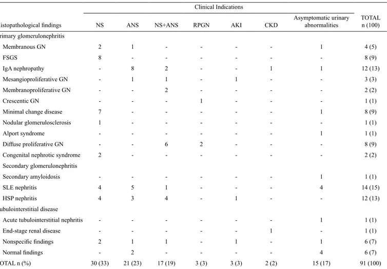
The histopathological diagnosis according to the clinical indications are shown in Table 2. The most frequent types of kidney diseases were systemic lupus erythematosus (SLE) nephritis in 14/91 patients (15%), IgA nephropathy (IgAN) and Henoch-Schonlein Purpura (HSP) nephritis both in 12/91 (13%), followed by focal segmental glomerulosclero-sis (FSGS), diffuse proliferative glomerulonephritis (DPGN), and minimal change disease (MCD) each in 8/91 patients (9%), normal findings in 6/91 patients (7%), nonspecific find-ings in 6/91 patients (7%), membranous glomerulonephritis (MGN) in 4/91 patients (4%), mesangioproliferative glomeru-lonephritis (MesGN) in 3/91 (3%), membranoproliferative glomerulonephritis (MPGN) in 2/91 (2%), congenital ne-phrotic syndrome (CNS) in 2/91 (2%), and others each in 1/91 patients (1%) (acute tubulointerstitial nephritis (ATIN), Alport syndrome, crescentic glomerulonephritis, nodular glomerulo-sclerosis, secondary amyloidosis, end-stage renal disease).
The most common diagnosis in the biopsies was glomerulone-phritis in 77/91 patients (84%). Primary glomeruloneglomerulone-phritis (GN) accounted for 65% (50/77) and secondary glomerulonephritis for Asymptomatic urinary abnormalities 17 (17)
35% (27/77). Common causes of primary GN were IgAN in 12/50 (24%), FSGS, DPGN and MCD each in 8/50 patients (16%). SLE nephritis and HSP nephritis were the most common causes of sec-ondary GN [14/27 (51%) and 12/27 (45%) respectively].
The histopathological findings in 6 cases of isolated he-maturia were: SLE nephritis in 3/6 (50%), IgAN, MGN and Alport syndrome each in 1/6 patients (~17%). Nine children with isolated proteinuria had the following histopathological findings: normal findings in 4/9 (45%) followed by MCD, secondary amyloidosis, SLE nephritis, ATIN and nonspecific findings each in 1/9 patients (11%).
DISCUSSION
Our study provides data regarding kidney disease pattern from a pediatric population in our region over a ten-year pe-riod. Our hospital is a tertiary referral hospital in our region,
playing a major role in healthcare management. At present, there is no national database of kidney biopsies in the country and this is the first reported kidney biopsy data from our popu-lation in this region.
We performed biopsies on 100 children with a success rate of 91% (insufficient specimen in 9 patients) and a complica-tion rate of 2% over ten-year period. Complicacomplica-tions seen in 2 patients were macroscopic hematuria and all recovered spon-taneously.
The sufficient biopsy material rate has a range of 69% to 100% in the different studies (9,10). The complication rates in different series may be as low as 2.6% or as high as 43% (9-15). In our biopsy series, the success and complication rates did not differ from most of the studies on children.
In our study, NS (33%) was the most common indication for kidney biopsy, in accordance with other studies in chil-dren (2,4,16,17). Other common indications were ANS (24%) and nephrotic-nephritic syndrome (18%). FSGS was the most TABLE 2. Comparison of histopathological findings according to clinical indications
Clinical Indications
Histopathological findings NS ANS NS+ANS RPGN AKI CKD Asymptomatic urinary abnormalities TOTAL n (100) Primary glomerulonephritis Membranous GN 2 1 - - - - 1 4 (5) FSGS 8 - - - 8 (9) IgA nephropathy - 8 2 - - 1 1 12 (13) Mesangioproliferative GN - 1 1 - 1 - - 3 (3) Membranoproliferative GN - - 2 - - - - 2 (2) Crescentic GN - - - 1 - - - 1 (1)
Minimal change disease 7 - - - 1 8 (9)
Nodular glomerulosclerosis 1 - - - 1 (1)
Alport syndrome - - - 1 1 (1)
Diffuse proliferative GN - - 6 2 - - - 8 (9)
Congenital nephrotic syndrome 2 - - - 2 (2)
Secondary glomerulonephritis
Secondary amyloidosis - - - 1 1 (1)
SLE nephritis 4 5 1 - - - 4 14 (15)
HSP nephritis 4 3 4 - 1 - - 12 (13)
Tubulointerstitial disease
Acute tubulointerstitial nephritis - - - 1 1 (1)
End-stage renal disease - - - 1 - 1 (1)
Nonspecific findings 2 1 1 - 1 - 1 6 (7)
Normal findings - 2 - - - - 4 6 (7)
TOTAL n (%) 30 (33) 21 (23) 17 (19) 3 (3) 3 (3) 2 (2) 15 (17) 91 (100)
GN: glomerulonephritis; FSGS: focal segmental glomerulosclerosis; SLE: systemic lupus erythematosus; HSP: Henoch-Schonlein Purpura; NS: nephrotic syndrome; ANS: acute nephritic syndrome; RPGN: rapidly progressing glomerulonephritis; AKI: acute kidney injury; CKD: chronic kidney disease
one center to another depending on age, ethnicity, geographi-cal region, patient characteristics and the indication for biop-sy. Chang et al. (22) reported that IgAN was the most common histopathological diagnosis in primary GN, while Printza et al. (1) defined FSGS as the most common primary GN. In our study, IgAN was the most common histopathological di-agnosis in children with primary GN (24%). Consistent with a local study, it was also the most common histopathological diagnosis in children presenting with ANS (38%) (4).
However, SLE nephritis was the leading the histopathologi-cal diagnosis and contributed 15% of cases in overall biop-sies. Since renal involvement is more frequent in children with SLE than in adults and represent 15-20% of all SLE patients, kidney biopsy is very important for prognostic and therapeutic approaches (1,23).
Systemic lupus erythematosus nephritis was also the most common diagnosis (51%) among secondary GN. The high proportion of SLE nephritis in our study is consistent with studies from Hong Kong (71%) and Pakistan (54%), but not consistent with the studies from other regions of our country (2,5). In a study of 614 children, Demircin et al. (4) found that amyloidosis was the most frequent histopathology in second-ary GN.
There were several limitations to our study. The population size was a relatively small sample from a single center even though it is a tertiary referral hospital. Additionally, data were collected retrospectively. Biopsy specimens were not evalu-ated by electron microscopy.
The pattern of kidney diseases in children is quite different among countries, even the regions of the country (1-8). Many differences in specific incidences of glomerulopathies can also be explained by confounding factors including genetic or environmental factors, race, frequency of infections, refer-ral bias, the non-availability of the necessary facilities (e.g., electron microscopy) or biopsy rate. We suggest that small numbers of our study population and the fact that as a tertiary centre, admitting mostly difficult cases might have affected our results.
In conclusion, our study presents the first epidemiologi-cal data regarding kidney disease pattern of kidney biopsies from the pediatric population of our region. IgAN as primary glomerulonephritis and SLE as secondary
glomerulonephri-School of Medicine.
Informed Consent: Written informed consent was obtained from
the parents of the patients who participated in this study.
Peer-review: Externally peer-reviewed.
Author contributions: Concept - N.Ö.; Design - N.Ö.;
Supervi-sion - N.Ö.; Resource - N.Ö., O.T.; Materials - N.Ö., G.Ç., U.U., H.G.; Data Collection and/or Processing - N.Ö.; Analysis and/or In-terpretation - N.Ö.; Literature Search - N.Ö.; Writing - N.Ö., G.Ç.; Critical Reviews - N.Ö., G.Ç.
Conflict of Interest: No conflict of interest was declared by the authors. Financial Disclosure: The authors declared that this study has
re-ceived no financial support.
REFERENCES
1. Printza N, Bosdou J, Pantzaki A, Badouraki M, Kollios K, Ghogha Ch, et al. Percutaneous ultrasound guided renal bi-opsy in children: a single centre experience. Hippokratia 2011;15:258-61.
2. Yuen LK, Lai WM, Lau SC, Tong PC, Tse KC, Chiu MC. Ten-year review of disease pattern from percutaneous renal biopsy: an experience from a paediatric tertiary renal centre in Hong Kong. Hong Kong Med J 2008;14:348-55.
3. Coppo R, Gianoglio B, Porcellini MG, Maringhini S. Frequency of renal diseases and clinical indications for renal biopsy in chil-dren (report of the Italian National Registry of Renal Biopsies in Children). Group of Renal Immunopathology of the Italian Society of Pediatric Nephrology and Group of Renal Immuno-pathology of the Italian Society of Nephrology. Nephrol Dial
Transplant 1998;13:293-7. [CrossRef]
4. Demircin G, Delibas A, Bek K, Erdogan O, Bülbül M, Baysun S, et al. A one-center experience with pediatric percutaneous renal biopsy and histopathology in Ankara, Turkey. Int Urol
Nephrol 2009;41:933-9. [CrossRef]
5. Lanewala A, Mubarak M, Akhter F, Aziz S, Bhatti S, Kazi JI. Pattern of pediatric renal disease observed in native renal biop-sies in Pakistan. J Nephrol 2009;22:739-46.
6. Piotto GH, Moraes MC, Malheiros DM, Saldanha LB, Koch VH. Percutaneous ultrasound-guided renal biopsy in children - safety, efficacy, indications and renal pathology findings:
14-year Brazilian university hospital experience. Clin Nephrol 2008;69:417-24. [CrossRef]
7. Ergin M, Yavaşcan Ö, Serdaroğlu E, Ergin I, Diniz AG, Ortaç R, et al. Dr. Behçet Uz Çocuk Hastanesi patoloji laboratuarında 2009-2010 yıllarında incelenen böbrek biopsilerinin klinik ve histopatolojik profili. Behcet Uz Cocuk Hast Derg 2011;1:51-7.
[CrossRef]
8. Taşlı F, Şahin T, Tanrısev M, Özkök G, Cirit M, Çolak H, et al. Böbrek hastalıkları tanısında böbrek biyopsileriyle alınan sonuçlar. Tepecik Eğit Hast Derg 2012;22:133-8.
9. Feneberg R, Schaefer F, Zieger B, Waldherr R, Mehls O, Scharer K. Percutaneous renal biopsy in children: a 27-year experience.
Nephron 1998;79:438-46. [CrossRef]
10. Simckes AM, Blowey DL, Gyves KM, Alon US. Success and safety of same-day kidney biopsy in children and adolescents.
Pediatr Nephrol 2000;14:946-52. [CrossRef]
11. Chesney DS, Brouhard BH, Cunningham RJ. Safety and cost effectiveness of pediatric percutaneous renalbiopsy. Pediatr
Nephrol 1996;10:493-5. [CrossRef]
12. Kamitsuji H, Yoshioka K, Ito H. Percutaneous renal biopsy in children: survey of pediatric nephrologists in Japan. Pediatr
Nephrol 1999;13:693-6. [CrossRef]
13. Hussain F, Watson AR, Hayes J, Evans J. Standards for renal biopsies: comparison of inpatient and day care procedures.
Pe-diatr Nephrol 2003;18:53-6. [CrossRef]
14. White RHR, Poole C. Day care renal biopsy. Pediatr Nephrol 1996;10:408-11. [CrossRef]
15. Davis ID, Oehlenschlager W, O’Riordan MA, Avner ED. Pe-diatric renal biopsy: should this procedure be performed in an outpatient setting? Pediatr Nephrol 1998;12:96-100. [CrossRef]
16. Yavaşcan Ö, Aksu N, Erdoğan H, Kara OD, Çerçi TT, Şen S, et al. Çocuklarda böbrek biyopsi sonuçlarının değerlendirilmesi: On yıllık bir süre. Turk Neph Dial Transpl 2005;14:195-201. 17. Paripović D, Kostić M, Kruščić D, Spasojević B, Lomić G,
Marković-Lipkovski J, et al. Indications and results of renal bi-opsy in children: a 10-year review from a single center in Serbia.
J Nephrol 2012;25:1054-9. [CrossRef]
18. International Study of Kidney Disease in Children. Nephrotic syndrome in children: Prediction of histopathology from clinical and laboratory characteristics at time of diagnosis. Kidney Int 1978;13:159-65. [CrossRef]
19. Gulati S, Sharma AP, Sharma RK, Gupta A. Changing trends of histopathology in childhood nephrotic syndrome. Am J Kidney
Dis 1999;34:646-50. [CrossRef]
20. Filler G, Young E, Geier P, Carpenter B, Drukker A, Feber J. Is there really an increase in non-minimal change nephrotic syndrome in children? Am J Kidney Dis 2003;42:1107-13.
[CrossRef]
21. Altuntaş Ü, Yıldız N, Benzer M, Gökçe İ, Bıyıklı N, Arıbal E, et al. Perkutan böbrek iğne biopsisi yapılan çocuklarda renal hematom değerlendirilmesinde ultrasonografi ve bilgisayarlı tomografinin karşılaştırılması. Türkiye Çocuk Hast Derg 2012;6:221-7.
22. Chang JH, Kim DK, Kim HW, Park SY, Yoo TH, Kim BS, et al. Changing prevalence of glomerular diseases in Korean adults: a review of 20 years of experience. Nephrol Dial Transplant 2009;24:2406-10. [CrossRef]
23. Sallmann S, Fiebig B, Hedrich CM, Heubner G, Gahr M. Sys-temic lupus erythematosus in children and adolescents. Z