Investigation on Tularemia in potential reservoirs’ of Anatolia*
Derya KARATAŞ YENİ
1, Müjgan İZGÜR
21
Veterinary Control Central Research Institute, Laboratory of Bakteriology, Ankara; 2 Ankara University, Faculty of Veterinary Medicine, Department of Microbiology, Ankara, Turkey.
Summary: Tularemia is a zoonotic infection caused by Francisella tularensis and has gained renewed importance since there has been a recent increase in the number of human cases in several countries across the world. In this study the existence of F.
tularensis in rodents and in water was investigated by culture and PCR techniques. Also, F. tularensis specific antibodies were
investigated by serological methods in sheep. At the end of the study, F. tularensis was isolated from one water sample by culture and PCR techniques, on the other hand, 27.6% seropositivity was detected in the blood samples of sheep.
Key words: Francisella tularensis, rodent, sheep, water, zoonoses.
Anadolu’nun potansiyel rezervuarlarında Tulareminin araştırılması
Özet: Tularemi, Francisella tularensis tarafından oluşturulan zoonotik bir enfeksiyondur ve dünya çapında bazı ülkelerde insan vakalarının artmasından dolayı yeniden önem kazanmıştır. Bu çalışmada rodentlerde ve suda F. tularensis'in varlığı kültür ve PCR teknikleriyle araştırılmıştır. Bunun yanı sıra, serolojik metodlarla koyunlarda F. tularensis spesifik antikorlarının varlığı araştırılmıştır. Çalışmanın sonunda, bir adet su numunesinden F. tularensis izole edilmiştir ve diğer taraftan koyun kan numunelerinde %27.6 seropozitiflik tespit edilmiştir.
Anahtar sözcükler: Francisella tularensis, koyun, rodent, su, zoonoz.
Introduction
Francisella tularensis is classified as a small
pleomorphic Gram-negative coccobacillus. Its isolation dates back to 1911 from ground squirrels found dying of a plague-like illness in Tulare County, CA, USA. Initially, this agent called Bacterium tularense, then eventually classified to a new genus named Francisella inspired from the name of the man who pioneered research on the organism (11).
The risk of laboratory-acquired infection with of F.
tularensis is an important issue that has to be dealt with
extreme care. While working with live bacteria in the laboratory, biosecurity level III precautions should be met. In addition, while working with suspected samples also, biosecurity level II precautions should be provided.
The number of residential areas endemic for tularemia has been increasing rapidly in Turkey and there are many such today. Besides, the reservoirs of the disease are still unknown. Consistent with the changes in ecologic balances, the number of tularemia cases in Turkey has been increasing. It is thought that, the increase in the rodent population after rainy seasons could be responsible for the increase in tularemia cases.
Transmission occurs usually via contact with infected animals, contaminated water and food, reservoir animals, wild animals and pets and by tick biting. Sheep are more sensitive to tularemia than other animals, but the disease can also be seen in other animals such as cats, dogs, pigs and horses. Rodents are generally believed to be the natural reservoir of the bacterium.
Postmortem examination is important when there is no clear information about the clinical symptoms of the tularemia in animals. During necropsy, enlarged spleen and white necrotic foci in the liver are noticed. When these findings are available, then it can be concluded that an animal sensitive to tularemia is in question (8). The aim of this study was to investigate the existence of F.
tularensis in rodents and in water by culture and PCR
techniques. In addition, spesific antibodies for F.
tularensis were investigated by serological methods in
sheep.
Materials and Methods
Sampling: A total of 445 samples, including
rodents, sheep blood sera were collected from 8 different residental areas in Turkey and 4 water samples from
* This study is reviewed from the PhD thesis of Derya KARATAŞ YENİ which is supported by General Directorate of Agricultural Research and Policies and performed with the permission of Ministry of Environment and Forests and Ethical Approval of Ministry of Food, Agriculture and Livestock.
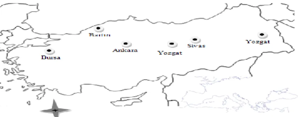
Ankara/Beypazarı. Details of the sampling period and the distribution of serum samples collected from these regions of Turkey is represented in Table 1 and Figure 1.
Table 1. Distribution of examined rodent, sheep blood serum and water samples according to the residental area.
Tablo 1. İncelenen rodent, koyun kanı ve su materyallerinin yerleşim birimlerine göre dağılımı.
Region Rodent Sheep Blood
Serum Water Van 3 - - Ankara-Beypazarı 3 101 4 Ankara-Altındağ - 46 - Bursa M. Kemalpaşa 10 159 - Zonguldak Kurtköy 3 20 - Ankara- Bala 7 75 - Sivas Gürün 9 - - Yozgat 5 - - Total 40 401 4
Strains and Reagents: For isolation of Francisella tularensis from rodent organs and from water, antibiotic
Francis medium was used. The medium was composed of brain heart infusion agar, dextrose (Difco) 1%, L-Cystein (Aldrich) 0.1%, sheep blood 8-9%, antibiotics (penicillin G 1ml/100 IU, Cyclohexamide L/100mg, Polymyxin B l/8x104) and Helicobacter supplement (8).
Francisella tularensis subsp. holarctica (NCTC
10857) strain was used as the positive control in the identification of the strains isolated from cultures, for testing the media and for molecular examinations of rodent and water samples. Francisella tularensis subsp.
tularensis (SCHU S4), another positive control strain,
was used for the subspecies determination in molecular examination. A tularemia antigen stained with safranine O which was prepared from Francisella tularensis subsp.
holarctica (NCTC 10857) vaccine strain was used in the
microagglutination test. Francisella tularensis antiserum was used as positive control (at 1/160 titer) in lam agglutination test, microagglutination test and in verifying the replicating colonies.
Primers used for determining the species and subspecies of the agents isolated are listed in Table 2 and 3.
Isolation: Isolation of the agents from the rodent and water samples: In order to detect the rodents that
were bacteremic with F. tularensis, liver and spleen samples from 40 rodents were inoculated into the Francis medium supplemented with antibiotics and defibrinated sheep blood. After inoculation processes, dishes were incubated at 37˚C for 10 days under 10% CO2
environment. Colony morphology, physical features and development times of forming colonies were evaluated (5,8). In order to isolate the agent from water, samples
Figure. 1. Distribution of serum samples collected from various regions of Turkey. Şekil 1. Türkiye’nin farklı bölgelerinden toplanan serum örneklerinin dağılımı.
Table 2. The nucleotide composition of primers, the specific gene areas they amplify, and the length of the PCR products. Tablo 2. Primerlerin baz dizileri bağlandıkları spesifik gen bölgeleri, PCR ürünlerinin uzunlukları.
Primer Name Primer Sequence Target gene Length of product Refererences
Tul 4-435 (F) 5`-GCT GTA TCA TCA TTT AAT AAA CTG CTG-3'
tul 4 420 bp [16]
Tul 4-863 (R) 5`-TTG GGA AGC TTG TAT CAT GGC ACT-3'
Table 3. Primer base groups and specific genes they amplify, PCR product lengths for the sub-species examinations used. Tablo 3. Alt tür tayininde kullanılan primerlerin baz dizileri bağlandıkları spesifik gen bölgeleri, PCR ürünlerinin uzunlukları.
Primer Name Primer Sequence Target gene Length of product References
RD1 (F) 5′-TTT ATA TAG GTA AAT GTT TTA CCT GTA CCA-3′
rd1 924 bp [3]
taken from 4 different sources at a volume of 500 ml were used. The water samples were filtered through a sterile cellulose acetate membrane filter (0.20 μm pore diameter). After filtration process, the filter was placed on the antibiotic and defibrinated sheep-blood-supplemented Francis medium with the inoculated side upwards (16). Colonies growing at the end of the incubation period were subjected to lam agglutination test with F. tularensis positive immune serum and confirmation of the isolates were performed with molecular techniques.
Microagglutination test: Francisella tularensis
specific agglutinins were examined by using
microagglutination test (MAT) with antigen (0.004% including spleen-O) prepared with F. tularensis strains (NCTC 10857) (5). First, 40 µl saline buffer was put into the first well of the U based plate and 25 µl of saline put into the other 6 wells of the plate. 25 µl of positive serum against tularemia was put into the 8th well (positive control 1/160 titer) and 10 µl of test serum (TS) into the first well. Next, 25 µl was transferred from the first well to the other. This dilution process performed till the 6th dilution. Last, 25 µl of dilute was taken out from the 6th well and dilution was completed. The 7th well did not receive any serum (negative control). Then all of the wells received 25 µl of stained antigen, thus 1/10-1/640 dilutions were prepared. Test plate was put into a humidified box and placed into the incubator. After incubation overnight at 37˚C, evaluation of the test was performed according to the positive, negative and antigen controls (4,5).
PCR analysis: For the purpose of molecular
diagnosis, conventional PCR (Polymerase Chain
Reaction) was used in water samples, rodent liver and spleen samples and to confirm isolates. Extraction, amplification, electrophoresis and visualization operations were performed respectively (8).
In order to confirm F. tularensis with molecular techniques; a two stage PCR method was used to define
the species and subspecies of the samples. First, the tul4 gene encoding a 17 kDa membrane protein was studied in order to direct the detection of genetic material from rodent samples and verify the culture-positive water samples. The band seen at 420 bp indicated Francisella spp. PCR (+) (13). Next, for differentiation of subspecies of F. tularensis, The Region of Difference 1 (RD1) target gene sequence was amplified from the culture positive water samples (3,8,13).
The serological and molecular studies of this research has been performed at the BSL-2 laboratory of Turkish Public Health Institution Administrative whereas isolation studies accomplished at the BSL-3 laboratory of the same Administrative.
Results
Bacterial isolation from rodent and water samples:
In total, 40 rodent samples were collected from 6 residential areas including Ankara Beypazarı, Sivas Gürün, Yozgat, Van and Bursa. In addition, 4 water samples were collected from Beypazarı (Ankara) to perform culture analysis. While no positive samples were detected by culture or PCR tests from rodents, one positive sample was detected from the 4 water samples and this was confirmed by PCR. Suspected colonies growing after incubation in culture media from water sample and isolation in Francis media were subjected to lam agglutination test with tularemia immune serum and this verified the presence of F. tularensis spp.
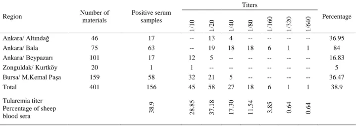
Serological Results: Serologic microagglutination
tests of 401 blood sera collected from 5 different regions (Ankara Bala, Beypazarı, Altındağ, Zonguldak Kurtköy, Bursa Mustafa Kemal Paşa) revealed the following results: Of the 401 sera, in 156 (38.9%) the antibody titer was between 1/10 and 1/640 while 111 (27.6%) exhibited 1/20 antibody titer which was also interpreted as a positive tularemia titer (Table 4).
PCR Results: Francisella spp. specific DNA could
not be detected in rodent samples. The results were in
Table 4. Results of microagglutination tests in sheep blood sera. Tablo 4. Koyun kan serumlarının mikroaglutinasyon test sonuçları.
Region Number of materials Positive serum samples Titers Percentage 1 /1 0 1 /2 0 1 /4 0 1 /8 0 1 /1 6 0 1 /3 2 0 1 /6 4 0 Ankara/ Altındağ 46 17 -- 13 4 -- -- -- -- 36.95 Ankara/ Bala 75 63 -- 19 18 18 6 1 1 84 Ankara/ Beypazarı 101 17 12 5 -- -- -- -- -- 16.83 Zonguldak/ Kurtköy 20 1 1 -- -- -- -- -- -- 5
Bursa/ M.Kemal Paşa 159 58 32 21 5 -- -- -- -- 36.47
Total 401 156 45 58 27 18 6 1 1 38.9 Tularemia titer Percentage of sheep blood sera 3 8 .9 2 8 .8 5 3 7 .1 8 1 7 .3 0 1 1 .5 4 3 .8 5 0 .6 4 0 .6 4
agreement with the culture findings. From the 4 water samples, 1 sample was positive in culture, gender-specific (tul4), after the primers went through PCR 420 bp of the band was seen so it indicated the presence of a
F. tularensis spp. (Fig 2). a band at the 924bp area
verifying that agent was F. tularensis subspecies
holarctica. Thus, culture isolation of water sample was
confirmed with PCR (Fig 3).
Figure 2. The image after PCR on the culture positive water samples of the gained agarose gel.
Resim 2. Kültür pozitif su örneğinin PCR sonrası elde edilen agaroz jel görüntüsü.
Fig 3. The image showing agarose gel after PCR for F.tularensis sub-species identification.
Resim 3. PCR sonrası F.tularensis alttür ayrımını gösteren agaroz jel görüntüsü.
Discussion and Conclusion
The natural reservoirs for tularemia are mostly rodents. The disease is fatal for these animals when they are infected. Sheep, goats, cattle and other farm animals in addition to cats, dogs, horses and other pets are random hosts for the bacterium. In this study, existence of F. tularensis in sheep and other potential reservoirs was investigated. Furthermore, water samples were examined for isolation purposes.
A study by Arata et al. (1) on a variety of animals in Iran, analyzed 3548 spleen samples collected from 4600 wild animals potentially associated with tularemia. No F.
tularensis isolates were obtained from any sample, but
the agglutination tests of sera from 100 sheep, 100 cattle and 39 wild mammals, revealed a positive reaction in 3 cattle, 8 sheep and a hedgehog. Özsan et al. (12), reported no isolation of F. tularensis from 1379 wild
animal samples. However, when they injected liver and spleen suspensions prepared from wild animals into guinea pigs, they observed seropositivity between 1/40-1/80 by agglutination tests. In the light of these studies, they concluded that these animals might be the potential hosts for F. tularensis.
In our study, all isolation and PCR analyses were negative. In total, 40 spleen and liver samples collected from rodents in various locations were analyzed. While collecting rodent samples, it was attempted to sample from locations where tularemia in humans has been reported. During the study, the information that the number of rodents had increased was received from residents. In agreement with previous reports, we also had difficulties to directly isolate F. tularensis from the animal samples (15).
The first water-based tularemia isolate was made by Karpoff Antoroff (9) in Russia. Somewhat later, Hussein Oz (6) performed the first isolation from Turkey. Even though it is unclear how the bacterium contaminates water, it is speculated that the water is contaminated with the agent from rodent carcasses or other contaminated materials (2). Since the pollution in water is sudden and highly diluted and due to late sampling time, the chance of agent isolation is low from water. During our study, information of an epidemic from Ankara Beypazarı location was obtained. According to this information, 4 water samples were taken from this location and isolation and conventional PCR studies were conducted. At the end of the tests, one water sample of the 4 was found to be positive by culture isolation. This isolate was confirmed by conventional PCR technique revealing that the agent was F. tularensis subsp. holarctica. This result is important and demonstrates that the agent can be spread by water.
In a study conducted in United States, Jellison and Kohls (7) detected an antibody titer above 1/20 in 25% of sera from 148 lambs and accepted this titer as positive for
F. tularensis. The researchers conducted another study
involving 283 ewes returning from grassland and found 28.3% seropositivity.
In the present study, in order to investigate tularemia in sheep serologically, blood samples were collected from sheep in the locations where disease had been observed in humans. Samples were collected from 401 sheep from Ankara-Altındağ, Ankara-Beypazarı,
Ankara-Bala, Bursa-Mustafakemalpaşa,
Zonguldak-Kurtköy districts. Of the 401 sera, 156 (38.9%) had antibody titers between 1/10 and 1/640 while 111 (27.6%) exhibited 1/20 antibody titer which were also accepted as indicative of tularemia. O'Toole et al. (10) studied the existence of F. tularensis in 3 different local herds of sheep between 1997 and 2007. The report describes 4 periods of tularemia in these herds. Herd owners reported that ticks were unusually numerous and
often present on the sheep during outbreaks. Tularemia presented as late-term abortions and death in lambs and, to a lesser extent, in ewes. The diagnosis was corroborated by bacterial isolation and, in individual cases, by serology, and PCR detection of F. tularensis. These studies represents an example that indicate the association of tularemia between humans and sheep. Also, it indicates that the bacterium can be transmitted to sheep from ticks. Parallel to these findings, during our study in Ankara Beypazarı region Macunköy countryside, an epidemic of tularemia broke out in humans. From the area 2 rodents, 4 water samples from a public use, and 90 blood samples from sheep were taken. The agent was not isolated from the rodents, but on the other hand, while there was a positivity in water at this location, low antibody titers were detected in sheep that were drinking the water of the location. These results suggested that the possible transmission routes of the agent might be ticks rather than water.
Among the selected areas, a positive tularemia titer was observed in of 84% of the sera was found in Bala region as highest. This percentage is the highest seropositivity ratio in lambs ever seen in Turkey. From this perspective, our study is thought to be important and therefore represents an indication of the widespread distribution of sheep to the disease, detection of disease level and identifies the association of tularemia between human and sheep. Şeyda (14), analyzed 1600 sheep sera from the Kars region by microagglutination tests and detected 1/20 or higher antibody titers in 50 sheep.
In this study, the existence of F. tularensis was investigated by culture and PCR in rodents and water and by serological methods in sheep in locations selected in parallel with human tularemia cases.
Since we have studied several aspects of the tularemia epidemiology including several residential areas, this study is the first example in veterinary medicine in Turkey. In addition, serological results obtained are among the highest agglutination titers observed in sheep anywhere in the world. However, it is not possible to explain how the disease is transmitted in the areas with high titers, without identifying the reservoirs and potential vectors of the bacterium. The isolation of the agent has not been successful in studies of rodents. In rodents, after the entry of the organism into the body, mortality is likely to occur in a short time. In wildlife, it is unlikely to find a dead rodent, and in materials from live and healthy rodents caught, the chance of isolation is very low as reported from previous studies. It is not possible to suggest that rodents are not potential reservoirs because there are no isolates. Water-associated positivity was found in areas where tularemia outbreaks took place. In this extensive study, the importance of these topics were emphasized by isolation trials from rodent and water samples, PCR tests and in
sheep by serological findings. A parallelism was observed in findings in isolation and PCR carried out. Therefore, the determination of the existence of the agent during a short time in the samples to be studied with PCR is very important.
In the studies to be carried out in future, periodical checks of samples from selected pilot locations, routine rodent control activities, and examination of humans seems to be important. Furthermore, considering water-associated transmission, concurrent studies in humans, animals and water will be very important in the epidemiological evaluation of tularemia. At the end of the study, since paralel findings was observed by both isolation and PCR methods, it can be concluded that it would be important for rapid diagnosis of the existence of agent in samples to be examined by PCR. In addition, serological findings exhibit the highest agglutination titers in sheep ever known worldwide researches. With these findings, it is aimed to emphasize the importance of tularemia which is emerging as a popular zoonotic disease.
Acknowledgements
I would like to express my gratitude to Turkish Public Health Institution Administrative staff (TPHI, Ankara) for providing standard strains, antigens and antisera and to Veterinary Control and Central Research Institute (VCCRI, Ankara) where the study carried out. This study was a part of corresponding author's Doctoral Dissertation, which supported by the TR Ministry of
Food, Agriculture and Animal Husbandry
(#TAGEM/HSYGAD/12/A02/P02/01).
References
1. Arata M, Chamsa A, Farhang-azad, Meščerjakova I, Neronov V, Saidi S (1973): First detection of tularemia in
domestic and wild mammals in Iran. Bull Wld Hlth Org,
49, 597-603.
2. Bow MR, Brown JH (1944): Water-Borne Tularemia in
Western Canada. Can MAJ, 50, 14-16.
3. Broekhuijsen M, Larsson P, Johansson A (2003):
Genome-wide DNA microarray analysis of Francisella tularensis strains demonstrate extensive genetic conservation within the species but identifies regions that are unique to the highly virulent F.tularensis subsp. tularensis. J Clin Microbiol, 41, 2924-2931.
4. Brown SL, Mckinney FT, Klein GC, Jones, WL (1980):
Evaluation of safranin-O stained antigen microagglutination test for Francisella tularensis antibodies. J Clin Microbiol, 11, 146-148.
5. Gürcan Ş (2009): Francisella tularensis and tularemia. In: Serology. Nobel Tıp Press, Turkey, İstanbul, pp, 161-168. 6. Husseın Y K, Oz T AV (1938): Turkische Zeitsch. F Hyg
und Exp Biol, Chapter 1.
7. Jellıson WL, Kohls GM (1950): Persistence of agglutinins
against Pasteurella tularensis in serums of naturally infected sheep. J Am Vet Med Assoc, 117,405-408.
8. Johansson A, Petersen J, Sjöstedt A (2007): Laboratory
diagnostics and discrimination of subspecies and strains
In: WHO Guidlines On Tularemia, pp, 27-34.
9. Karpoff S P, Antonoff N I (1936): The Spread Of
Tularemia Through Water, As A New Factor in its Epidemiology. J Bact, 32, 243.
10. O’toole D, Williams E S, Woods LW, Mills K, Boerger-Fıelds A, Montgomery DL, Jaeger Edwards WH, Christensen D, Marlatt W (2008): Tularemia in range
sheep: an overlooked syndrome? J Vet Diagn Invest, 20,
508-513.
11. Oyston CF (2008): Francisella tularensis: unravelling the
secrets of an intracellular pathogen. Journal of Medical
Microbiology, 57,921–930
12. Özsan K, Fazlı A, Aktan M, Beyoğlu K, (1976):
Brucellosis, tularemia and borreliosis isolated from wild animals captured in Ankara, Konya, Urfa and Nevsehir provinces in Turkey. Mikrobiyol Bul, 10,414-421.
13. Sjöstedt A, Kuoppa K, Johansson T, Sandström G (1992): The 17 kDa lipoprotein and encoding gene of
Francisella tularensis LVS are conserved in strains of Francisella tularensis. Microb Pathog, 13, 243–249.
14. Şeyda T (1996): The serological and culturel studies on the incidence of Tularemia in founded in Kars area. Kafkas Üniv Veteriner Fak Derg, 2,49-60.
15. Tekin M, Çam O H, Acar G, Kaytancı E, Hanege F M (2009): Tularemia case with a neck mass. Göztepe Tıp Dergisi, 26,46-50.
16. Ulu Kılıç A, Kılıç S, Şencan İ, Şentürk GÇ, Gürbüz Y, Tütüncü E E, Çelebi B, Kıcıman Ö, Ergönül Ö (2011):
A water-borne tularemia outbreak caused by Francisella tularensis subspecies holarctica in Central Anatolia Region. Mikrobiyol Bul, 45,234-247.
Geliş tarihi: 26.08.2014/ Kabul tarihi: 17.11.2014
Address for correspondence:
Dr. Derya Karataş Yeni
Etlik Veterinary Control Central Research Institute, Laboratory of Bakteriology,
Etlik, Ankara, Turkey.